This document discusses rapidly progressive glomerulonephritis (RPGN) and crescentic glomerulonephritis. It describes the main immunopathologic categories of crescentic glomerulonephritis, which include immune complex crescentic glomerulonephritis, anti-glomerular basement membrane (GBM) disease, and pauci-immune crescentic glomerulonephritis associated with antineutrophil cytoplasmic antibodies (ANCA). Immune complex crescentic glomerulonephritis is most common in children, while pauci-immune crescentic glomerulonephritis is most common in adults. The pathology involves formation of
1. CHAPTER 31 — PRIMARY GLOMERULAR DISEASE 1071
inflammation (see Fig. 31.30). This pathologic feature can
be seen on light, IF, and electron microscopy.1127–1129
It is the
result of focal rupture of glomerular capillary walls that allows
inflammatory mediators and leukocytes to enter Bowman’s
space, where they induce epithelial cell proliferation and
macrophage influx and maturation, which together produce
cellular crescents (Fig. 31.34).1130–1132
Kidney diseases other than crescentic glomerulonephritis
can cause the signs and symptoms of RPGN. Two examples
are acute thrombotic microangiopathy and atheroembolic
kidney disease. Although acute tubular necrosis and acute
tubulointerstitial nephritis may cause rapid loss of kidney
function and oliguria, these processes typically do not cause
dysmorphic erythrocyturia, erythrocyte cylindruria, or sub-
stantial proteinuria.
A small minority of all patients with glomerulonephritis
develops RPGN, except patients with anti-GBM disease and
ANCA disease who have a high frequency of crescents. The
incidence of rapidly progressive glomerulonephritis has been
estimated to be as low as 7 cases/million population per
year.675,1133
The three major immunopathologic categories of
crescentic glomerulonephritis have different frequencies in
different age groups (Table 31.8).1126–1128,1134
In a patient who
has RPGN clinically and in whom crescentic glomerulone-
phritis is identified by light microscopy in a kidney biopsy
specimen, the precise diagnostic categorization of the disease
requires the integration of clinical, serologic, immunohisto-
logic, and electron microscopic data (Fig. 31.35).
Immune complex crescentic glomerulonephritis is caused
by immune complex localization within glomeruli. It is the
most common cause of RPGN in children (see Table 31.8).1127
The major clinical differential diagnosis in children is
hemolytic uremic syndrome, which also can cause rapid loss
of kidney function, hypertension, hematuria, and proteinuria.
glomerulonephritis. In regard to causative factors, patients
with immunotactoid glomerulonephritis were statistically more
likely to have an underlying lymphoproliferative disease, a
monoclonal spike on serum protein electrophoresis, and
hypocomplementemia.1118
Fibrillary glomerulonephritis with associated pulmonary
hemorrhage has been reported anecdotally.1119
One patient
with immunotactoid glomerulopathy also had extrarenal
deposits in both the liver and bone.1120
TREATMENT
At this time, there is no convincingly effective form of treat-
ment for patients with fibrillary glomerulonephritis or
immunotactoid glomerulopathy.1105
The dismal prognosis in
patients with either of these diseases has prompted physicians
to search for some immunosuppressive form of treatment.
Fully 40% to 50% of patients with these diseases develop
ESKD within 6 years of presentation.1097,1098,1100,1102
Efforts at
treatment with glucocorticoids or alkylating agents such as
cyclophosphamide have typically shown no response or, at
best, some amelioration of proteinuria.1121
In our experience,
prednisone therapy alone has had no benefit. One small
case series (three patients) reported significant improvement
in proteinuria in response to rituximab, either alone or in
combination with corticosteroids, or tacrolimus.1122
In fibrillary
glomerulonephritis and other forms of glomerulonephritis
associated with chronic lymphocytic leukemia or other forms
of lymphocytic lymphoma, there has been a report of improve-
ment in a minority of patients treated with chlorambucil.
Thus, it is possible that the treatment of the underlying
malignancy, if present, may improve the glomerulonephri-
tis.1106
Of note, a recent case series and review of previously
published large case series have revealed a role for rituximab
in patients with fibrillary glomerulonephritis if administered
early in the disease process with a preserved eGFR.1123
The recurrence rate of fibrillary glomerulonephritis after
kidney transplantation is unclear. One report has described
recurrent disease in three of four patients who had received
five transplants.1124
In a larger case series, recurrent disease
occurred in none of five patients with fibrillary glomerulo-
nephritis, but in five of seven patients with monoclonal
gammopathy and fibrillary deposits.1125
RAPIDLY PROGRESSIVE
GLOMERULONEPHRITIS AND
CRESCENTIC GLOMERULONEPHRITIS
NOMENCLATURE AND CATEGORIZATION
The term “rapidly progressive glomerulonephritis” (RPGN)
refers to a clinical syndrome characterized by a rapid loss of
kidney function, often accompanied by oliguria or anuria
and features of glomerulonephritis, including dysmorphic
erythrocyturia, erythrocyte cylindruria, and glomerular
proteinuria.1126
Aggressive glomerulonephritis that causes
RPGN usually has extensive crescent formation.1127
For this
reason, the clinical term “rapidly progressive glomerulone-
phritis” is sometimes used interchangeably with the pathologic
term “crescentic glomerulonephritis.” Crescentic glomeru-
lonephritis is the most aggressive structural phenotype in
the continuum of injury that results from glomerular
Fig. 31.34 Light micrograph showing a cellular crescent in Bowman’s
space. The underlying glomerular tuft is delineated by the glomerular
basement membranes. (Periodic acid–Schiff stain, ×500.)
2. 1072 SECTION V — DISORDERS OF KIDNEY STRUCTURE AND FUNCTION
Table 31.8 Relative Frequency of Immunopathologic Categories of Crescentic Glomerulonephritis In Different
Age Groups (%)a
Immunopathologic Category
Age in Years
All Ages
(n = 632)
1−20
(n = 73)
21−60
(n = 303)
>60
(n = 256)
Antiglomerular basement membrane CGN 15 12 15 15
Immune complex CGN 24 45 35 6
Pauci-immune CGNb
60 42 48 79
Other 1 0 30 0
a
CGN is defined as the presence of crescents in >50% of glomeruli. Frequency is determined with respect to age in patients whose kidney
biopsy specimens were evaluated at the University of North Carolina Nephropathology Laboratory. Note the very high frequency of
pauci-immune disease (usually antineutrophil cytoplasmic antibody [ANCA]−associated) in older adults.
b
Approximately 90% associated with ANCA.
CGN, Crescentic glomerulonephritis.
Data from Jennette JC, Nickeleit V: Anti-glomerular basement membrane glomerulonephritis and Goodpasture’s syndrome. In Jennette
JC, Olson JL, Silva FG, D’Agati V, eds. Heptinstall’s pathology of the kidney. 7th ed. Wolters Klewer: Philadelphia; 2015:657−684.
Immunoglobulin- and Complement-Mediated Glomerulonephritis
Anti-GBM disease Immune complex GN C3 glomerulopathy ANCA disease
ANCA GN MPA
DDD C3 GN
Anti-GBM
GN
Goodpasture’s
syndrome
GPA EGPA
Immune
complex MPGN
Membranous
GN
Lupus
nephritis
IgA
vasculitis
IgA
nephropathy
Postinfectious
acute GN
Fibrillary
GN
Other immune
complex GN
Linear GBM
immunoglobulin
IF staining
Granular IF staining
for immunoglobulin
and C3
Granular IF staining
for C3 and little or
no immunoglobulin
Paucity of IF staining
for immunoglobulin
and complement
No
systemic
vasculitis
Vasculitis
with no
granulomas,
or asthma
Dense
deposits
in GBMs
No dense
deposits
in GBMs
No lung
hemorrhage
Lung
hemorrhage
Vasculitis with
granulomas,
no asthma
Vasculitis
with
granulomas
and asthma
Thick
capillaries and
hypercellularity
Thick
capillaries, no
hypercellularity
Systemic
lupus
erythematosus
Dominant
IgA and
vasculitis
Dominant
IgA and no
vasculitis
Acute strep
or staph
infection
~20 nm
fibrils
Other
features
Fig. 31.35 Algorithm for the diagnostic classification of glomerulonephritis that is known or suspected of being mediated by antibodies and
complement. Note that the integration of light microscopy, immunofluorescence (IF) microscopy, electron microscopy, laboratory data, and
clinical manifestations is required to diagnose glomerulonephritis (GN) precisely. ANCA, Antineutrophil cytoplasmic autoantibody; DDD, dense
deposit disease; EGPA, eosinophilic granulomatosis with polyangiitis; GBM, glomerular basement membrane; GPA, granulomatosis with
polyangiitis; MPA, microscopic polyangiitis.
3. CHAPTER 31 — PRIMARY GLOMERULAR DISEASE 1073
PATHOLOGY
Light Microscopy
The light microscopic appearance of crescentic immune
complex glomerulonephritis depends on the underlying
category of glomerulonephritis. For example, in their most
aggressive expressions, MPGN, acute postinfectious glomeru-
lonephritis, or proliferative glomerulonephritis, including
IgA nephropathy, can all have crescent formation.*
This underlying phenotype of immune complex glomeru-
lonephritis is recognized best in the intact glomeruli or
glomerular segments. Immune complex–mediated glomeru-
lonephritis and C3 glomerulopathy usually have varying
combinations of capillary wall thickening and endocapillary
hypercellularity in the intact glomeruli. This is in contrast
to anti-GBM glomerulonephritis and ANCA glomerulone-
phritis, which tend to have surprisingly few alterations in
intact glomeruli and segments, in spite of the severe necrotiz-
ing injury in involved glomeruli and segments. In glomerular
segments adjacent to crescents in immune complex glomeru-
lonephritis, there usually is some degree of necrosis with
karyorrhexis; however, the necrosis is rarely as extensive as
that typically seen with anti-GBM or ANCA glomerulonephritis.
In addition, there is less destruction of Bowman’s capsule
associated with crescents in immune complex glomerulone-
phritis, as well as less pronounced periglomerular tubuloin-
terstitial inflammation. Crescents in immune complex
glomerulonephritis have a higher proportion of epithelial
cells to macrophages than crescents in anti-GBM or ANCA
glomerulonephritis, which may be related to the less severe
disruption of Bowman’s capsule and thus less opportunity
for macrophages to migrate in from the interstitium.1138
Immunofluorescence Microscopy
IF microscopy, as well as electron microscopy, provides the
evidence that crescentic glomerulonephritis is immune
complex-mediated or complement-mediated versus anti-GBM
The presence of microangiopathic hemolytic anemia and
thrombocytopenia are indicators that the rapid loss of kidney
function is more likely caused by hemolytic uremic syndrome
than crescentic glomerulonephritis. Pauci-immune crescentic
glomerulonephritis, which shows little or no evidence of the
localization of immune complex or anti-GBM antibodies in
glomeruli, is usually associated with the presence of ANCAs
and is the most common cause for RPGN and crescentic
glomerulonephritis in adults, especially older adults (Table
31.9; see Table 31.8).1126,1134–1136
In most patients, pauci-immune
crescentic glomerulonephritis is a component of a systemic
small-vessel vasculitis, such as GPA or MPA; however, some
patients have renal-limited (primary) disease.1127,1137
Anti-GBM
disease is the least frequent cause of crescentic glomerulo-
nephritis (see Tables 31.8 and 31.9).1126,1127,1134,1135
IMMUNE COMPLEX–MEDIATED AND C3
GLOMERULOPATHY CRESCENTIC
GLOMERULONEPHRITIS
EPIDEMIOLOGY
Most patients with immune complex–mediated crescentic
glomerulonephritis have clinical or pathologic evidence of
a specific category of primary glomerulonephritis, such as
IgA nephropathy, postinfectious glomerulonephritis, or
MPGN, or they have glomerulonephritis that is a component
of a systemic immune complex disease, such as SLE, cryo-
globulinemia, or IgA vasculitis. A minority of patients with
immune complex crescentic glomerulonephritis, however,
do not have patterns of immune complex localization that
readily fit into these specific categories of immune complex
glomerulonephritis.1138
Immune complex crescentic glomerulonephritis accounts
for most crescentic glomerulonephritides in children, but
for only a minority of crescentic glomerulonephritis in older
adults (see Table 31.8). The higher frequency in children
and young adults reflects a similar trend in other types of
immune complex glomerulonephritides, such as IgA
nephropathy, PSGN, MPGN, DDD, and lupus nephritis.
Table 31.9 Frequency of Immunopathologic Categories of Glomerulonephritis in Kidney Biopsy Specimens
Evaluated by Immunofluorescence Microscopya
Immunohistology
All Proliferative
Glomerulonephritis
(n = 1093)
Any Crescents
(n = 540)
>50% Crescents
(n = 195)
Arteritis in Biopsy
(n = 37)
Pauci-immune (<2+ Ig) 45% (496/1093) 51% (227/540) 61% (118/195)b
84% (31/37)
Immune complex (≥2+ Ig) 52% (570/1093) 44% (238/540) 29% (56/195) 14% (5/37)c
Anti-GBM 3% (27/1093) 5% (25/540)d
11% (21/195) 3% (1/37)e
a
Based on the analysis of over 3000 consecutive nontransplant renal biopsy specimens evaluated at the University of North Carolina
Nephropathology Laboratory.
b
Of 77 patients, 70 (91%) tested positive for antineutrophil cytoplasmic antibody (ANCA), (44 for perinuclear ANCA [P-ANCA] and 26
cytoplasmic ANCA [C-ANCA]).
c
Four patients had lupus and one had poststreptococcal glomerulonephritis.
d
Three of 19 patients (16%) tested positive for ANCA (2 for P-ANCA and 1 for C-ANCA).
e
This patient also tested positive for P-ANCA (myeloperoxidase ANCA).
GBM, Glomerular basement membrane; IF, immunofluorescence.
Modified from Jennette JC, Nickeleit V: Anti-glomerular basement membrane glomerulonephritis and Goodpasture’s syndrome. In
Jennette JC, Olson JL, Silva FG, D’Agati V, eds. Heptinstall’s pathology of the kidney. 7th ed. Wolters Klewer: Philadelphia;
2015:657−684.
*References 359–363, 697, 703, 831, 1058, 1133, and 1139.
4. 1074 SECTION V — DISORDERS OF KIDNEY STRUCTURE AND FUNCTION
mesangial cells. The activated cells also release soluble media-
tors, such as cytokines and chemokines. If the resultant inflam-
mation is contained internally to the GBM, a proliferative
or membranoproliferative phenotype of injury ensues, with
only endocapillary hypercellularity. However, if the inflam-
mation breaks through capillary walls into Bowman’s space,
extracapillary hypercellularity (crescent formation) results.
Complement activation has often been considered a major
mediator of injury in immune complex glomerulonephritis;
however, experimental data also indicate the importance of
Fc receptors in immune complex-mediated injury.1141,1142
For
example, mice deficient for the Fcγ R1 and Fcγ RIII receptors
have a markedly reduced tendency to develop immune
complex glomerulonephritis.1143,1144
TREATMENT
The therapy for crescentic immune complex glomerulone-
phritis is influenced by the nature of the underlying category
of immune complex glomerulonephritis. For example, acute
PSGN with 50% crescents might not prompt the same therapy
as IgA nephropathy with 50% crescents. However, there have
been an inadequate number of controlled prospective studies
to guide therapy for most forms of crescentic immune complex
glomerulonephritis. Some nephrologists extrapolate from
the lupus nephritis experience and choose to treat patients
with crescentic immune complex disease with immunosup-
pressive drugs that they would not use if the glomerular
lesions appeared less aggressive. For the minority of patients
who have idiopathic immune complex crescentic glomeru-
lonephritis, the most common treatment is immunosuppres-
sive therapy with pulse methylprednisolone, followed by
prednisone at a dosage of 1 mg/kg daily tapered over the
second to third month to an alternate-day regimen until
completely discontinued.675,1145–1147
In patients with a rapid
decline in kidney function, cytotoxic agents, with or without
plasma exchange, in addition to corticosteroids, may be
considered. As with anti-GBM and ANCA disease, immuno-
therapy should be initiated as early as possible during the
course of crescentic immune complex glomerulonephritis
to reduce the likelihood of reaching the irreversible stage
of advanced scarring. There is evidence, however, that
crescentic glomerulonephritis with an underlying immune
complex proliferative glomerulonephritis is less responsive
to aggressive immunosuppressive therapy than is anti-GBM
or ANCA crescentic glomerulonephritis.1058,1138
ANTI–GLOMERULAR BASEMENT MEMBRANE
GLOMERULONEPHRITIS
EPIDEMIOLOGY
Anti-GBM disease accounts for about 10% to 20% of crescentic
glomerulonephritides.675
This disease is characterized by
circulating antibodies to the GBM (anti-GBM) and deposition
of IgG or, rarely, IgA along the GBM (see also Chapter
32).675,1138,1148–1160
Anti-GBM antibodies may be eluted from
kidney tissue samples from patients with anti-GBM disease,
which allows verification that the antibodies are specific to
the GBM.675,1154,1158
The antibodies eluted from kidney tissue
bind to the same epitope of type IV collagen as the circulating
anti-GBM antibodies from the same patient.1161
Anti-GBM disease occurs as a renal-limited disease (anti-
GBM glomerulonephritis) and as a pulmonary-renal vasculitic
antibody-mediated or ANCA-mediated. The pattern and com-
position of immunoglobulin and complement staining depend
on the underlying category of immune complex glomerulo-
nephritis or C3 glomerulopathy that has induced crescent
formation.360,702,1140
For example, crescentic glomerulonephritis
with predominantly mesangial IgA-dominant deposits is indica-
tive of crescentic IgA nephropathy, C3-dominant deposits
with peripheral bandlike configurations suggest crescentic
MPGN, coarsely granular capillary wall deposits raise the
possibility of crescentic postinfectious glomerulonephritis, and
finely granular IgG-dominant capillary wall deposits suggest
crescentic MN. The latter may be a result of concurrent
anti-GBM disease, which also causes linear GBM staining
beneath the granular staining, or concurrent ANCA disease,
which can be documented serologically. About 25% of all
patients with crescentic immune complex glomerulonephritis
are ANCA-positive, whereas fewer than 5% of patients with
noncrescentic immune complex glomerulonephritis are ANCA
positive. This suggests that the presence of ANCAs in patients
with immune complex glomerulonephritis may predispose
to a disease that is more aggressive.
Electron Microscopy
As with the findings by IF microscopy, the findings by electron
microscopy in patients with crescentic immune complex
glomerulonephritis depend on the type of immune complex
disease that has induced crescent formation. The hallmark
ultrastructural finding is immune complex– type, electron-
dense deposits. These can be mesangial, subendothelial,
intramembranous, subepithelial, or any combination of these.
The pattern and distribution of deposits may indicate a
particular phenotype of primary crescentic immune complex
glomerulonephritis, such as postinfectious, membranous,
membranoproliferative, or DDD.360,702,1140
Ultrastructural
findings also may suggest that the disease is secondary to
some unrecognized systemic process. For example, endothelial
tubuloreticular inclusions suggest lupus nephritis, and
microtubular configurations in immune deposits suggest
cryoglobulinemia.
In all types of crescentic glomerulonephritis, breaks in
GBMs usually can be identified if sought carefully, especially
in glomerular segments adjacent to crescents. Dense fibrin
tactoids occur in thrombosed capillaries, in sites of fibrinoid
necrosis, and in the interstices between the cells in crescents.
In general, the extent of fibrin tactoid formation in areas of
fibrinoid necrosis is less conspicuous in crescentic immune
complex glomerulonephritis than in crescentic anti-GBM or
ANCA glomerulonephritis.
PATHOGENESIS
Crescentic glomerulonephritis is the result of a final common
pathway of glomerular injury that results in crescent for-
mation. Multiple causes and pathogenic mechanisms can
lead to the final common pathway, including many types
of immune complex disease. The general dogma is that
immune complex localization in glomerular capillary walls
and mesangium, by deposition, in situ formation, or both,
activates multiple inflammatory mediator systems.211,1126,1127
This includes humoral mediator systems, such as the coagula-
tion system, kinin system, and complement system, as well
as inflammatory cells, such as neutrophils, monocytes and
macrophages, lymphocytes, platelets, endothelial cells, and
5. CHAPTER 31 — PRIMARY GLOMERULAR DISEASE 1075
anti-GBM glomerulonephritis have also been reported.1156,1178
Linear staining for both κ and λ light chains typically accom-
panies the staining for γ heavy chains. Linear staining for
γ heavy chains alone indicates γ heavy-chain deposition
disease. Most specimens with anti-GBM glomerulonephritis
have discontinuous linear to granular capillary wall staining
for C3, but a minority show little or no C3 staining. Linear
staining for IgG may also occur along distal tubular basement
membranes.1159
The linear IgG staining of GBMs frequently seen in patients
with diabetic glomerulosclerosis and the less intense linear
staining seen in older patients with hypertensive vascular
disease, must not be confused with that in anti-GBM disease.
Clinical data and light microscopic findings should help make
this distinction. Serologic confirmation should always be
obtained to substantiate the diagnosis of anti-GBM disease.
Serologic testing for ANCAs should be ordered simul-
taneously because one-quarter to one-third of patients
with anti-GBM disease are also ANCA-positive. This may
modify the prognosis and likelihood of systemic small-vessel
vasculitis.1179,1180
Light Microscopy
At the time of biopsy, 97% of patients with anti-GBM disease
have some degree of crescent formation, and 85% have
crescents in 50% or more of glomeruli (Table 31.10).1127,1174
On average, 77% of glomeruli have crescents. Glomeruli
with crescents typically have fibrinoid necrosis in adjacent
glomerular segments. Nonnecrotic segments may look entirely
normal by light microscopy or may have slight infiltration
by neutrophils or mononuclear leukocytes. This differs from
crescentic immune complex glomerulonephritis and C3
glomerulopathy, which typically have capillary wall thickening
and endocapillary hypercellularity in the intact glomeruli.
Special stains that outline basement membranes, such as
Jones’ silver methenamine or periodic acid–Schiff stain,
often demonstrate focal breaks in GBMs in areas of necrosis
and also show focal breaks in Bowman’s capsule. The most
severely injured glomeruli have global glomerular necrosis,
syndrome (Goodpasture syndrome).675,1138,1148–1160,1162
The
incidence of anti-GBM disease has two peaks with respect to
age. The first peak is in the 2nd and 3rd decades of life, and
anti-GBM disease in this age group shows a higher frequency
of pulmonary hemorrhage (Goodpasture syndrome). The
second peak is in the 6th and 7th decades, and this later
onset disease is more common in women, who more often
have renal-limited disease. Interestingly, anti-GBM autoanti-
bodies were detected in multiple serum samples from the
Department of Defense Serum Repository before diagnosis
in a case-control study involving 30 patients with anti-GBM
disease and 30 healthy controls (50% vs. 0%; P < .001),1163
which suggests the development of the autoimmune response
prior to onset of disease.
Genetic susceptibility to anti-GBM disease is associated
with HLA-DR2 specificity.1164
More detailed analysis of the
association with HLA-DR2 has revealed a link with the DRB1
alleles, DRB1*1501 and DQB*0602.1165–1169
Further refinement
of this association has shown that polymorphic residues in
the second peptide-binding region of the HLA class II antigen
segregated with disease, supporting the hypothesis that the
HLA association in anti-GBM disease reflects the ability of
certain class II molecules to bind and present anti-GBM
peptides to helper T (TH) cells.1165
This concept is further supported by mouse models of
anti-GBM disease in which crescentic glomerulonephritis
and lung hemorrhage are restricted to only certain major
histocompatibility complex (MHC) haplotypes, despite the
ability of mice of all haplotypes to produce antibodies to the
α3-NC1 (“noncollagenous”) domain of type IV collagen.1170
Analyses of gene expression in the kidneys of mouse strains
susceptible to anti-GBM antibody-induced nephritis, compared
with those of control strains, have revealed that 20% of the
underexpressed genes in these mice belonged to the kallikrein
gene family, which encodes serine esterases implicated in the
regulation of inflammation, apoptosis, redox balance, and
fibrosis.1171
Antagonizing the kallikrein pathway by blocking the
bradykinin receptors B1 and B2 augmented disease, whereas
bradykinin administration reduced the severity of anti-GBM,
antibody-induced nephritis in a susceptible mouse strain.
Nephritis-sensitive mouse strains had kallikrein haplotypes
that were distinct from those of control strains, including
several regulatory polymorphisms. These results suggest that
kallikreins are protective disease-associated genes in anti-
GBM antibody-induced nephritis.1171
Whether these findings
pertain to susceptibility to or severity of anti-GBM disease in
humans in unknown. It should also be noted that another
genetic association study of 48 Chinese patients with anti-GBM
disease compared with 225 matched healthy controls revealed
a genetic association of an FCγRIIB polymorphism (I232T)
with disease susceptibility.1172
This same polymorphism has
been identified in patients with SLE and is thought to alter
this inhibitory receptor responsible for the maintenance of
B cell tolerance and activation thresholds.1173
PATHOLOGY
Immunofluorescence Microscopy
The pathologic finding of linear staining of the GBMs for
immunoglobulin is indicative of anti-GBM glomerulonephritis
(Fig. 31.36).1155,1158,1159,1174–1177
The immunoglobulin is pre-
dominantly IgG; however, rare patients with IgA-dominant,
Fig. 31.36 Immunofluorescence micrograph of a portion of a glomerulus
with antiglomerular basement membrane (anti-GBM) glomerulonephritis
showing linear staining of GBMs for immunoglobulin G (IgG). (Fluorescein
isothiocyanate anti-IgG stain, ×500.)
6. 1076 SECTION V — DISORDERS OF KIDNEY STRUCTURE AND FUNCTION
circumferential cellular crescents, and extensive disruption
of Bowman’s capsule.
The acute necrotizing glomerular lesions and the cellular
crescents evolve into glomerular sclerosis and fibrotic cres-
cents, respectively.1174
If the kidney biopsy specimen is obtained
several weeks into the course of anti-GBM disease, the only
lesions may be these chronic sclerotic lesions. There may be a
mixture of acute and chronic lesions; however, the glomerular
lesions of anti-GBM glomerulonephritis tend to be more in
synchrony than those of ANCA glomerulonephritis, which
more often show admixtures of acute and chronic injury.
Tubulointerstitial changes are commensurate with the
degree of glomerular injury. Glomeruli with extensive necrosis
and disruption of Bowman’s capsule typically have intense
periglomerular inflammation, including occasional multi-
nucleated giant cells. There also is focal tubular epithelial
acute simplification or atrophy, focal interstitial edema and
fibrosis, and focal interstitial infiltration of predominantly
mononuclear leukocytes. There are no specific changes in
arteries or arterioles. If necrotizing inflammation is observed
in arteries or arterioles, the possibility of concurrent anti-GBM
and ANCA disease should be considered.
Electron Microscopy
The findings by electron microscopy reflect those seen by light
microscopy.1174,1181
In acute disease, there is focal glomerular
necrosis with disruption of capillary walls. Bowman’s capsule
also may have focal gaps. Leukocytes, including neutrophils
and monocytes, often are present at sites of necrosis, but are
uncommon in intact glomerular segments. Fibrin tactoids,
which are electron-dense curvilinear accumulations of polym-
erized fibrin, accumulate at the sites of coagulation system
activation, including sites of capillary thrombosis, fibrinoid
necrosis, and fibrin formation in Bowman’s space (Fig.
31.37). Cellular crescents contain cells with ultrastructural
Table 31.10 Frequency of Crescent Formation in Various Glomerular Diseasesa
Disease
Patients With
Crescents (%)
Patients With
≥50% Crescents
Average No. of Glomeruli
With Crescents (%)
Anti-GBM glomerulonephritis 97 85 77
ANCA-associated glomerulonephritis 90 50 49
Immune complex−mediated glomerulonephritis
Lupus glomerulonephritis (classes III and IV) 56 13 27
IgA vasculitis (formerly Henoch-Schönlein purpura
glomerulonephritis)b
61 10 27
IgA nephropathyb
32 4 21
Acute postinfectious glomerulonephritisb
33 3 19
Fibrillary glomerulonephritis 23 5 26
Type I membranoproliferative glomerulonephritis 24 5 25
Membranous lupus glomerulonephritis (class V) 12 1 17
Membranous glomerulonephritis (nonlupus) 3 0 15
a
Based on analysis of over 6000 native kidney biopsy specimens evaluated at the University of North Carolina Nephropathology Laboratory.
In general, diseases in which crescents are most often seen also have the largest percentage of glomeruli involved by crescents when
they are present.
b
Because more severe cases of immunoglobulin A nephropathy and postinfectious glomerulonephritis are more often evaluated by kidney
biopsy, the extent of crescent involvement is higher in the patients included in this table than in the general group of patients with these
diseases.
ANCA, Antineutrophil cytoplasmic antibody; GBM, glomerular basement membrane; GN, glomerulonephritis; IgA, immunoglobulin A.
Modified from Jennette JC: Rapidly progressive and crescentic glomerulonephritis. Kidney Int. 2003;63:1164–1177.
Fig. 31.37 Electron micrograph of a portion of a glomerular capillary
wall and adjacent urinary space from a patient with antiglomerular
basement membrane (anti-GBM) glomerulonephritis. Note the fibrin
tactoids within a capillary thrombus (straight arrow) and in Bowman’s
space (curved arrow) between the cells of a crescent. Also note the
absence of immune complex–type, electron-dense deposits in the
capillary wall. (×6000.)
7. CHAPTER 31 — PRIMARY GLOMERULAR DISEASE 1077
141 encompasses the EB epitope, recognized by the autoan-
tibodies of only a small number of patients.1198
In a large
cohort of Chinese patients,1199
the levels of antibody against
EA and EB were strongly correlated with each other. Antibody
levels against α3, EA, and EB correlated with serum creatinine
level and with death or ESKD at 1 year, but not with gender,
age, presence of ANCAs, or hemoptysis. Interestingly, a more
recent study has found that autoantibodies against EA and
EB are crucial for kidney dysfunction; multivariate Cox regres-
sion analysis revealed that autoantibody reactivity to EB was
an independent risk factor for renal failure (HR, 6.91;, P =
.02).1200
The stimuli and mechanism(s) leading to the forma-
tion of autoantibodies remain unclear, as is the mechanism
whereby the normally hidden target epitopes become acces-
sible to circulating autoantibodies.
About one-third of patients with anti-GBM–Goodpasture
syndrome also have circulating ANCAs, with most being to
MPO (MPO-ANCA).1180,1193,1196,1201,1202
In a study of a large
cohort of Chinese patients with anti-GBM disease, with or
without ANCAs, no differences in reactivity to the EA, EB,
and S2 epitopes (a recombinant construct expressing the
nine amino acid residues critical for the anti-GBM epitope)1203
were detected between patients with anti-GBM plus ANCA
compared with anti-GBM alone.1204
The mechanism whereby
some patients develop both anti-GBM and ANCA is unknown.
It has been speculated that in such patients, ANCA may
appear first and cause damage to the GBM, thus exposing
the normally hidden target epitopes of anti-GBM antibodies.
The coexistence of ANCA in patients with anti-GBM antibodies
is associated with small-vessel vasculitis in organs in addition
to the lung and kidney. In experimental models, the presence
of antibodies to MPO aggravate experimental anti-GBM
disease.1180,1205,1163
Some patients with X-linked Alport syndrome (XLAS)
develop anti-GBM antibodies and glomerulonephritis post-
transplantation. The main structural component of mature
GBM is type IV collagen, expressed as a heterotrimer com-
posed of three α chains, α3, α4, and α5,1206,1207
with a central
collagenous domain and noncollagenous (NC) domain at
the N- and C-terminal ends. Self-reactive B cells are negatively
regulated at different stages of B cell development. Deletion,
anergy (functional inactivation), and receptor editing are
some of the mechanisms for B cell tolerance.1208
Patients
with XLAS lack the network of α3α4α5 in the GBM; therefore,
B cells specific for epitopes in α3α4α5-NC1 cannot undergo
tolerance. Unlike the autoantibodies seen in anti-GBM disease
that are directed to the NC1 domain of the α3 chain of type
IV collagen, the anti-GBM alloantibodies that cause post-
transplantation nephritis in some patients with XLAS are
directed against conformational epitopes in the NC1 domain
of α5(IV) collagen only, which is normally expressed in the
allograft but absent in the recipient. Allograft-eluted alloan-
tibodies mainly targeted two epitopes accessible in the
α3α4α5-NC1 hexamers of human GBM, unlike the sequestered
α3-NC1 epitopes of anti-GBM autoantibodies.1209
A number of animal models of anti-GBM disease have
been developed over the years, based on the immunization
of animals with heterologous or homologous GBM.1210
Alternatively, anti-GBM antibody-induced injury can be
produced passively by the intravenous injection of heterolo-
gous anti-GBM antibodies.1210
This leads to two phases of
injury. The first, or so-called heterologous phase, occurs in
features of macrophages and epithelial cells. An important
negative observation is the absence of immune complex–type,
electron-dense deposits. These occur only in specimens from
patients with anti-GBM disease patients who have concurrent
immune complex disease. Glomerular segments that do not
have necrosis may appear remarkably normal, with only focal
effacement of visceral epithelial foot processes. There may
be slight lucent expansion of the lamina rara interna, but
this is an inconstant and nonspecific feature. In chronic
lesions, amorphous and banded collagen deposition distorts
or replaces the normal architecture.
PATHOGENESIS
The landmark studies opening the way to an understanding
of the pathogenesis of anti-GBM disease were those of Lerner
and colleagues.1154
In these studies, antibodies eluted from
kidneys of patients with Goodpasture syndrome and injected
into monkeys led to the induction of glomerulonephritis,
proteinuria, renal failure, and pulmonary hemorrhage, along
with intense staining of the GBM for human IgG.
The antigen to which anti-GBM antibodies react was initially
found to be in the collagenase-resistant part of type IV col-
lagen, the so-called noncollagenous domain, or NC1
domain.1182–1184
The antigenic epitopes found in the NC1
domain are in a cryptic form, as evidenced by the fact that
little reactivity is found against the native hexameric structure
of the NC1 domain. However, when the hexameric NC1
domain is denatured and dissociates into dimers and mono-
mers, the reactivity of antibodies increases 15-fold.1184
About
90% of anti–type IV collagen antibodies are directed against
the α3 chain of type IV collagen.1146,1185
The Goodpasture
epitopes in the native autoantigen are sequestered within
the NC1 hexamers of the α3α4α5(IV) collagen network and
are a feature of the quaternary structure of two distinct subsets
of α3α4α5(IV) NC1 hexamers. Goodpasture antibodies breach
only the quaternary structure of hexamers containing only
monomer subunits, whereas hexamers composed of both
dimer and monomer subunits (D-hexamers) are resistant to
autoantibodies under native conditions.1186,1187
The epitopes
of D-hexamers are structurally sequestered by dimer reinforce-
ment of the quaternary complex.1187
Extensive work over the
past several decades that has focused on elucidating
autoantibody-specific epitopes along the quaternary structure
of the α3α4α5(IV) NC1 hexamer has reinforced the paradigm
that this disease process is an autoimmune conformeropa-
thy.1188
It is presumed that environmental factors, such as
exposure to hydrocarbons,1189
tobacco smoke,1190
and endog-
enous oxidants1191
can also expose the cryptic Goodpasture
epitopes. In patients with anti-GBM disease who do not have
antibodies to the classic epitope on the α3 chain, antibodies
to entactin have been detected.1192
A small percentage of
patients with anti-GBM disease may also have limited reactivity
with the NC1 domains of the α1 or α4 chains of type IV
collagen. These additional reactivities seem to be more
frequent in patients with anti-GBM–mediated glomerulone-
phritis alone.1193
Most patients with anti-GBM disease express antibodies
to two major conformational epitopes (EA and EB) located
within the carboxy-terminal noncollagenous (NC1) domain
of the α3 chain of type IV collagen.1194–1197
The immunodomi-
nant target epitope, EA, is encompassed by α3-NC1 residues
17 to 31. A homologous region at α3 NC1 residues 127 to
8. 1078 SECTION V — DISORDERS OF KIDNEY STRUCTURE AND FUNCTION
Although anti-GBM disease is considered a prototypical
antibody-mediated glomerulonephritis, several lines of evi-
dence have indicated an important role for T cells in the
initiation or pathogenesis of this disease. A role for T cells
in the autoimmune response is suggested by the increased
susceptibility to the disease associated with the presence of
HLA class II antigens DRB1*1501 and DQB*0602.1165–1169
Further evidence of the involvement of T cell activation in
the development of the autoimmune response to the NC1
domain of the α3 chain of type IV collagen comes from
studies of T cell proliferation in response to other monomeric
components of the GBM1220
and synthetic oligopeptides.1221
The transfer of CD4+
T cells specific to a recombinant GBM
antigen into syngeneic rats resulted in a crescentic glomeru-
lonephritis, without linear anti-GBM IgG deposition.1222
Furthermore a single nephritogenic T cell epitope of type
IV collagen α3-NC1 was demonstrated to induce glomerulo-
nephritis in WKY rats.1223
More recently, CD4+ T cell clones
generated from HLA-DRB*1501 transgenic mice immunized
with a peptide corresponding to amino acids 3136–3146 of
the NC1 domain of α3(IV) were capable of transferring disease
into HLA-DRB*1501 transgenic mice.1224
Interestingly, cross-
reactive peptides from human infection–related microbes
could be identified that also induced severe proteinuria and
moderate to severe glomerulonephritis in immunized rats.1225
One peptide derived from Clostridium botulinum also induced
pulmonary hemorrhage.1225
On immunization of mice with α3-NC1 domains of type IV
collagen, the development of glomerulonephritis and lung
hemorrhage depends on certain MHC haplotypes and the
ability of mice to mount a TH1 response.1170
The role of
T cells in this model was further documented by the fact
that the passive transfer of lymphocytes or antibodies from
nephritogenic strains to syngenetic recipients led to the
development of nephritis, whereas the passive transfer of
antibodies to T cell receptor–deficient mice failed to do so.1170
CD4+
CD25+
regulatory T cells may play an important role
in regulating the immune response in anti-GBM disease.
Thus, the transfer of regulatory T cells into mice that were
previously immunized with rabbit IgG, and before an injection
of anti-GBM rabbit serum, significantly attenuated the develop-
ment of proteinuria and dramatically decreased glomerular
damage. On histologic analysis, there was reduced infiltration
of CD4+
T cells, CD8+
T cells, and macrophages, but the
deposition of immune complexes was not prevented.1226
In
humans, the action of regulatory T cells may explain, in part,
the uncommon occurrence of disease relapses and the
eventual disappearance of anti-GBM antibodies in patients,
even without the use of immunosuppressant medications.1227
Thus analyses of peripheral blood mononuclear cells from
patients with Goodpasture syndrome have revealed the
emergence of GBM-specific CD25+
regulatory T cells in
the convalescent period, whereas they were undetected at
the time of presentation.1228
The role of complement in the pathogenesis of anti-GBM
disease is evidenced by the deposition of C3 along the GBM.
The role of complement activation has been examined largely
in studies of passive injection of heterologous antibodies to
GBM. Investigations using this model have suggested that
the terminal components of the complement system are not
involved in the pathogenesis of disease.1229
Results of further
studies in rabbits that are congenitally deficient in the sixth
the first 24 hours and is mediated by direct deposition of
the heterologous antibodies on the GBM, with subsequent
recruitment of neutrophils. This is usually followed by an
autologous phase, depending on the host’s immune response
to the heterologous immunoglobulin bound to the GBM.1210
The rat model of anti-GBM disease induced by the injection
of heterologous anti-GBM antibodies has permitted the study
of the roles of various inflammatory mediators in the develop-
ment of anti-GBM disease.1211–1214
Thus in Wistar-Kyoto (WKY)
rats injected with a rabbit antiserum to rat GBM, impairing
leukocyte recruitment and monocyte-macrophage glomerular
infiltrate by blocking the chemokine C-X-C motif ligand 16
(CXCL16) with a polyclonal anti-CXCL16 antiserum in the
acute inflammatory or progressive phase of established
glomerulonephritis, has significantly attenuated glomerular
injury and improved proteinuria.1215
Similarly, the depletion
of CD8+
cells has prevented the initiation and progression
of anti-GBM crescentic glomerulonephritis. In the same animal
model, treatment with an antibody to perforin resulted in
a significant reduction in the amount of proteinuria, frequency
of glomerular crescents, and number of glomerular monocytes
and macrophages, although the number of glomerular CD8+
cells was not changed.1216
These results suggest that CD8+
cells play a role in glomerular injury as effector cells, in part
through a perforin-granzyme-mediated pathway.
The more recent development of analogous murine models
of anti-GBM disease opens the way for more specific evalua-
tions of the inflammatory processes with the use of strains of
mice with specific gene knockouts.1170
For example, the role
of protease-activated receptor 2 (PAR-2) in kidney inflam-
mation has been studied using PAR-2–deficient (PAR-2–/–
)
mice.1217
PAR-2 is a cellular receptor expressed predominantly
on epithelial, mesangial, and endothelial cells in the kidney
and on macrophages. PAR-2 is activated by serine proteases
and coagulation factors VIIa and Xa. In the kidney,
PAR-2 induces endothelium-dependent and endothelium-
independent vasodilation of afferent renal arteries and renal
mesangial cells proliferation in vitro. Glomerulonephritis
was induced in mice by the intravenous injection of sheep
antimouse GBM globulin. In this model, PAR-2–deficient
mice had reduced crescent formation, proteinuria, and serum
creatinine level compared with wild type mice, but this was
not associated with a difference in the glomerular accumula-
tion of CD4+
T cells or macrophages or with the number of
proliferating cells in glomeruli. These results demonstrate a
proinflammatory role for PAR-2 in crescentic glomerulone-
phritis that is independent of effects on glomerular leukocyte
recruitment and mesangial cell proliferation.
The theory of autoantigen complementarity states that
the initiator of an autoimmune response is not necessarily
the autoantigen or its mimic, but is instead an exogenous
or endogenous peptide that is antisense or complementary
to the autoantigen. This theory has been applied to a
WKY rat model of anti-GBM disease. Rats immunized with
a complementary peptide corresponding to the critical
immunodominant epitope of the α3 chain of type IV collagen
developed crescentic glomerulonephritis within 8 weeks, and
sera from patients with anti-GBM disease were found to have
antibodies to the complementary peptide of the α3 chain of
type IV collagen, suggesting a role for autoantigen comple-
mentarity in anti-GBM disease. This was also previously
implicated in ANCA vasculitis.1218,1219
9. CHAPTER 31 — PRIMARY GLOMERULAR DISEASE 1079
environmental exposures to hydrocarbons1243–1246
or upper
respiratory tract infections.1247
Occupational exposure to
petroleum-based mineral oils is essentially a risk factor for
the development of anti-GBM antibodies.1248
The association
of pulmonary hemorrhage with environmental exposures and
infection raises the theoretic possibility that they expose the
cryptic antigen in the alveolar basement membrane, thereby
allowing its recognition by circulating anti-GBM antibodies.
LABORATORY FINDINGS
Kidney involvement by anti-GBM disease typically causes an
acute nephritic syndrome with hematuria that includes
dysmorphic erythrocytes and red blood cells casts. Although
nephrotic-range proteinuria may occur, full nephrotic syn-
drome is rarely seen.1159,1172,1174,1177,1241
The diagnostic laboratory finding in anti-GBM disease is
the detection of circulating antibodies to GBM, specifically
to the α3 chain of type IV collagen. These antibodies are
detected in approximately 95% of patients by immunoassays
using various forms of purified or recombinant substrates.1249
The anti-GBM antibodies are most often of the IgG1 subclass,
but may also be of the IgG4 subclass, with the latter being
more often seen in females.1250
TREATMENT
The standard treatment for anti-GBM disease is intensive
plasmapheresis combined with corticosteroids and cyclophos-
phamide.1145,1237,1251–1254
Plasmapheresis consists of removal of
2 to 4 L of plasma and its replacement with a 5% albumin
solution, continued on a daily basis until circulating antibody
levels become undetectable. In patients with pulmonary
hemorrhage, clotting factors should be replaced by administer-
ing fresh-frozen plasma at the end of each treatment.
Prednisone should be administered starting at a dose of
1 mg/kg of body weight for at least the first month and then
tapered to alternate-day therapy during the second and third
months of treatment. Cyclophosphamide is administered
orally (2 mg/kg/day, adjusted with consideration for the
degree of impairment of kidney function and white blood
cell count) for 8 to 12 weeks. The role of high-dose intrave-
nous methylprednisolone pulses remains unproven in the
treatment of anti-GBM disease.1255–1259
Nonetheless, the urgent
nature of the clinical process prompts some nephrologists
to administer methylprednisolone (7 mg/kg daily for 3
consecutive days) as part of induction therapy in this and
other forms of crescentic glomerulonephritis.
When the regimen of aggressive plasmapheresis with
corticosteroids and cyclophosphamide is used, patient survival
is approximately 85%, with 40% progression to ESKD.1145,1236–1241
These results are better than those achieved before the
introduction of plasmapheresis, when patient survival was
less than 50% with an almost 90% rate of ESKD. In a study at
the Hammersmith Hospital in the United Kingdom, Gaskin
and Pusey have demonstrated that aggressive plasmapheresis,
even in patients with severe renal insufficiency, may have an
ameliorative effect and provide improved long-term patient
and renal survival.1260
In that cohort, among patients who had
a creatinine concentration of 500 μmol/L or more (>5.7 mg/
dL) at presentation but did not require immediate dialysis,
patient and renal survival were 83% and 82% at 1 year
and 62% and 69% at last follow-up, respectively. The renal
prognosis of patients who presented with dialysis-dependent
component of complement have also suggested that the
terminal components of complement do not play a major
part in the pathogenesis of the disease, except in leukocyte-
depleted animals.1230,1231
The role of complement cascade
activation in a murine model of heterologous anti-GBM
previously led to conflicting results in regard to the role of
complement activation in this model.1232
More recent studies
involving the same model, using mice completely deficient
of complement components C3 or C4, have revealed a greater
protective effect of C3 deficiency, more than C4 deficiency.
Both protective effects could be overcome if the dose of
nephritogenic antibodies was increased.1233
To evaluate the role of complement activation and of Fcγ
receptors further, an attenuated mouse model of anti-GBM
was developed using a subnephritogenic dose of rabbit
antimouse GBM antibody, followed 1 week later with an
injection of mouse monoclonal antibody against rabbit IgG,
which resulted in albuminuria.1234
In this model, albuminuria
was absent in Fcγ chain–deficient mice and reduced in
C3-deficient mice, which indicates a role for both Fcγ receptors
and complement. C1q- and C4-deficient mice did develop
proteinuria, which is suggestive of involvement of the alterna-
tive complement pathway.1234
The role of Fcγ receptors is
also evidenced by the occurrence of severe lung hemorrhage
in mice deficient in the inhibitory Fcγ 2b receptor that were
treated with bovine type IV collagen.1235
Conclusions about the pathogenesis of human anti-GBM
disease from animal models must be tempered because animal
models may not accurately replicate human disease.
CLINICAL FEATURES AND NATURAL HISTORY
The onset of renal anti-GBM disease is typically characterized
by an abrupt, acute glomerulonephritis, with severe oliguria
or anuria. There is a high risk of progression to ESKD if
appropriate therapy is not instituted immediately. Prompt
treatment with plasmapheresis, corticosteroids, and cyclo-
phosphamide results in patient survival of approximately
85% and renal survival of approximately 60%.1145,1236–1240
Rarely, the disorder has a more insidious onset, in which
patients remain essentially asymptomatic until the develop-
ment of uremic symptoms and fluid retention.675,1162,1177,1241
The onset of disease may be associated with arthralgias, fever,
myalgias, and abdominal pain; however, neurologic distur-
bances and gastrointestinal complaints are rare.
Goodpasture syndrome is characterized by the presence of
pulmonary hemorrhage concurrent with glomerulonephritis.
The usual pulmonary manifestation is severe pulmonary
hemorrhage, which may be life threatening; however, patients
may have milder disease, which can be focal. The absence of
hemoptysis does not rule out diffuse alveolar hemorrhage.
For patients with early or focal disease, a high level of
suspicion is necessary to establish the diagnosis, especially
in the presence of unexplained anemia. The diagnosis may
be aided by measurements showing an increased diffusing
capacity of carbon monoxide and by findings on computed
tomography of the chest. Ultimately, the diagnostic evalua-
tion of alveolar hemorrhage usually includes bronchoscopic
examination and bronchoalveolar lavage.1242
This approach
also allows exclusion of airway sources of bleeding and possible
associated infections. In patients with anti-GBM disease, the
occurrence of pulmonary hemorrhage is far more common
in smokers in than nonsmokers1243
and may be associated with
10. 1080 SECTION V — DISORDERS OF KIDNEY STRUCTURE AND FUNCTION
small-vessel vasculitis, is the most common category of RPGN
in adults, especially older adults (see Table 31.8). The disease
has a predilection for Caucasians compared with African
Americans (see Table 31.7). There are no gender differences
(see Table 31.7).
PATHOLOGY
Light Microscopy
The light microscopic appearance of ANCA-associated
pauci-immune crescentic glomerulonephritis is indistinguish-
able from that of anti-GBM crescentic glomerulonephri-
tis.360,701,1128,1138,1269–1272
Renal-limited (primary) pauci-immune
crescentic glomerulonephritis also is indistinguishable from
pauci-immune crescentic glomerulonephritis that occurs as a
component of a systemic small-vessel vasculitis, such as GPA
(formerly Wegener granulomatosis), microscopic polyangiitis
(MPA), or EGPA, formerly Churg-Strauss syndrome). As
illustrated in Fig. 31.30, ANCA glomerulonephritis and anti-
GBM glomerulonephritis most often manifest as crescentic
glomerulonephritis.
At the time of biopsy, approximately 90% of kidney biopsy
specimens with ANCA-associated pauci-immune glomerulo-
nephritis have some degree of crescent formation, and
approximately half of the specimens have crescents involving
50% or more of glomeruli (see Tables 31.9 and 31.10). Over
90% of specimens have focal segmental to global fibrinoid
necrosis (Fig. 31.38). As with anti-GBM disease, the intact
glomerular segments often have no light microscopic abnor-
malities. The most severely injured glomeruli not only have
extensive necrosis of glomerular tufts but also have extensive
lysis of Bowman’s capsule, with resultant periglomerular
inflammation. The periglomerular inflammation contains
varying mixtures of neutrophils, eosinophils, lymphocytes,
monocytes, and macrophages, including occasional multi-
nucleated giant cells. This periglomerular inflammation area
renal failure was poor—92% of patients had ESKD at 1 year.
All patients who required immediate dialysis and whose kidney
biopsy specimens had crescents involving 100% of glomeruli
remained dialysis dependent.1261
The major prognostic marker for the progression to ESKD
is the serum creatinine level at the time of initiation of treat-
ment. Patients with a serum creatinine concentration above
7 mg/dL are unlikely to recover sufficient kidney function to
discontinue renal replacement therapy.1157
At issue is whether
and for how long aggressive immunosuppression should persist
in dialysis-dependent patients. Aggressive immunosuppression
and plasmapheresis are warranted in patients with pulmo-
nary hemorrhage. Aggressive immunosuppression should
be withheld in patients with disease limited to the kidney,
whose kidney biopsy specimens show widespread glomerular
and interstitial scarring and who have a serum creatinine
concentration of more than 7 mg/dL at presentation. In
such patients, the risks of therapy outweigh the potential
benefits. In patients who have an elevated serum creatinine
level, yet whose biopsy specimens show active crescentic
glomerulonephritis, aggressive treatment should continue for
at least 4 weeks. If there is no restoration of kidney function
by 4 to 8 weeks, and in the absence of pulmonary bleeding,
immunosuppression should be discontinued.
Patients who have both circulating anti-GBM antibodies
and ANCA may have a better chance of recovery of kidney
function than patients with anti-GBM antibodies alone. In
these patients, immunosuppressive therapy should not be
withheld, even with serum creatinine levels above 7 mg/dL,
because the concomitant presence of ANCA was associated
with a more favorable renal outcome in some studies,1259,1262
although not in all.1263
In a retrospective analysis comparing
patients with anti-GBM autoantibodies, MPO-ANCA, and both,
so-called double-positive patients and those with anti-GBM
autoantibodies had significantly higher serum creatinine levels
at presentation (10.3 ± 5.6 and 9.6 ± 8.1 mg/dL, respectively)
than patients with MPO-ANCA alone (5.0 ± 2.9 mg/dL).
One-year renal survival was better in patients with MPO-ANCA
alone (63%) than in the double- positive group (10.0%; P =
.01) and the anti-GBM group (15.4%; P = .17).1263
Once remission of anti-GBM disease has been achieved
with immunosuppressive therapy, recurrent disease occurs
only rarely.1264–1267
Similarly, the recurrence of anti-GBM
disease after kidney transplantation is also rare, especially
when transplantation is delayed until after the disappear-
ance or substantial diminution of anti-GBM antibodies in
the circulation.1268
PAUCI-IMMUNE CRESCENTIC
GLOMERULONEPHRITIS
EPIDEMIOLOGY
In pauci-immune crescentic glomerulonephritis, the char-
acteristic feature of the glomerular lesion is focal necrotizing
and crescentic glomerulonephritis, with little or no glomerular
staining for immunoglobulins by IF microscopy.1127,1149,1174,1252,1254
Pauci-immune crescentic glomerulonephritis usually is a
component of a systemic small-vessel vasculitis; however, some
patients have renal-limited (primary), pauci-immune cres-
centic glomerulonephritis.1127,1137,1179,1269
ANCA-associated
vasculitis is also discussed in Chapter 32. Pauci-immune
crescentic glomerulonephritis, including that accompanying
Fig. 31.38 Light micrograph showing segmental fibrinoid necrosis
(segmental bright red staining) in a glomerulus from a patient with
antineutrophil cytoplasmic autoantibody–associated pauci-immune
crescentic glomerulonephritis. (Masson trichrome stain, ×300.)
11. CHAPTER 31 — PRIMARY GLOMERULAR DISEASE 1081
Glomerular capillary wall or mesangial staining usually
accompanies immunoglobulin staining and is present in
occasional specimens that do not have immunoglobulin
staining. There is irregular staining for fibrin at sites of
intraglomerular fibrinoid necrosis and capillary thrombo-
sis and in the interstices of crescents. Foci of glomerular
necrosis and sclerosis also may have irregular staining for C3
and IgM.
Electron Microscopy
The findings by electron microscopy are indistinguishable
from those described earlier for anti-GBM glomerulonephri-
tis.1181
Specimens with pure pauci-immune crescentic glo-
merulonephritis have no or only a few immune complex–type,
electron-dense deposits. Foci of glomerular necrosis have
leukocyte influx, breaks in GBMs, and fibrin tactoids in
capillary thrombi and sites of fibrinoid necrosis.
PATHOGENESIS
The pathogenesis of pauci-immune crescentic glomerulone-
phritis is currently not fully understood, but there is strong
evidence that ANCA IgG is a major pathogenic factor.1275–1277
In the absence or paucity of immune complex deposition
in glomeruli or other vessels, classic mechanisms of immune
complex–mediated damage are not implicated in the patho-
genesis of pauci-immune crescentic glomerulonephritis. On
the other hand, the substantial accumulation of polymor-
phonuclear leukocytes at sites of vascular necrosis has led
to examination of the role of neutrophil activation in this
disease. A large body of in vitro data has implicated a patho-
genic role for ANCA based on the demonstration that these
autoantibodies activate normal human polymorphonuclear
leukocytes.1272,1275,1278–1280
For anti-MPO autoantibodies, anti-PR3 autoantibodies, or
autoantibodies to other neutrophil antigens contained in
the azurophilic granules to interact with their corresponding
antigens, either the antibodies must penetrate the cell or
those antigens must translocate to the cell surface. Indeed,
small amounts of cytokine (e.g., TNF-α, IL-1) at concentrations
too low to cause full neutrophil activation are capable of
inducing such a translocation of ANCA antigens to the cell
surface.1281
This translocation of ANCA antigens to the cell
surface has been demonstrated in vivo on the neutrophils
of patients with GPA and in patients with sepsis.1282–1284
Patients
with ANCA disease aberrantly express PR3 and MPO genes,
and this expression correlates with disease activity.1285
Despite
the fact that these genes exist on different chromosomes,
their expression appears coordinately upregulated during
disease activity and downregulated during remission. Epi-
genetic changes occur as a result of increased unmethylated
DNA at the MPO and PR3 loci, and as a result of loss of
recruitment of histone methylase PRC2 (polycomb recessive
complex 2) by RUNX3 (Runt-related transcription factor 3)
in both MPO and PR3 genes with depressed gene transcription.
In addition, JMJD3 (Jumonji domain-containing protein 3)
appears to be expressed in these patients, which further
diminishes histone H3K27me3 methylation status.1286
In
further support of epigenetic modifications contributing to
autoantigen gene expression and disease state in ANCA
vasculitis, measurement of gene-specific DNA methylation
of the MPO and protein-coding proteinase 3 (PRTN3) genes
in leukocytes of patients with ANCA vasculitis over their
may have a granulomatous appearance, especially when the
glomerulus that was the nidus of inflammation has been
destroyed or is not in the plane of section. This granulomatous
appearance is a result of the periglomerular reaction to
extensive glomerular necrosis and is not specific for a par-
ticular category of necrotizing glomerulonephritis.
This pattern of injury can be seen with anti-GBM glomeru-
lonephritis, renal-limited pauci-immune crescentic glomeru-
lonephritis, and crescentic glomerulonephritis secondary to
MPA, GPA and EGPA. Necrotizing granulomatous inflam-
mation that is not centered on a glomerulus, but rather is
in the interstitium or centered on an artery, raises the pos-
sibility of GPA or EGPA. The presence of arteritis in a biopsy
specimen that has pauci-immune crescentic glomerulone-
phritis indicates that the glomerulonephritis is a component
of a more widespread vasculitis, such as MPA, GPA, or EGPA.
The acute necrotizing glomerular lesions evolve into
sclerotic lesions. During completely quiescent phases, a kidney
biopsy specimen may have only focal sclerotic lesions that
may mimic FSGS. ANCA-associated glomerulonephritis is
also often characterized by many recurrent bouts of exacerba-
tion. Therefore, combinations of active acute necrotizing
glomerular lesions and chronic sclerotic lesions often occur
in the same kidney biopsy specimen.
Immunofluorescence Microscopy
By definition, the distinguishing pathologic difference between
pauci-immune crescentic glomerulonephritis and anti-GBM
and immune complex crescentic glomerulonephritis is the
absence or paucity of glomerular staining for immunoglobu-
lins. How pauci-immune is pauci-immune crescentic glomeru-
lonephritis? One basis for categorizing the disorder as
pauci-immune crescentic glomerulonephritis is to determine
whether the patient is likely to be ANCA-positive, which
increases the likelihood of certain systemic small-vessel vas-
culitides.365,1269,1273,1274
The likelihood of positivity for ANCA
is inversely proportional to the intensity of glomerular
immunoglobulin staining by IF microscopy in a specimen
with crescentic glomerulonephritis.1272
The likelihood of
positive results on an ANCA serologic assay is approximately
90% if there is no staining for immunoglobulin, approximately
80% if there is trace to 1+ staining (on a scale of 0–4+),
approximately 50% if there is 2+ staining, approximately
30% if there is 3+ staining, and less than 10% if there is 4+
staining. Thus, even patients with definite evidence for
immune complex–mediated glomerulonephritis have a higher
than expected frequency of ANCA, but the highest frequency
is in patients with little or no evidence for immune complex–
or anti-GBM– mediated disease.
The presence of ANCA at a higher than expected frequency
in immune complex disease is intriguing and raises the
possibility that ANCA contributes to the pathogenesis of not
only pauci-immune crescentic glomerulonephritis, but also
the most severe examples of immune complex disease.365
Considering this issue from a different perspective, approxi-
mately 25% of patients with idiopathic immune complex
crescentic glomerulonephritis (i.e., immune complex glo-
merulonephritis that does not fit well into one of the categories
of primary or secondary immune complex disease) are
ANCA-positive, compared with less than 5% of patients who
have idiopathic immune complex glomerulonephritis with
no crescents.365
12. 1082 SECTION V — DISORDERS OF KIDNEY STRUCTURE AND FUNCTION
of FcγRII1313,1314
) appear to influence the severity of ANCA
vasculitis.
In addition to the Fc receptor–mediated mechanism,
substantial data support a role for the F(ab’)2 portion of the
antibody molecule in leukocyte activation. ANCA F(ab’)2
portions induce oxygen radical production1306
and the
transcription of cytokine genes in normal human neutrophils
and monocytes. Microarray gene chip analysis has shown
that ANCA IgG and ANCA-F(ab’)2 stimulate the transcription
of a distinct subset of genes, some unique to whole IgG,
some unique to F(ab’)2 fragments, and some common to
both.1315
It is most likely that F(ab’)2 portions of ANCA are
capable of low-level neutrophil and monocyte activation.1306
The Fc portion of the molecule almost certainly causes
leukocyte activation once the F(ab’)2 portion of the immu-
noglobulin has interacted with the antigen, either on the
cell surface or in the microenvironment.1291
The signal
transduction pathways of F(ab’)2 and Fc receptor activation
through a specific p21ras (Kristen-ras) pathway have also
been elucidated.1316
Given the pathogenicity of ANCA, there has been consider-
able current effort directed toward identifying specific
epitope(s) on MPO that are recognized by ANCAs in an
effort to begin to identify therapeutic avenues to eliminate
this interaction in vivo. Using a highly sensitive epitope
excision and mass spectrometry approach, investigators from
the United States, the Netherlands, and Australia have identi-
fied autoantibodies to specific epitopes of MPO in sera from
patients with active disease that differed from those in
remission. Furthermore, this same study reported what may
be a seminal finding in that pathogenic MPO-ANCA were
found in patients with ANCA-negative disease. These were
detected after IgG purification that eliminated ceruloplasmin,
the natural inhibitor of MPO, contamination from serum.1317
The role of T cells in the pathogenesis of pauci-immune
necrotizing small-vessel vasculitis or glomerulonephritis,
although suspected,1318,1319
is somewhat less well defined. Such
a role is suggested by the presence of CD4+
T cells in granu-
lomatous1320
and active vasculitic lesions1321–1325
and by some
correlation of the levels of soluble markers of T cell activation
with disease activity,1320,1326
specifically, soluble IL-2 receptor
and sCD3.1327,1328
Much is known about T cell responsivity in
ANCA disease, including the recognition of PR3 and MPO
by T cells.1329,1330
The proportion of regulatory T cells in
ANCA patients increases, although these regulatory T cells
seem defective in their inability to suppress proliferation of
effector cells in cytokine production. In addition, the percent-
age of T cells secreting IL-17 increases in the periphery, and
serum levels of TH17-associated cytokine IL-23 correlate with
the propensity for disease activity.1331
Although yet to be
replicated and validated, gene expression profiling of purified
CD8+
T cells from patients with ANCA-associated vasculitis
has identified a signature associated with poor prognosis
and that included an expanded CD8+
memory T cell popula-
tion.1332
A separate study examining purified CD4+ T cells
from patients with ANCA-associated vasculitis has found an
increased frequency of regulatory T cells with decreased
suppressive function in patients with active disease, as well
as a second T cell population that was resistant to regulatory
T cell suppression.1333
It has long been proposed that patients with ANCA-
associated vasculitis and glomerulonephritis have a genetic
disease course has revealed that patients with active disease
have hypomethylation of MPO and PRTN3 and increased
autoantigen expression, whereas DNA methylation during
times of remission increased.1287
Furthermore, patients with
increased DNA methylation at the PRTN3 promoter had a
significantly greater probability of a relapse-free period
(P < .001), regardless of ANCA serotype, suggesting the
potential use of these types of measurements as bioindicators
of disease activity.1287
Regardless of whether the antigen is expressed on the
surface of the cell as a consequence of cytokine stimulation
or gene expression, in the presence of circulating ANCAs,
the interaction of the autoantibody with its externalized
antigen results in full activation of the neutrophil, which
leads to the respiratory burst and degranulation of primary
and secondary granule constituents.1288,1289
The current
hypothesis stipulates that ANCAs induce a premature
degranulation and activation of neutrophils at the time of
their margination and diapedesis, which leads to the release
of lytic enzymes and toxic oxygen metabolites at the site of
the vessel wall, thereby producing a necrotizing inflammatory
injury. This view has been supported by in vitro studies
demonstrating that neutrophils activated by ANCAs lead to
the damage and destruction of human umbilical vein endo-
thelial cells in culture.1290–1292
Not only does neutrophil degranulation cause direct
damage of the endothelium, but ANCA antigens released
from neutrophils and monocytes enter endothelial cells and
cause cell damage. PR3 can enter the endothelial cells by a
receptor-mediated process1293–1295
and result in the production
of IL-81296
and chemoattractant protein-1. PR3 also induces
an apoptotic event from both proteolytic and nonproteolytic
mechanisms.1297,1298
Interestingly, PR3-mediated apoptosis
appears to be in part related to cleavage of the cell cycle
inhibitor p21CIP1/WAF1
and nuclear factor-kappa B (NF-
κB).1299,1300
Similarly, MPO enters endothelial cells by an
energy-dependent process1301
and transcytoses intact endo-
thelium to localize within the extracellular matrix. There,
in the presence of the substrates H2O2 and NO2
-
, MPO catalyzes
the nitration of tyrosine residues on extracellular matrix
proteins,1302
which results in the fragmentation of extracellular
matrix protein.1303
It also appears that endothelial cells inhibit
superoxide generation by ANCA-activated neutrophils and
that serine proteases may play a more important role than
reactive oxygen species as mediators of endothelial injury
during ANCA-associated vasculitis.1304
Neutrophil activation by ANCA is likely mediated by
both the antigen-binding portion of the autoantibodies
(F[ab’]2) and by the engagement of their Fc fraction to Fc
gamma receptors on the surface of neutrophils.1137,1291,1305,1306
Human neutrophils constitutively express the IgG recep-
tors FcγRIIa and FcγRIIIb.1307
ANCAs have been shown to
engage both types of receptors.1291,1308
Engagement of the
Fc receptors results in a number of neutrophil activation
events, including respiratory burst, degranulation, phago-
cytosis, cytokine production, and upregulation of adhesion
molecules.1309
In particular, FcγRIIa engagement by ANCAs
appears to increase neutrophil actin polymerization in
neutrophils, which leads to distortion in their shape and
possibly decreases their ability to pass through capillaries (the
primary site of injury in ANCA vasculitis).1310
Furthermore,
polymorphisms of the FcγRIIIb receptors1311,1312
(but not
13. CHAPTER 31 — PRIMARY GLOMERULAR DISEASE 1083
the transfer of T cell–enriched splenocytes (>99% T cells)
did not cause glomerular crescent formation or vascular
necrosis. These data do not support a pathogenic role for
anti-MPO T cells in the induction of acute injury.1343
Fur-
thermore, the role of genetic predisposition was investigated
by inducing disease in 13 inbred mouse strains from the
Collaborative Cross; however, a dominant quantitative trait
locus was not identified, suggesting that differences in severity
are likely polygenic in nature and possibly related to envi-
ronmental milieu as well.1344
Using the same model described previously, a previously
unsuspected role of complement activation was demonstrated.
Glomerulonephritis and vasculitis were abolished with the
administration of cobra venom factor and failed to develop
in mice deficient in complement factors C5 and B, whereas
C4-deficient mice developed disease comparable with that
in wild-type mice.1345
These results indicate that the alternative
complement pathway is required for disease induction, but
not the classic or lectin pathways. Using this same mouse
model, glomerulonephritis was completely abolished or
markedly ameliorated by treating the mice with a C5-inhibiting
monoclonal antibody either 8 hours before or 1 day after
disease induction with anti-MPO IgG and lipopolysaccha-
ride.1346
Thus, anti-C5 had a dramatic therapeutic effect on
this mouse model of ANCA vasculitis. These results have
been corroborated by in vitro experiments demonstrating
that blockade of the C5a receptor on human neutrophils
abrogated their stimulation.1347
More recent work has confirmed the immunopathogenetic
importance of the alternative complement pathway in that
blockade of C5a receptor (C5aR) activity protects against
disease development. Mice expressing human C5aR were
protected from anti-MPO autoantibody-induced disease when
given an oral small-molecule antagonist of human C5aR called
“CCX168.”1348
In contrast, using the same mouse model, mice
deficient in complement factor 6 were not protected from
disease, thereby supporting the concept that formation of
the membrane attack complex is not necessary for disease
development.1348
In aggregate, these results suggest an important role for
complement activation in the pathogenesis of ANCA vasculitis
and have implications for possible future therapeutic interven-
tions using blockers of the complement cascade. Although yet
to be confirmed, there is also preliminary evidence for this
in humans, because abnormal levels of C3a, C5a, and soluble
C5b-9 in plasma and urine have been identified in patients
with active disease.1349,1350
A randomized, placebo-controlled
clinical trial, recruited adults with newly diagnosed or relaps-
ing ANCA-associated vasculitis treated with cyclophosphamide
or rituximab, as well as placebo plus corticosteroids, avacopan
(oral anti-C5a receptor antagonist, 30 mg orally bid) with
reduced prednisone (20 mg orally daily) or avacopan (30 mg
orally bid) without steroids.1351
The primary outcome was the
proportion of patients receiving a more than 50% reduction
in the Birmingham Vasculitis Activity Score (BVAS) by week
12 and no worsening in any body system; 67 patients were
enrolled. Clinical response at week 12 was achieved in 14
of 20 (70%) of the placebo group, 19 of 20 (86.4%) of the
avacopan plus reduced-dose steroids group, and 17 of 21
(81%) in the avacopan without prednisone group (difference
from control 11.0%; two-sided 90% CI, –11.0% to 32.9%;
P = .01 for noninferiority). Adverse events were similar
predisposition for disease. The first ever GWAS was performed
using DNA samples from 1233 patients in the United
Kingdom, with ANCA-associated vasculitis and 5884 controls,
and a replication cohort of 1454 Northern European case
patients and 1666 controls.1334
Interestingly, genetic associa-
tions were made most strongly with respect to ANCA serotype
rather than disease phenotype in that patients with PR3-ANCA
had significant associations with HLA-DP and genes encoding
α1-antitrypsin (SERPINA1, the endogenous inhibitor of PR3)
and PR3 (PRTN3) itself (P = 6.2 × 10−89
, P = 5.6 × 10⁻¹²
and
P = 2.6 × 10⁻⁷
, respectively). Patients with MPO-ANCA had
a significant genome-wide association with HLA-DQ (P = 2.1
× 10−8
). Of note, ANCA-associated vasculitis is notably rare
in African Americans; however, an association of the
HLA-DRB1*15 alleles with PR3-ANCA-positive disease has
been found, conferring a 73.3-fold higher risk in African
American patients than in community-based controls.1335
Interestingly, the DRB1*1501 allelic variant, which is of
Caucasian descent, was found in 50% of African American
patients, whereas the DRB1*1503, of African descent, was
underrepresented in this group. A recent GWAS of 1986
patients with GPA or MPA in North America identified risk
alleles associated with HLA-DPB1, SERPINA1, PTPN22, and
PRTN3 loci.1336
Further establishment of a pathogenetic link between ANCA
and the development of pauci-immune necrotizing glomeru-
lonephritis and small-vessel vasculitis has greatly benefited
from the development of animal models of this disease. Early
models of disease were based on the finding of circulating
anti-MPO antibodies in 20% of female MRL/lpr mice1337
and
in an inbred strain of mice, SCG/Kj, derived from the MRL/
lpr mice and BXSB strains that develop a severe form of
crescentic glomerulonephritis and systemic necrotizing
vasculitis.1338
Anti-MPO antibodies have been isolated from
these strains of mice. Treatment of rats with mercuric chloride
has led to the development of widespread inflammation,
including necrotizing vasculitis in the presence of anti-MPO
antibodies and anti-GBM antibodies.1339
A more convincing
model has indicated a pathogenetic role for ANCA. Aggrava-
tion of a mild, anti-GBM–mediated glomerulonephritis in
rats, when the animals were previously immunized with
MPO.1205
suggests that minor proinflammatory events could
be driven to severe necrotizing processes in the presence of
ANCA.
More compelling models for ANCA small-vessel vasculitis
now exist. MPO-deficient mice were immunized with murine
MPO, and splenocytes from these mice were transferred to
immunoincompetent recombination-activating gene (Rag2)–
deficient mice.1340
This resulted in the development of
anti-MPO autoantibodies, severe necrotizing and crescentic
glomerulonephritis and, in some animals, vasculitis in the
lung and other organ systems. In a separate but similar set
of experiments, anti-MPO antibodies alone were transferred
into Rag2–/–
mice and induced pauci-immune necrotizing
and crescentic glomerulonephritis.1340
These studies indicate
that anti-MPO antibodies cause pauci-immune necrotizing
disease. The glomerulonephritis induced by anti-MPO antibod-
ies is aggravated by the administration of lipopolysaccharide
(LPS).1341
Conversely, the disease is abrogated when the
neutrophils of anti-MPO–recipient mice are depleted by a
selective antineutrophil monoclonal antibody.1342
In experi-
ments to assess the role of T cells using this animal model,
14. 1084 SECTION V — DISORDERS OF KIDNEY STRUCTURE AND FUNCTION
both renal-limited and vasculitis-associated pauci-immune
crescentic glomerulonephritis are considered, this category
of crescentic glomerulonephritis is the most common cause
of RPGN in adults.1127,1133,1179,1361,1362
When the disorder is
part of a systemic vasculitis, patients have pulmonary-renal,
dermal-renal, or a multisystem disease. Frequent sites of
involvement are the eyes, ears, sinuses, upper airways, lungs,
gastrointestinal tract, skin, peripheral nerves, joints, and
central nervous system. The three major ANCA-associated
syndromes are MPA, GPA, and EGPA.1270,1363,1364
Even when
patients have no clinical evidence of extrarenal manifestations
of active vasculitis, systemic symptoms consisting of fever,
fatigue, myalgias, and arthralgias are common.
Most patients with ANCA-associated pauci-immune
necrotizing glomerulonephritis have RPGN with rapid loss
of kidney function associated with hematuria, proteinuria,
and hypertension. However, some patients follow a more
indolent course of slow decline in function and less active
urine sediment. In the latter group of patients, episodes of
focal necrosis and hematuria resolve with focal glomerular
scarring. Subsequent relapses result in cumulative damage
to glomeruli.
Note that patients who have only pauci-immune crescentic
glomerulonephritis at presentation may later develop signs
and symptoms of systemic disease, with involvement of
extrarenal organ systems.1365
An autopsy study was conducted
in deceased patients with ANCA-associated vasculitis. This
study revealed the widespread presence of glomerulonephritis,
but also demonstrated the finding of clinically silent extrarenal
vasculitis. It was found that 8% of patients died from septic
infections or progressive recurrent vasculitis.1365
No studies currently available specifically examine the
prognostic factors of pauci-immune crescentic glomerulo-
nephritis in the absence of extrarenal manifestations of
disease. In studies addressing the question of prognosis of
patients with ANCA-related small-vessel vasculitis in
general,1273,1365,1366
the presence of pulmonary hemorrhage
was the most important determinant of patient survival. With
respect to the risk of ESKD, the most important predictor
of outcome is the entry serum creatinine level at the time
of initiation of treatment.1366
This parameter remained the
most important predictive factor of renal outcome in a
multivariate analysis that corrected for variables such as the
presence or absence of extrarenal disease. Treatment resis-
tance and progression to ESKD is also predicted by longer
disease duration and vascular sclerosis on kidney biopsy
specimens—presence of glomerular sclerosis, interstitial
infiltrates, tubular necrosis, and atrophy1367
—and the presence
of clinical markers of chronic disease, including cumulative
organ damage (measured by the vasculitis damage index).1368
A finding of vascular sclerosis on the biopsy was also found
to be an independent predictor of treatment resistance1369
and may be a reflection of chronic kidney damage due to
hypertension or other atherosclerotic processes, with ANCA-
associated nephritis providing an additional insult.
The impact of kidney damage as a predictor of resistance
emphasizes the importance of early diagnosis and prompt
institution of therapy. It is important to note that although
the entry serum creatinine level is the most important predic-
tor of renal outcome, there is no threshold of kidney dysfunc-
tion beyond which treatment is deemed futile, because more
than 50% of patients who have a GFR less than 10 mL/min
across groups.1351
In addition, the phase 3 ADVOCATE
clinical trial is underway to assess the efficacy of CCX168
(anti-C5a receptor antagonist) in a large group of patients with
ANCA-associated glomerulonephritis (www.clinicaltrials.gov,
NCT02994927). It is also important to note that all evidence
for the role of the alternative complement pathway emanates
from a model of anti-MPO autoantibody-mediated disease.
and there is no direct evidence in mice or humans that
this also applies to the anti-PR3 autoantibody-mediated
disease.
The pathogenic role of anti-MPO antibodies has been
documented in a second animal model, in which rats immu-
nized with human MPO developed antirat-MPO antibodies
and necrotizing and crescentic glomerulonephritis, as well
as pulmonary capillaritis.1352
Using intravital microscopy,
elegant studies have shown that anti-MPO–activated neutro-
phils undergo margination and diapedesis along the vascular
wall.1352,1353
These two animal models have documented that
anti-MPO antibodies are capable of causing necrotizing and
crescentic glomerulonephritis and a widespread systemic
vasculitis.
A model of anti-PR3–induced vascular injury was developed
in PR3-neutrophil elastase-deficient mice in which the passive
transfer of murine antimouse PR3 was associated with a
stronger localized cutaneous inflammation, and perivascular
infiltrates were observed around cutaneous vessels at the
sites of intradermal injection of TNF-α.1343,1354
In summary,
these animal studies have documented that both anti-MPO
and PR3 antibodies are capable of causing disease.
As is true for most autoimmune responses, the inciting
events in the breakdown of tolerance and the generation of
anti-MPO or anti-PR3 antibodies are not known. Although
genetic predispositions1355
and environmental exposure to
foreign pathogens,1356
notably to silica,1357,1358
have been
implicated, no direct link between these exposures and the
formation of ANCAs has been established. A serendipitous
finding in ANCA vasculitis has led to a theory of autoantigen
complementarity.1218,1359
This theory rests on evidence that
proteins transcribed and translated from the sense strand
of DNA bind to proteins that are transcribed and translated
from the antisense strand of DNA.1360
It has been demonstrated
that some patients with PR3-ANCA harbor antibodies to an
antigen complementary to the middle portion of PR3.1330
These anticomplementary PR3 antibodies form an antiidio-
typic pair with PR3-ANCA. Moreover, cloned complementary
PR3 proteins bind to PR3 and function as a serine proteinase
inhibitor. Preliminary data have suggested that the comple-
mentary PR3 antigens are found on a variety of microbes,
some of which have been associated with ANCA vasculitis
and have also been found in the genome of some patients
with both PR3-ANCA and MPO-ANCA.1218
Although these
studies need to be confirmed and expanded to determine
the source of the complementary PR3 antigen and their role
(if any) in inducing vasculitis, these observations may provide
a promising avenue for the detection of the proximate cause
of the ANCA autoimmune response.
CLINICAL FEATURES AND NATURAL HISTORY
Most patients with pauci-immune necrotizing crescentic
glomerulonephritis and ANCA have glomerular disease as
part of a systemic small-vessel vasculitis. The disease is clinically
limited to the kidney in about one-third of patients.1361
When
15. CHAPTER 31 — PRIMARY GLOMERULAR DISEASE 1085
sensitivity from 81% to 91%.365,1383
However, tests still do not
provide the necessary sensitivity, specificity, and predictive
power to allow their use as the basis for initiating or altering
cytotoxic therapy.
The positive predictive value (PPV) of a positive ANCA
test result (i.e., the percentage of patients with a positive
result who have pauci-immune crescentic glomerulone-
phritis) depends on the signs and symptoms of disease in
the patient who is being tested. The signs and symptoms
indicate the pretest likelihood of pauci-immune crescentic
glomerulonephritis (predicted prevalence), which greatly
influences predictive value. The PPV of a positive ANCA
result in a patient with classic features of RPGN is 95%.365
In patients with hematuria and proteinuria, the PPV of a
positive ANCA result is 84% if the serum creatinine level
is more than 3 mg/dL, 60% if the serum creatinine level
is 1.5 to 3.0 mg/dL, and only 29% if the serum creatinine
level is less than 1 mg/dL.1384
Although the PPV is not good
in this last setting, the negative predictive value is greater
than 95%, and thus a negative result can allay any concerns
that the patient has early or mild pauci-immune necrotizing
glomerulonephritis.
Urinalysis findings in pauci-immune crescentic glomeru-
lonephritis include hematuria with dysmorphic red blood
cells, with or without red cell casts, and proteinuria. The
proteinuria ranges from 1 g of protein/24 hours to as much
as 16 g of protein/24 hours.1365,1385
The serum creatinine
concentration usually is elevated at the time of diagnosis
and rising, although a minority of patients have relatively
indolent disease. The erythrocyte sedimentation rate and
C-reactive protein level are elevated during active disease.
Serum complement component levels are typically within
normal limits.
Whether a kidney biopsy is essential for the management
of ANCA-associated pauci-immune glomerulonephritis
depends on a number of factors, including the diagnostic
accuracy of ANCA testing, pretest probability of finding
pauci-immune glomerulonephritis, value of knowing the
activity and chronicity of the renal lesions, and risk associated
with immunotherapy for ANCA-associated pauci-immune
necrotizing glomerulonephritis. Based on a study of 1000
patients with proliferative and/or necrotizing glomerulone-
phritis and a positive test for PR3-ANCA or MPO-ANCA, the
PPV of ANCA testing was found to be 86%, with a false-positive
rate of 14% and a false-negative rate of 16%. Considering
the serious risks inherent in treatment with high-dose
corticosteroids and cytotoxic agents, it is prudent to confirm
the diagnosis and characterize the activity and chronicity of
ANCA-associated pauci-immune crescentic glomerulonephritis
by kidney biopsy, unless the patient is too ill to tolerate the
procedure.1384
TREATMENT
Data on the treatment of ANCA-positive pauci-immune
necrotizing and crescentic glomerulonephritis have been
derived from studies of ANCA-associated vasculitis, including
GPA and MPA. There are scant data specifically addressing
the treatment of patients with renal-limited pauci-immune
necrotizing glomerulonephritis. The treatment of pauci-
immune crescentic glomerulonephritis (with or without
systemic vasculitis) is still based primarily on varying regimens
of corticosteroids and cyclophosphamide.1366,1386,1387
at presentation reach remission and experience a substantial
improvement in kidney function.1370
Therefore, aggressive
immunosuppressive therapy is warranted in all patients with
newly diagnosed disease.1369
However, the risk of progression
to ESKD is also determined by the change in GFR within
the first 4 months of treatment. In the absence of other
disease manifestations, the decision to continue immunosup-
pressive therapy in patients with a sharply declining GFR
should be weighed against the diminishing chance of renal
recovery.1369
Relapses of ANCA small-vessel vasculitis occur in up to
40% of patients. Based on a large cohort study, the risk of
relapse appears to be predicted by the presence of PR3-ANCA
(as opposed to MPO-ANCA) and the presence of upper
respiratory tract or lung involvement.1369
Patients with glo-
merulonephritis alone who have predominantly MPO-ANCA
belong to the subgroup of patients with a relatively low risk
of relapse, with a rate of relapse rate of about 25% at a
median of 62 months.
Pauci-immune necrotizing glomerulonephritis and small-
vessel vasculitis may recur after kidney transplantation.1371,1372
The rate of recurrence for ANCA small-vessel vasculitis in
general, including pauci-immune necrotizing glomerulone-
phritis alone, is about 20%.1373
The rate of recurrence in the
subset of patients who have pauci-immune necrotizing glo-
merulonephritis alone, without systemic vasculitis is unknown,
but may be lower than 20%. A positive ANCA test result at
the time of transplantation does not seem to be associated
with an increased risk of recurrent disease.
LABORATORY FINDINGS
Approximately 80% to 90% of patients with pauci-immune
necrotizing and crescentic glomerulonephritis have circulat-
ing ANCA.365,1273,1276,1363,1374–1376
On indirect IF microscopy of
alcohol-fixed neutrophils, ANCAs cause two patterns of stain-
ing, perinuclear (P-ANCA) and cytoplasmic (C-ANCA).1276,1376
The two major antigen specificities for ANCA are MPO and
PR3.1269,1376–1379
Both proteins are found in the primary granules
of neutrophils and the lysosomes of monocytes. With rare
exceptions, anti-MPO autoantibodies produce a P-ANCA
pattern of staining on indirect IF microscopy, whereas anti-PR3
autoantibodies produce a C-ANCA pattern of staining.
About two-thirds of patients with pauci-immune necrotizing
crescentic glomerulonephritis, without clinical evidence of
systemic vasculitis, will have MPO-ANCA or P-ANCA, and
approximately 30% have PR3-ANCA or C-ANCA.1270,1380
The
relative frequency of MPO-ANCA to PR3-ANCA is higher in
patients with renal-limited disease than in patients with MPA
or GPA.1270
A small percentage of patients will harbor both
MPO- and PR3-ANCA; however, this likely represents primarily
patients who have been exposed to levamisole-adulterated
cocaine.1381,1382
As mentioned previously, about one-third of patients with
anti-GBM disease and approximately 25% of patients with
idiopathic immune complex crescent glomerulonephritis test
positive for ANCAs; therefore, ANCA positivity is not com-
pletely specific for pauci-immune crescentic glomerulone-
phritis.365
Maximal sensitivity and specificity with ANCA testing
is achieved when both IF and antigen-specific assays are
performed. Antigen-specific assays may be ELISA or radioim-
munoassays. A variety of commercial tests are now available,
and their diagnostic specificity ranges from 70% to 90% and
16. 1086 SECTION V — DISORDERS OF KIDNEY STRUCTURE AND FUNCTION
cyclophosphamide arm (HR. 0.50; CI, 0.26–0.93; P = .029)
although kidney function was similar by the end of the study
(P = .82), as were adverse events.
This RCT confirms that the two cyclophosphamide regimens
are associated with similar remission induction rates and
time to remission induction, with the pulse cyclophosphamide
regimen resulting in about 50% of the cumulative medication
dose of the oral regimen and a significantly lower rate of
leukopenia. The long-term results would suggest that the
daily oral cyclophosphamide regimen portends less relapse
risk, and there was a trend toward this in the original study.
At this point, clinicians must weigh the risks and benefits of
either regimen to determine which is most appropriate, and
this decision may likely be based more heavily now on the
level of patient compliance.
The length of cyclophosphamide therapy has changed
significantly, largely based on the results of a large controlled
trial in which patients who attained complete remission with
cyclophosphamide after 3 months of therapy were randomly
assigned to switch to azathioprine or to continue taking
cyclophosphamide for a total of 12 months. After 12 months,
both groups received azathioprine maintenance therapy for
an additional year.1211
Changing to azathioprine after 3 months
of cyclophosphamide treatment appeared to be as effective
as receiving oral cyclophosphamide for 12 months followed
by 12 months of azathioprine, based on kidney function and
frequency of relapse. It is noteworthy that patients whose
PR3-ANCA titers remained positive at the time of the switch
had about a twofold increased risk of subsequent relapse
compared with patients whose ANCA titers had reverted
to negative.1392
In three relatively small RCTs addressing the role of
plasmapheresis in the treatment of ANCA-associated vasculitis
and glomerulonephritis,1393–1395
plasmapheresis was not found
to provide any added benefit over immunosuppressive treat-
ment alone in patients with renal-limited disease or patients
with mild to moderate kidney dysfunction. However, the use
of plasmapheresis in addition to immunosuppressive therapy
appears to be beneficial in the subset of patients who require
dialysis at the time of presentation.1395,1396
In a study performed
by the European vasculitis study group (MEPEX trial) of 137
patients with a new diagnosis of severe biopsy-confirmed
ANCA-associated glomerulonephritis, the use of plasma
exchange was found to be superior to pulse methylprednisolone
in producing recovery of kidney function in patients with
severe kidney dysfunction at the time of entry into the study
(serum creatinine level >5.8 mg/dL).1397
Long-term follow-up
of these patients did not show a significant difference in the
proportion of patients free of ESKD or death; however, the
small number of patients limited the power to detect differ-
ences.1398
Because of the clinically observed increased risk
of severe bone marrow suppression with the use of cyclo-
phosphamide in patients receiving dialysis, such treatment
should be pursued with extreme caution.
Patients who eventually are able to discontinue dialysis
usually do so within 3 to 4 months of initiation of therapy.1370,1388
For this reason, continuing immunosuppressive therapy
beyond 4 months in patients who are still undergoing dialysis
is unlikely to be of added benefit (unless they continue to
have extrarenal manifestations of vasculitis). In a retrospec-
tive analysis of 523 patients with ANCA vasculitis followed
over a median of 40 months, 136 patients reached ESKD.1399
In view of the potential explosive and fulminant nature
of this disease, induction therapy should be instituted using
pulse methylprednisolone at a dose of 7 mg/kg/day for 3
consecutive days in an attempt to halt the aggressive, destruc-
tive, inflammatory process. This is followed by the institution
of daily oral prednisone, as well as cyclophosphamide, either
orally or intravenously. Prednisone is usually started at a
dosage of 1 mg/kg/day for the first month, tapered to an
alternate-day regimen, and then discontinued by the end of
the fourth to fifth month. When a regimen of monthly
intravenous doses of cyclophosphamide is used, the starting
dose should be about 0.5 g/m2
and should be adjusted upward
to 1 g/m2
based on the 2-week leukocyte count nadir.1387,1388
A regimen based on daily oral cyclophosphamide should
begin at a dose of 2 mg/kg/day1386
and should be adjusted
downward, as needed, to keep a nadir leukocyte count above
3000 cells/mm3
.
The optimal form of cyclophosphamide therapy (daily
oral vs. intravenous pulse) has been the subject of investiga-
tion. In general, the intravenous regimen allows for an
approximately twofold lower cumulative dose of cyclophos-
phamide than the oral regimen and is associated with a
significant decrease in the rate of clinically significant
neutropenia and other complications. In a metaanalysis of
three RCTs, the rate of relapse associated with pulse cyclo-
phosphamide was not statistically higher than the rate of
relapse with a daily oral regimen, but the intravenous pulse
regimen was associated with a statistically higher rate of
remission and lower rates of leucopenia and infections.1389
The final outcomes (death or ESKD) were no different for
the two dosing regimens.
A large RCT (CYCLOPS) of pulse versus daily oral cyclo-
phosphamide for the induction of remission was conducted
that included 149 patients with newly diagnosed generalized
ANCA vasculitis with kidney involvement.1390
Patients were
randomly assigned to receive pulse cyclophosphamide,
15 mg/kg every 2 weeks times 3, then every 3 weeks, or daily
oral cyclophosphamide, 2 mg/kg/day. Cyclophosphamide
therapy was continued for 3 months beyond the time of
remission. All patients were then switched to azathioprine
(2 mg/kg/day orally) until month 18. All patients received
prednisolone, starting at 1 mg/kg orally, followed by a taper.
Patients with a serum creatinine level more than 500 μmol/L
(5.7 mg/dL) were excluded from the study; 79% of patients
achieved remission by 9 months (median time to remission
was 3 months for both groups). The two treatment groups
did not differ in time to remission or proportion of patients
who achieved remission at 9 months (88.1% in the pulse
group vs. 87.7% in the daily oral group). The GFR did not
differ between the two groups at any time point. By 18 months,
13 patients in the pulse group and 6 in the daily oral group
had experienced a relapse (HR, 2.01; CI, 0.77–5.30). Absolute
cumulative cyclophosphamide dose in the daily oral group
was almost twice that in the pulse group (15.9 vs. 8.2 g,
respectively; P < .001). The pulse group had a lower rate of
leukopenia (HR, 0.41; CI, 0.23–0.71), but the frequency of
serious infections was not statistically different between the
two treatment groups. The long-term results of this trial were
reported with a median duration of 4.3 years follow-up and
data from 90% of patients in the original trial.1391
There was
no difference in survival between the two groups; however,
risk of relapse was significantly lower in the daily oral
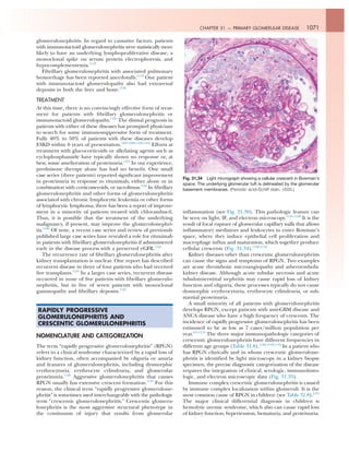
![1072 SECTION V — DISORDERS OF KIDNEY STRUCTURE AND FUNCTION
Table 31.8 Relative Frequency of Immunopathologic Categories of Crescentic Glomerulonephritis In Different
Age Groups (%)a
Immunopathologic Category
Age in Years
All Ages
(n = 632)
1−20
(n = 73)
21−60
(n = 303)
>60
(n = 256)
Antiglomerular basement membrane CGN 15 12 15 15
Immune complex CGN 24 45 35 6
Pauci-immune CGNb
60 42 48 79
Other 1 0 30 0
a
CGN is defined as the presence of crescents in >50% of glomeruli. Frequency is determined with respect to age in patients whose kidney
biopsy specimens were evaluated at the University of North Carolina Nephropathology Laboratory. Note the very high frequency of
pauci-immune disease (usually antineutrophil cytoplasmic antibody [ANCA]−associated) in older adults.
b
Approximately 90% associated with ANCA.
CGN, Crescentic glomerulonephritis.
Data from Jennette JC, Nickeleit V: Anti-glomerular basement membrane glomerulonephritis and Goodpasture’s syndrome. In Jennette
JC, Olson JL, Silva FG, D’Agati V, eds. Heptinstall’s pathology of the kidney. 7th ed. Wolters Klewer: Philadelphia; 2015:657−684.
Immunoglobulin- and Complement-Mediated Glomerulonephritis
Anti-GBM disease Immune complex GN C3 glomerulopathy ANCA disease
ANCA GN MPA
DDD C3 GN
Anti-GBM
GN
Goodpasture’s
syndrome
GPA EGPA
Immune
complex MPGN
Membranous
GN
Lupus
nephritis
IgA
vasculitis
IgA
nephropathy
Postinfectious
acute GN
Fibrillary
GN
Other immune
complex GN
Linear GBM
immunoglobulin
IF staining
Granular IF staining
for immunoglobulin
and C3
Granular IF staining
for C3 and little or
no immunoglobulin
Paucity of IF staining
for immunoglobulin
and complement
No
systemic
vasculitis
Vasculitis
with no
granulomas,
or asthma
Dense
deposits
in GBMs
No dense
deposits
in GBMs
No lung
hemorrhage
Lung
hemorrhage
Vasculitis with
granulomas,
no asthma
Vasculitis
with
granulomas
and asthma
Thick
capillaries and
hypercellularity
Thick
capillaries, no
hypercellularity
Systemic
lupus
erythematosus
Dominant
IgA and
vasculitis
Dominant
IgA and no
vasculitis
Acute strep
or staph
infection
~20 nm
fibrils
Other
features
Fig. 31.35 Algorithm for the diagnostic classification of glomerulonephritis that is known or suspected of being mediated by antibodies and
complement. Note that the integration of light microscopy, immunofluorescence (IF) microscopy, electron microscopy, laboratory data, and
clinical manifestations is required to diagnose glomerulonephritis (GN) precisely. ANCA, Antineutrophil cytoplasmic autoantibody; DDD, dense
deposit disease; EGPA, eosinophilic granulomatosis with polyangiitis; GBM, glomerular basement membrane; GPA, granulomatosis with
polyangiitis; MPA, microscopic polyangiitis.](data:image/gif;base64,R0lGODlhAQABAIAAAAAAAP///yH5BAEAAAAALAAAAAABAAEAAAIBRAA7)