
Recommandé
Contenu connexe
Tendances
Tendances (20)
En vedette
En vedette (20)
Similaire à Dr. B Ch 10_lecture_presentation
Similaire à Dr. B Ch 10_lecture_presentation (20)
Plus de TheSlaps
Plus de TheSlaps (20)
Dr. B Ch 10_lecture_presentation
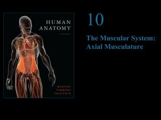
- 1. © 2012 Pearson Education, Inc. 10 The Muscular System: Axial Musculature PowerPoint® Lecture Presentations prepared by Steven Bassett Southeast Community College Lincoln, Nebraska
- 2. Introduction • The skeletal muscle of the body can be subdivided into: • Axial musculature • Muscles that position the head and vertebral column • Muscles that move the rib cage • Roughly 60 percent of the skeletal muscles in the body are axial muscles. • Appendicular musculature • Muscles that stabilize or move the appendicular skeleton © 2012 Pearson Education, Inc.
- 3. The Axial Musculature • The axial muscles can be placed into four groups based on location or function • Muscles of the head and neck • Muscles of the vertebral column • Oblique and rectus muscles • Muscles of the pelvic floor © 2012 Pearson Education, Inc.
- 4. © 2012 Pearson Education, Inc. Epicranial aponeurosis Temporoparietalis Trapezius Clavicle Deltoid Pectoralis major Biceps brachii (short head) Biceps brachii (long head) Frontal belly of occipitofrontalis Temporoparietalis (reflected) Temporalis Sternocleidomastoid Sternum Serratus anterior Latissimus dorsi External oblique Rectus abdominis Linea alba Omohyoid Acromion Figure 10.1 Superficial Skeletal Muscles, Anterior View (Part 1 of 2)
- 5. Figure 10.2 Superficial Skeletal Muscles, Posterior View (Part 1 of 2) © 2012 Pearson Education, Inc. Epicranial aponeurosis Trapezius Deltoid Infraspinatus Teres minor Teres major Occipital belly of occipitofrontalis Sternocleidomastoid Rhomboid major Triceps brachii (long head) Latissimus dorsi Triceps brachii (lateral head)
- 6. The Axial Musculature Muscles of the Head and Neck Can be subdivided into several groups: Muscles of facial expression: All inervated by Facial Nerve (CN VII) See table 10.1 for the name and function of the muscles. Extraocular muscles Muscles of mastication Muscles of the tongue: see table 10.4 for more detail. Muscles of the pharynx Anterior muscles of the neck: see table 10.6 for more detail. © 2012 Pearson Education, Inc.
- 7. Figure 10.3a Muscles of the Head and Neck, Part I Frontal belly of occipitofrontalis Epicranial Corrugator supercilii Temporalis (temporoparietalis removed) Orbicularis oculi Nasalis Zygomaticus minor Zygomaticus major Orbicularis oris Platysma Mentalis (cut) © 2012 Pearson Education, Inc. Anterior view Thyroid cartilage of the larynx aponeurosis Temporoparietalis (cut and reflected) Temporalis Procerus Levator labii superioris Masseter Buccinator Depressor anguli oris Depressor labii inferioris Sternal head of sternocleidomastoid Clavicular head of sternocleidomastoid Trapezius Clavicle Platysma (cut and reflected) Risorius
- 8. Figure 10.4a Muscles of the Head and Neck, Part II Epicranial aponeurosis Frontal belly of occipitofrontalis Procerus Orbicularis oculi Nasalis Levator labii superioris Zygomaticus minor Levator anguli oris Zygomaticus major Orbicularis oris Mentalis (cut) Depressor labii inferioris © 2012 Pearson Education, Inc. A diagrammatic lateral view Omohyoid Platysma (cut and reflected) Depressor anguli oris Temporoparietalis (cut and reflected) Temporalis Occipital belly of occipitofrontalis Masseter Buccinator Sternocleidomastoid
- 9. Table 10.1 Muscles of Facial Expression (Part 1 of 5) © 2012 Pearson Education, Inc. A&P Flix: Buccinator
- 10. Table 10.1 Muscles of Facial Expression (Part 2 of 5) © 2012 Pearson Education, Inc.
- 11. Table 10.1 Muscles of Facial Expression (Part 3 of 5) © 2012 Pearson Education, Inc.
- 12. Table 10.1 Muscles of Facial Expression (Part 4 of 5) © 2012 Pearson Education, Inc.
- 13. Table 10.1 Muscles of Facial Expression (Part 5 of 5) © 2012 Pearson Education, Inc.
- 14. Figure 10.5a Extra–ocular Muscles Muscles on the lateral surface of the right eye © 2012 Pearson Education, Inc. Maxilla Inferior oblique Inferior rectus Lateral rectus Optic nerve Frontal bone Superior rectus Superior oblique Levator palpebrae superioris Trochlea (ligamentous sling)
- 15. Figure 10.5b Extra–ocular Muscles Muscles on the medial surface of the right eye © 2012 Pearson Education, Inc. Trochlea Levator palpebrae superioris Superior rectus Superior oblique Medial rectus Inferior rectus Optic nerve
- 16. The Axial Musculature • Extra-ocular Muscles • Eye movements • Lateral rectus: rotates the eye laterally • Medial rectus: rotates the eye medially • Superior rectus: rotates the eye upward • Inferior rectus: rotates the eye downward • Superior oblique: rotates the eye downward and laterally • Inferior oblique: rotates the eye upward and laterally © 2012 Pearson Education, Inc.
- 17. Figure 10.5c Extra–ocular Muscles Superior rectus Lateral rectus Inferior oblique Anterior view of the right eye showing the orientation of the extra-ocular muscles and the directions of eye movement produced by contractions of the individual muscles © 2012 Pearson Education, Inc. Trochlea Superior oblique Medial rectus Inferior rectus
- 18. The Axial Musculature • Muscles of Mastication • Masseter • Temporalis • Pterygoids © 2012 Pearson Education, Inc. A&P Flix: Masseter A&P Flix: Temporalis
- 19. Figure 10.6a Muscles of Mastication Superior temporal line The temporalis and masseter are prominent muscles on the lateral surface of the skull. The temporalis passes medial to the zygomatic arch to insert on the coronoid process of the mandible. The masseter inserts on the angle and lateral surface of the mandible. © 2012 Pearson Education, Inc. Temporalis Zygomatic arch Capsule of temporomandibular joint Masseter
- 20. Figure 10.6b Muscles of Mastication Lateral pterygoid Medial pterygoid © 2012 Pearson Education, Inc. Mandible The location and orientation of the pterygoid muscles can be seen after removing the overlying muscles, along with a portion of the mandible.
- 21. Table 10.3 Muscles of Mastication © 2012 Pearson Education, Inc.
- 22. The Axial Musculature • Muscles of the Tongue • Genioglossus • Hyoglossus • Palatoglossus • Styloglossus © 2012 Pearson Education, Inc.
- 23. Figure 10.7 Muscles of the Tongue © 2012 Pearson Education, Inc. Styloid process Palatoglossus Styloglossus Genioglossus Hyoglossus Hyoid bone Mandible (cut)
- 24. Table 10.4 Muscles of the Tongue © 2012 Pearson Education, Inc.
- 25. The Axial Musculature • Muscles of the Pharynx • Pharyngeal constrictors • Superior constrictor • Middle constrictor • Inferior constrictor © 2012 Pearson Education, Inc.
- 26. Figure 10.8a Muscles of the Pharynx © 2012 Pearson Education, Inc. Lateral view Superior pharyngeal constrictor Stylopharyngeus Palatopharyngeus Middle pharyngeal constrictor Inferior pharyngeal constrictor Esophagus Tensor veli palatini Levator veli palatini
- 27. The Axial Musculature • Muscles of the Pharynx (continued) • Laryngeal elevators • Palatopharyngeus • Stylopharyngeus © 2012 Pearson Education, Inc.
- 28. Figure 10.8a Muscles of the Pharynx © 2012 Pearson Education, Inc. Lateral view Superior pharyngeal constrictor Stylopharyngeus Palatopharyngeus Middle pharyngeal constrictor Inferior pharyngeal constrictor Esophagus Tensor veli palatini Levator veli palatini
- 29. The Axial Musculature • Muscles of the Pharynx (continued) • Palatal muscles • Levator veli palatini • Tensor veli palatini © 2012 Pearson Education, Inc.
- 30. Figure 10.8a Muscles of the Pharynx © 2012 Pearson Education, Inc. Lateral view Superior pharyngeal constrictor Stylopharyngeus Palatopharyngeus Middle pharyngeal constrictor Inferior pharyngeal constrictor Esophagus Tensor veli palatini Levator veli palatini
- 31. Table 10.5 Muscles of the Pharynx © 2012 Pearson Education, Inc.
- 32. Figure 10.9ab Anterior Muscles of the Neck Mandible Muscles that form the floor of the Mylohyoid oral cavity, superior view Anterior view of neck muscles Digastric Anterior belly Posterior belly Sternocleidomastoid (cut) Superior belly Inferior belly Clavicle Omohyoid Cut heads of sternocleidomastoid © 2012 Pearson Education, Inc. Mylohyoid (cut and reflected) Geniohyoid Stylohyoid Hyoid bone Thyrohyoid Thyroid cartilage of larynx Cricothyroid Sternothyroid Sternohyoid Clavicular head Sternal head Sternocleido-Sternum mastoid Genioglossus (cut) Mylohyoid Geniohyoid Mandible Hyoid bone
- 33. Table 10.6 Anterior Muscles of the Neck © 2012 Pearson Education, Inc.
- 34. The Axial Musculature Muscles of the Vertebral Column Back muscles form three distinct layers: Superficial—move the neck Intermediate—extend the vertebral column Deep—interconnect vertebrae Extrinsic muscles—those in the superficial and intermediate layers Intrinsic (or true) muscles—those in the deepest layer; in turn, these intrinsic muscles are arranged in superficial, intermediate, and deep layers Vertebral muscles are also divided into: Extensors: ilicostalis, longissimus, spinalis Flexors: longus capitis, longus colli, quadratus lumborum © 2012 Pearson Education, Inc.
- 35. Figure 11.2 Superficial and Deep Muscles of the Neck, Shoulder, and Back © 2012 Pearson Education, Inc. SUPERFICIAL DEEP Sternocleidomastoid Cut edge of right trapezius Trapezius Scapular spine Deltoid Infraspinatus Teres minor Teres major Triceps brachii Erector spinae muscle group (see Figure 10.10b) Latissimus dorsi Thoracolumbar fascia External oblique Iliac crest Gluteus medius Gluteus maximus Semispinalis capitis Splenius capitis Levator scapulae Supraspinatus Rhomboid minor (cut and reflected) Serratus posterior (superior) Rhomboid major (cut and reflected) Serratus anterior Latissimus dorsi (cut and reflected) Serratus posterior (inferior) External oblique Internal oblique Latissimus dorsi (cut and reflected)
- 36. Figure 10.10b Muscles of the Vertebral Column © 2012 Pearson Education, Inc. Longissimus capitis (cut) Spinalis cervicis Middle scalene Semispinalis cervicis Posterior scalene Longissimus cervicis Semispinalis thoracis Multifidus Quadratus lumborum Semispinalis capitis Splenius Longissimus capitis Longissimus cervicis Iliocostalis cervicis Iliocostalis thoracis Longissimus thoracis Spinalis thoracis Iliocostalis lumborum Erector spinae muscles Thoracodorsal fascia Posterior view of superficial (right) and deeper (left) muscles of the vertebral column
- 37. Figure 10.10d Muscles of the Vertebral Column © 2012 Pearson Education, Inc. Longus capitis Longus colli Slips of anterior scalene C1 C2 C3 C4 C5 C6 C7 T1 T2 T3 Rib 1 Rib 2 Anterior scalene Middle scalene Posterior scalene Muscles on the anterior surfaces of the cervical and superior thoracic vertebrae Anterior scalene
- 38. The Axial Musculature Oblique and Rectus Muscles The muscles of the oblique and rectus groups lie between the vertebral column and the ventral midline. The oblique muscles can compress underlying structures or rotate the vertebral column, depending on whether one or both sides are contracting. The rectus muscles are important flexors of the vertebral column, acting in opposition to the erector spinae. The abdominal muscles are: External oblique (the most superficial muscle) Internal oblique Rectus abdominis Transversus abdominis (the deepest muscle) All abdominal muscles can compress the abdomen. Abdominal cavity is separated from Thoracic cavity by Diaphragm. Diaphragm is innervated by Phrenic nerve. © 2012 Pearson Education, Inc.
- 39. Figure 11.4 Muscles That Position the Pectoral Girdle, Part II (Part 1 of 2) Trapezius Subclavius Pectoralis major (cut and reflected) Pectoralis minor Internal intercostals External intercostals © 2012 Pearson Education, Inc. Levator scapulae Pectoralis minor (cut) Coracobrachialis Serratus anterior Short head Long head Biceps brachii T12
- 40. Figure 10.12b The Diaphragm © 2012 Pearson Education, Inc. Inferior vena cava Central tendon of diaphragm Diagrammatic superior view Xiphoid process Rectus abdominus Transversus thoracis Thoracic aorta T10 Trapezius Spinal cord Costal cartilages External oblique Diaphragm External intercostal Esophagus Serratus anterior Internal intercostal Latissimus dorsi Serratus posterior (inferior) Erector spinae group
- 41. Figure 11.5 Superficial and Deep Muscles of the Trunk and Proximal Limbs © 2012 Pearson Education, Inc. SUPERFICIAL DEEP Platysma Deltoid Pectoralis major Serratus anterior External oblique Rectus sheath Aponeurosis of external oblique Superficial inguinal ring Tensor fasciae latae Sartorius Sternocleidomastoid Deltoid (cut and reflected) Pectoralis minor Subscapularis Pectoralis major (cut and reflected) Coracobrachialis Biceps brachii (short and long heads) Teres major Trapezius Subclavius Serratus anterior Internal intercostal External intercostal Internal oblique (cut) External oblique (cut and reflected) Rectus abdominis Transversus abdominis Gluteus medius Iliopsoas Pectineus Adductor longus Gracilis Rectus femoris Latissimus dorsi
- 42. Figure 10.11b The Oblique and Rectus Muscles © 2012 Pearson Education, Inc. Rectus abdominis Linea alba Rectus sheath External oblique Transversus abdominis Internal oblique Psoas major Quadratus lumborum L3 Diagrammatic horizontal section through the abdominal region Latissimus Thoracolumbar dorsi fascia
- 43. Figure 10.11a The Oblique and Rectus Muscles Anterior view of the trunk showing superficial and deep members of the oblique and rectus groups, and the sectional plane shown in part (b) © 2012 Pearson Education, Inc. Internal intercostal External intercostal External oblique (cut) Internal oblique Cut edge of rectus sheath External oblique Serratus anterior Tendinous inscription Rectus abdominis Linea alba
- 44. The Axial Musculature • Muscles of the Perineum and Pelvic Diaphragm • Main functions • Support the organs of the pelvic cavity • Flex the joints of the sacrum and coccyx • Control the movement of material through the urethra and anus © 2012 Pearson Education, Inc.
- 45. Figure 10.13b Muscles of the Pelvic Floor Testis Urethra (connecting segment removed) Superficial transverse perineal © 2012 Pearson Education, Inc. SUPERFICIAL DEEP Inferior view, male External urethral sphincter No differences between deep musculature in Pubococcygeus Iliococcygeus Coccygeus male and female Pelvic diaphragm UROGENITAL TRIANGLE ANAL TRIANGLE Bulbospongiosus Ischiocavernosus Anus External anal sphincter Gluteus maximus
- 46. Figure 10.13a Muscles of the Pelvic Floor Anus © 2012 Pearson Education, Inc. SUPERFICIAL DEEP Inferior view, female Ischiocavernosus Bulbospongiosus Vagina Superficial transverse perineal Gluteus maximus Urethra External urethral sphincter Deep transverse perineal Urogenital diaphragm Central tendon of perineum Pubococcygeus Iliococcygeus Levator ani External anal sphincter Sacrotuberous ligament Coccygeus
- 47. Table 10.9 Muscles of the Perineum © 2012 Pearson Education, Inc.
- 48. Table 10.10 Muscles of the Pelvic Diaphragm © 2012 Pearson Education, Inc.