

Recommandé
Contenu connexe
Tendances
Tendances (20)
En vedette
En vedette (20)
Similaire à Pedia pulp therapy
Similaire à Pedia pulp therapy (20)
Plus de IAU Dent
Plus de IAU Dent (20)
Dernier
Dernier (20)
Pedia pulp therapy
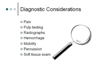
- 2. Clinical Assessment Extent of lesion location, color Mobility R/O root resorption Soft tissue swelling Lymphadenopathy Sensitivity to percussion reliable in primary teeth Pulp exposure hemorrhagic v necrotic Pulp testing Electrical thermal
- 3. Assessment of Pain Types of Pain Pulp Status Spontaneous Nocturnal Irreversible: Non-vital treatment Constant • Thermal • Chemical Reversible: Vital treatment • Intermittent
- 5. Reliability of Pulp Testing Teeth Primary Young p Mature p Electrical --- + + Thermal + + ++ Percussion ++ + + • No single diagnostic test is reliable
- 11. RADIOGRAPHIC ASSESSMENT • Proximity of caries • Assess the periodontal ligament • Furcational pathology. This is due to the accessory canals and foramina present on the pulp chamber floor of primary molars. • Internal and external root resorption Differentiate between root pathology and normal physiologic root resorption. Always check the antemere on a radiograph.
- 12. Radiographic Criteria for Health Pulp • Adequate periodontal support • No decalcified lesions or root fractures • No internal/external resorption or • Radiolucency • Integrity of lamina dura
- 14. Moss et al. 1965: • accessory canals in furcation area • no vital pulp tissue with interradicular bone loss • increased porosity of pulpal floor when infected Wrbas et al. 1997: • 77.5% of mandibular primary molars had accessory canals in floor of chamber
- 17. Histologic Components of Primary Pulp Lymph vessels Blood vessels Nerve tissue Collagenous fibers Fibroblasts Histologically similar to young permanent pulp Defense cells macrophages, neutrophils Lymphocytes Odontoblasts Odonto-/osteoclasts
- 18. Pulp response in primary molars with large proximal caries? • Does occlusal caries induce similar pulp response to proximal caries? • Pulp response in primary molars with large proximal caries?
- 20. Indirect Pulp Capping • Indications – Deep carious lesion – Incomplete caries removal – No pulp exposure • Objectives (AAPD) – Complete seal, preserve vitality, no post-tx signs/symptoms, formation of tertiary dentin, – No evidence of pathology/resorption
- 21. Pulp Capping Agents • Ca(OH)2 still widely used • ZOE - chronic inflammation • Mineral trioxide aggregate (MTA)
- 22. Technique of indirect pulp cap • LA & RD application. • Caries removal without pulp exposure. • CaOH application seal the cavity • Evaluation after 6-8 weeks – Maintenance of pulp vitality – Normal radiograph • Permanent restoration
- 23. Direct Pulp Cap • Indications – small mechanical or traumatic exposure in primary or permanent tooth • Contraindicated for (carious) exposure in primary teeth – persistent inflammation – internal resorption - related to high cellularity – calcific metamorphosis • Pulpal inflammation occurs before exposure. • Pulpal inflammation quickly becomes irreversible
- 24. Direct Pulp Cap
- 25. A pulpotomy is defined as the surgical removal of the entire coronal pulp pre-sumed to be partially or totally inflamed and quite possibly infected, leaving intact the vital radicular pulp within the canals. The aim is to relieve pain due to pulpalgia and leave the vital pulp in roots for its completion , if incomplete (apexogenesis ) DEFINITION
- 26. Pulpotomy for Primary Teeth • Indications (AAPD) –infected coronal tissue can be amputated –remaining radicular tissue vital (clinically and radiographically) • Objectives (AAPD) –preserve vitality of radicular pulp –no adverse signs or symptoms –no harm to succedaneous teeth
- 27. Pulpotomy: Clinical Indications • Mechanical or carious exposure • Inflammation limited to coronal pulp • Absence of spontaneous pain (?) • Absence of swelling or alveolar abscess formation • Restorable tooth
- 28. Pulpotomy Contraindications • History of unprovoked pain (?) • Presence of fistula or swelling • Evidence of necrotic pulp • Uncontrolled pulpal hemorrhage • Periapical or bifurcation radiolucency • Pathologic resorption • Dystrophic calcification • More than 1/3 root resorption
- 29. contraindication
- 30. The ideal dressing material / medicaments for radicular pulp • Non-toxic, non-mutagenic &non-carcinogenic. • Biocompatible. • Dimensionally stable. • Bactericidal. • Harmless to pulp & surrounding structures. • Promote healing of the radicular pulp. • Not interfere with physiologic process of root resorption.
- 31. classification Pulpotomy Partial pulpotomy complete pulpotomy calcium hydroxide formocresol pulpotomy pulpotomy
- 32. CLASSIFICATION Non vital pulpotomy Vital pulpotomy Beechwood cresol Devitalization Formocresol Preservation Regeneration
- 33. Vital pulpotomy technique Devitalization Preservation 1. Glutaraldehyde 2. Ferric sulphate Regeneration 1. Bone morphogenetic protein 2. Dentin chips Single sitting – Formocresol Electrosurgery Laser Two stage – Gysi triopaste Easlick’s formaldehyde Paraform devitalising pastePreservation
- 34. Categories of Medicaments Fixatives FMC, glutaraldehyde Mineralizing and/or bacteriostatic agents Ca(OH)2, tricalcium phosphate Palliative sealers ZOE Obturators mineral trioxide aggregate Coagulants ferric sulfate, aluminum chloride Antibiotics/Antimicrobials erythromycin, others Tissue healing agents collagen, BMP Glucocorticoids corticosteroids
- 35. Actions of Formocresol • Composition (Buckley’s Solution ) • 19% formaldehyde, • 35% cresol in vehicle of • 15% glycerin and water • Fixation with progressive fibrosis – acidophilic zone: fixation – pale staining zone: atrophy – broad zone of inflammatory cells • Bactericidal - biggest benefit? • No dentin bridging
- 38. Formocresol Pulpotomy Success Rates • Range of success rates in literature: • 62-100% depending on study and criteria used • Clinical>Radiographic>Histological • Formocresol pulpotomies may be empirical clinical successes, but histologically they are failures to one degree or another
- 39. Effects on Succedaneous Teeth • Pruhs et al. (1977) • Rolling and Poulsen (1978) • It is possible that enamel defects in premolars were caused by inflammation prior to the pulpotomy
- 40. Concerns About Formocresol • Systemic Toxicity • Myers et al 1983 • Damage to Permanent Successor • Pruhs et al 1977. • Rolling and Poulson 1978 • Mutagenic • Ranly 1984
- 41. Dilution of Formocresol • 1:5 dilution – – 1 part FMC, 4 parts vehicle (3 parts glycerin, 1 part distilled water) • Histology and clinical success comparable to full strength • Neither produces ideal histology • Long-term clinical success of 1:5 still questioned by some
- 42. Two-Appointment Pulpotomy. Indications 1 Evidence of sluggish or profuse bleeding at the amputation site 2 Difficult-to-control bleeding 3 Slight purulence in the chamber but none at the amputation site 4 Thickening of the periodontal ligament 5 A history of spontaneous pain without other contraindications. Contraindications. (1) Nonrestorable (2) Soon To Be Exfoliated (3) Necrotic.
- 43. Procedure 1. The steps are the same as for the one-appointment procedure 2. A cotton pellet moistened with diluted formocresol is sealed into the chamber for 5 to 7 days with a durable temporary cement. 3. At the second visit, the temporary filling and cotton pellet are removed and the chamber is irrigated with hydrogen peroxide. 4. A ZOE cement base is placed. 5. The tooth is restored with a stainless steel crown.
- 44. Partial pulpotomy (pulp curretage ) It is removal of coronal pulp tissue up to the level of healthy pulp. This process is also known as partial pulpotomy. INDICATIONS : -- when zones of inflammation has extended more than 2 mm. in an apical direction but has not reached root pulp. Eg. A traumatic exposure (a few days post injury in a large young pulp)
- 45. TECHNIQUE 1. Area is anaesthetised and isolated 2. A 2 mm. deep cavity is prepared into pulp using sterile diamond bur and copius water coolent 3. Excess blood is removed by saline & small cotton pelletes 4. Calcium hydroxide is placed onto cavity 5. Sealed with ZOE reinforced IRM restoration. IT IS RARELY SUCESSFUL AND HENCE HAS NO CLINICAL SIGNIFICANT.
- 46. REASONS FOR FAILURE Pulp is highly vascular so, even with slightest infection in any corner of pulp , the whole of it gets infected very quickly. Its practically impossible to remove one part of coronal pulp without disturbing the other parts of it in pulp chamber.
- 47. DEVITALIZATION PULPOTOMY It is two stage procedure involving the use of paraformaldehyde to fix the entire coronal & radicular pulp tissue. The medicament used have a devitalizing, mummifying, & bactericidal action.
- 48. TECHNIQUE First appointment :- Same as formocresol pulpotomy but place the paraformaldehyde paste in cotton pellete over the exposure & seal the tooth for 1 to 2 weeks. Formaldehyde gas liberates from the paraformaldehyde permeates through the coronal and radicular pulp, fixing the tissue. Second appointment :- pulpotomy is carried out with the help of local anaesthesia
- 49. ELECROSURGICAL PULPOTOMY Given by mack & dean (1933 ) It is a non chemical devitalizaton technique. Electrocautery carbonizes and heat denatures the pulp & bacterial contamination After amputation of coronal pulp,the pulp stumps are cauterized through this method
- 50. Laser pulpotomy Jeng-fen liu et al in 1999 studied the effect on Nd:YAG laser for pulpotomy in primary teeth
- 51. Glutaraldehyde by kopel (1979 ) Advantages over formocresol 1. Superior fixative property 2. Self limiting penetration 3. Low antigenicity 4. Low toxicity 5. Elimination of cresol 2-5 % concentration
- 52. Ferric sulphate It forms a metal protein clot at the surface of the pulp stump and this act as a barrier to irritating components of the sub-base
- 53. Potential Problems With Ferric Sulphate • In cases where diagnosis is not accurate and the remaining pulp is inflamed – Not a fixative – Will not be as forgiving as formoc