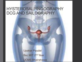
2. Introduction
• Radiological investigation of uterus and
uterine tube following an injection of
contrast medium into the cervix through
the vagina .
• Usually done within 4 to 10 days of
menstrual onset.
• First described by RUBIN and CAREY in
1914
2
3. General anatomy-uterus
• Hollow, muscular, pear shaped organ, flattened
anteroposteriorly.
• Lies in the pelvic cavity between the urinary
bladder and rectum.
• Anteversion and anteflexion, almost
perpendicular to vagina.
• Diameter-7.5cm*5cm*2.5cm
• Weight-30-40 gm
• Has three parts : fundus ,body and cervix(neck)
3
5. • Fundus: dome-shaped, above the opening of the uterine
tube
• Body: main part, narrowest inferiorly at the internal os where
it is continuous with the cervix.
• cervix (neck): protrudes through the anterior wall of the
vagina, opening into at the external os.
• Wall consists of three layers : perimetrium,
myomertium and endometrium.
5
6. • Perimetrium: Anteriorly lies over the fundus and body and
folds on upper surface of the urinary bladder forming
vesicouterine pouch.
• Posteriorly covers fundus, body and cervix then folds on
the rectum forming retrouterine pouch(of douglas).
• Laterally only fundus is covered.
• Myometrium: thickest layer of tissue, mass of smooth muscle
fibres interlaced with areolar tissue, blood vessels and
nerves
• Endometrium: consists of columnar epithelium, large no. of
mucus-secreting tubular glands. Consists of two layer:
functional layer and basal layer.
6
7. • Upper 2/3rd of the cervical canal is lined with mucous
membrane. Lower down however the mucosa changes,
becoming stratified squamous epithelium.
• Arterial supply: by the uterine arteries, branches of the
internal iliac arteries.
• Venous drainage: uterine vein into the internal iliac veins
• Lymph drainage: deep and superficial lymph vessels drain
lymph from the uterus and uterine tubes to the aortic
lymph nodes.
• Nerve supply: parasympathetic fibres from the sacral outflow
and sympathetic fibres from the lumbar outflow.
7
8. • Supporting structures: broad ligament, round ligament,
uterosacral ligament, transverse cervical (cardinal)
ligaments and pubocervical ligament.
Uterine tubes (fallopian)
• Are 10cm long.
• Extend from the sides of the uterus between the body and
the fundus
• End of each tube has finger like projection called fimbriae.
• Longest is the overial fimbriae.
8
9. • Each uterine tube consists of interstitial part, isthmus,
ampulla and infundibulum.
Structure:
• Outer covering of peritoneum(broad ligament).
• Middle layer of smooth muscle and lined with ciliated
epithelium
• Blood supply, lymph drainage and nerve
supply:as for the uterus.
9
11. Indications
• Infertility
• Recurrent miscarriages
• Patency of uterine tube in case of sterility.
• Abnormality of uterus-congenital or fibroid.
• T.B. of female genital tract.
• Congenital abnormalities such as:
unicornuate, bicornuate, septed
• Pathology of uterus and uterine tubes.
• Post tuboplasty and tube patency
11
12. contraindications
• Pregnancy
• Virgin patient
• Acute or chronic vaginal infection.
• Acute per-vaginal bleeding.
• Permulent discharge on inspection of cervix.
• Immediately post menstruation
• Recent dilation and curettage or abortion (use of oily CM
because of the risk of intravasation).
• Hypersensitivity to iodinated CM.
12
13. Contrast media
• Oily CM is no longer recommended only in case of recent
dilation of the uterus.
• HOCM or LOCM can be used.
• Non-ionic dimer has lower incidence and decreased
severity of delayed pain.
• Dose:10-20ml
13
14. Equipments
• High power x-ray generator.
• Fluoroscopic unit with IITV system.
• Vaginal speculum.
• Valsellum forceps
• Uterine cannula and syringe, leech-wilkinson cannula, olive
or 8-F pediatric foleys catheter.
• Uterine sound
14
16. Patient preparation
• Must abstain from intercourse between the 4th
and 10th days in a patient with regular 28- days
cycle.
• The part must be shaved.
• Apprehensive patient may need premedication.
• Patient must micturate just before examination.
• Patient should take light diet.
• Take understood consent from patient and
explain the procedure
16
17. Role of radio-technologist
• Proper position of patient.
• Cooperate with gynecologist and other staff
during procedure.
• Keep the side marker.
• Operate fluoroscopy during procedure.
• Take the image in correct position and time.
• Check the radiograph.
• Minimize the radiation dose to patient, doctor and
himself .
17
18. Radiation protection
• 10 days rule should apply.
• Pregnancy rule should follow.
• Use fluoroscopy only when required.
• Avoid repeat radiograph.
• Use lead apron, thyroid protection,
lead barrier for doctor, radiographer
and other assist staff.
18
19. Preliminary film
• Coned PA view of the pelvic cavity in supine
position.
• Centering position : on the mid line 2.5cm below
the ASIS.
• Cassette: 8”X10” OR 10X12
19
20. Technique
• Reassurance to the patient and lies in supine on the
table.
• Knees are flexed, legs abducted and heels together
(lithotomy position)
• Using aseptic technique the gynaecologist inserts a
speculum and cleans the vagina and cervix with
chlorhexidine.
• The anterior lip of cervix is steadied with the vulsellum
forceps and the cannula is inserted into the cervical
canal.
• If a foleys catheter is used, there is usually no need to
grasp the cervix with the vulsellum forceps.
20
21. • Then balloon is inflated by the use of air or normal
saline.
• Air bubbles have to be expelled from syringe and
cannula.
• CM is injected slowly under intermittent
fluoroscopic control.
• Spasm of the uterine cornu may be relieved by
inhalation of octylnitrite.
21
23. Filming
• Use under couch tube (PA view)
• 1st film: as the uterine (fallopian) tube begin to
fill.
• 2nd film: when the peritoneal spill has occurred
with all instruments removed.
23
24. Complications
• Due to CM: allergic phenomenon.
• Due to technique: pain may be during
I. Using the vulsellum forceps.
II. Insertion of cannula.
III. Tubal distension proximal to a block.
IV. Distension of uterus if there is tubal spasm.
V. Peritoneal irritation up to 2 weeks.
• Bleeding from the trauma to uterus or cervix.
• Transient nausea, vomiting and headache.
• infection
24
25. After care
• Sanitary pad is placed over the part.
• Reassurance the patient.
• Patient must be advised , there may be per vaginal
bleeding for 1-2 days and pain may persist for upto
2 weeks.
• Antibiotic is suggested.
• Analgesic is suggested.
25
27. ANATOMY
Lacrimal system comprises of:
Lacrimal gland, secretes tear
Lacrimal sac
Nasolacrimal duct, through which tears pass
into the nasal cavity.
Lacrimal gland lies anteriorly in the upper
outer quadrant of the orbit
About the size and shape of an almond, is
nestled in a shallow fossa of the frontal bone in
the superolateral corner of the orbit.
About 12 short ducts lead from the lacrimal
gland to the surface of the conjunctiva
27
28. The gland communicates with the lacrimal
sac via the lacrimal canaliculi
On the margin of each eyelid near the
medial commissure is a tiny pore, the
lacrimal punctum, is the opening to a short
lacrimal canal, which leads to the lacrimal
sac in the medial wall of the orbit.
A nasolacrimal duct is a membranous canal
approx 2 cm long extending from lower part
of the lacrimal sac to the nasal cavity
opening at the level of inferior concha
28
30. INDICATIONS
Epiphora to demonstrate the presence and extent of
obstruction
Obstruction may be due to:
congenital obstructions
supernumerary canaliculi
lacrimal fistula or diverticula
concretions (dacryoliths)
neoplastic or inflammatory processes
post treatment changes
31. CONTRAINDICATION
None
A small quantity of local anesthesia is dropped into the inner canthus of eye prior
to cannulation of the punctum lacrimalia
32. EQUIPMENT
Undercouch image intensifier with digital imaging
equipment to facilitate production of subtracted image
Dedicated skull unit with focal spot size 0.3 mm to facilitate
macroradiography
33. CONTRAST MEDIA
Oil based contrast media ,Lipiodol produces higher quality
images of
the lacrimal sac than water-soluble dye
Low Osmolar CM [LOCM], 300mgI/ml
Dose: 0.5– 2 ml
Oil-soluble contrast media should not be utilized in the
suspicion of tumors, traumatism or fistulae, considering the
risk of leakage and permanence in the subcutaneous
tissue for many years, inducing the formation of
granulomas
35. PROCEDURE
The lacrimal sac is massaged to express its contents prior
to injection of the contrast medium.
The lower eyelid is everted to locate the lower canaliculus
at the medial end of the lid.
The lower canaliculus is dilated and the cannula or
catheter is inserted.
The lower lid should be drawn laterally during insertion to
straighten the bend in the canaliculus, and so avoid
perforation by the cannula.
The contrast medium is injected, and radiographs are
taken immediately afterwards (or during the injection if a
catheter is used).
37. AFTER CARE
The eye is covered for approx. 1 hr after the examination to
prevent ingress of foreign material
Pt is usually kept in the department for about half an hour
after the examination until the effects of the LA is worn out.
42. ANATOMY
Three pairs of salivary glands:
Parotid gland
Submandibular gland
Sublingual gland
Situated adjacent to the oral cavity
secrete saliva which contains enzyme ptylase, aid to
digestion
43.
44. PAROTID GLAND
Largest salivary gland
Lie below the zygomatic arch infront of and below the ear
has a large single duct (Stenson's) that runs forwards
crossing the masseter muscle, turning inwards at its
anterior border to pierce the buccinator muscle and then
opening into the mouth on a papilla opposite the second
upper molar tooth.
45. SUBMANDIBULAR GLAND
Paired and lie on either side of neck forming the part of the
soft tissues on the medial margin of the mandible between
the mandible and hyoid bone extends.
A single duct (Wharton's) emerges from the deep surface
of the gland, runs forward, medially and upwards beneath
the mucous membrane of the floor of the mouth and opens
at small papilla at the base of frenulum of the tongue.
46. SUBLINGUAL GLAND
the smallest and lies anteriorly in the floor of the mouth on
the surface of the mylohyoid muscle and opens into the
mouth through a number of duct(duct of Rivinus.)
Ducts within all of these glands are evenly distributed and
gently tapered.
Ducts may open adjacent to frenulum of the tongue , or
may joint to form a single duct (Bartholin’s duct)which
empties into the submandibular glands.
47. INDICATIONS
Pain
Recurrent salivary gland enlargement
Duct obstruction
To localize diverticula ,strictures, calculi
To determine extent of Fistula
Foreign body
Inflammatory lesions and Tumor
49. CONTRAST MEDIA
Oil based or water soluble contrast medium
Dose:1 to 2 ml
Because oil is immiscible with the watery tear secretion, an
oil-based, iodinated contrast medium is employed in
examinations of the nasolacrimal duct system.
Either a compound with low viscosity or an ethiodized oil
may be used after the medium has been warmed to body
temperature to further reduce its viscosity.
50. EQUIPMENT
skull unit using macro radiography techniques
Silver cannula or 18 G blunt needle and polythene
catheter (Connecting tube.]
Syringe –3 0r 5 ml
Adhesive tape, Cotton swabs , Gauze pads,Sterile gloves
Lemon
Overhead light
Fluoroscopic unit with spot film device.
51. PRELIMINARY FILMS
Parotid gland
PA view centered at the level of angle of mandible
Lateral, centered to the angle of the mandible.
Lateral oblique, centered to the angle of the mandible,
and with the tube angled 200 cephalad.
52. Submandibular gland
Inferosuperior using an occlusal film. This is a useful
view to show calculi.
Lateral, with the floor of the mouth depressed by a
wooden spatula.
Lateral oblique, centred 1 cm anterior to the angle of the
mandible, and with the tube angled 200 cephalad.
53. TECHNIQUE
Duct orifice is located and sprayed with topical anaesthesia
If they are not visible, a sialogogue (e.g. citric acid) is placed in the mouth to
promote secretion from the gland, and so render the orifice visible.
The orifice of the duct is dilated with the silver wire probe and the cannula or
polythene catheter is introduced into the duct.
Up to 2 ml of contrast medium are injected.
If the cannula method is used, films are taken immediately after the injection.
The catheter method has the advantage that films can be taken during the
injection, with the catheter in situ, and that both sides can be examined
simultaneously.
54. FILMING
Immediate - the same views as for the preliminary films
are repeated. The occlusal film for the submandibular
gland may be omitted, as this is only to demonstrate
calculi.
Post-secretory - the same views are repeated 5 min after
the administration of a sialogogue. The purpose of this is to
demonstrate sialectasis.
62. REFERENCES
Diagnostic radiography by Glenda J Bryan
A guide to radiological procedure by Stephen Chapman
and Richard Nakielny.
Clarks special procedure in Diagnostic imaging
Various websites.
63. ?????
• 1)Describe HSG
• 2)List some indications and contraindications of HSG.
• 3)Describe is DCG
• 4)List some indications of DCG
• 5)why oil based CM is not used in DCG though it produces
higher quality images?
• 6)what is sailography?
• 7)List some indication and contraindication of sailography
• 8)Mention filming sequences used in sailography of parotid
gland.
63