SlideShare une entreprise Scribd logo
1  sur  213
Télécharger pour lire hors ligne
1
NOTES
for
“Value Added Course -2023”
MEDICAL TECHNIQUES
Advanced Techniques for Diagnosis
&
Molecular and Microbial Techniques
Co-ordinated by: Dr. Sunil Kumar & A Vamsi Kumar
Assistant professor – Dept of MLT
(April - May 2023)
2
Table of Contents
SYLLABUS................................................................................................................................................3
LECTURE PLAN ........................................................................................................................................4
1. FLOW CYTOMETRY .............................................................................................................................6
Flow cytometry Multiple Choice Questions with Answers...................................................................9
Flow cytometry Assignment Questions with Answers........................................................................12
FLOW CYTOMETRY CASE STUDIES .......................................................................................................15
2. HEMOCYTOMETRY: PRINCIPLES AND APPLICATIONS......................................................................17
Hemocytometry Multiple Choice Questions .......................................................................................20
Hemocytometry Assignment questions with answers........................................................................23
HEMOCYTOMETRY CASE STUDIES .......................................................................................................26
CHROMATOGRAPHY MULTIPLE CHOICE QUESTIONS ..........................................................................31
Chromatography Assignment questions with answers.......................................................................33
CHROMATOGRAPHY CASE STUDIES.....................................................................................................36
4. ELECTROPHORESIS: TYPES, PRINCIPLES AND APPLICATION...........................................................39
Electrophoreses Multiple choice questions.........................................................................................41
Electrophoreses Assignment questions with answers........................................................................44
ELECTROPHORESES CASE STUDIES.......................................................................................................47
5. PCR AND TRANSILLUMINATOR: THEORY AND ITS APPLICATIONS TO BIOMEDICAL FIELD.............49
PCR Multiple choice questions.............................................................................................................52
PCR ASSINGMENT QUESTIONS WITH ANSWERS................................................................................54
PCR CASE STUDIES................................................................................................................................57
Inoculation & Isolation of microbes Multiple choice questions ........................................................60
Inoculation & Isolation of microbes Assignment question with Answers..........................................62
INOCULATION & ISOLATION OF MICROBES CASE STUDIES.................................................................64
3
SYLLABUS
Lecture
No.
Topic Reading Material /
Reference
No. of
Hours
Name of
the Expert
handling
the Topic
1. Advance
techniques for
diagnosis
1. Flowcytometery: Principles
and applications.
2. Hemocytometery: Principles
and applications.
3. Chromatography: Types,
Principles and application
4. Electrophoresis: Types,
Principles and application
15
Sunil.e12102@
cumail.in
2. Molecular and
microbial
Techniques
1. PCR and transillumnator:
Theory and its applications to
biomedical field.
2. Inoculation and isolation of
Microorganism from the
different type of samples.
15
Attuluri.e13404
@cumail.in
4
LECTURE PLAN
Lecture
No.
Topic Reading Material / Reference Date & Time
1 Introduction to
Advanced Techniques
for Diagnosis
Basic introduction to diagnostic
techniques
15-04-2023
12:00PM
2 Flow Cytometry:
Principles
Flow Cytometry: A Basic Introduction
(Book)
15-04-2023
2:00PM
3 Flow Cytometry:
Applications
Flow Cytometry Applications in Cell
and Molecular Biology (Review)
15-04-2023
2:00PM
4 Hemocytometry:
Principles
Hemocytometer: Protocol, Calculation,
and Calibration (Article)
22-04-2023
12:00PM
5 Hemocytometry:
Applications
Hemocytometry: Methods and
Applications (Review)
22-04-2023
2:00PM
6 Chromatography: Types Introduction to Chromatography (Book) 22-04-2023
3:00PM
7 Chromatography:
Principles
Chromatographic Separation Methods
(Book)
23-04-2023
12:00PM
8 Chromatography:
Applications
Applications of Chromatography in Life
Science (Review)
23-04-2023
2:00PM
9 Electrophoresis: Types Introduction to Gel Electrophoresis
(Article)
23-04-2023
3:00PM
10 Electrophoresis:
Principles
Principles and Methods of
Electrophoresis (Book)
29-04-2023
12:00 PM
11 Electrophoresis:
Applications
Applications of Electrophoresis in
Biomedical Research (Review)
29-04-2023
02:00 PM
12 Introduction to
Molecular and
Microbial Techniques
Molecular Biology Techniques: A
Classroom Laboratory Manual (Book)
29-04-2023
03:00 PM
13 PCR: Theory PCR: Principles, Procedures, and
Applications (Book)
30-04-2023
12:00 PM
14 PCR: Applications to
Biomedical Field
PCR Applications in Medical
Diagnostic: An Overview (Review)
30-04-2023
02:00 PM
15 Transilluminator:
Theory
Gel Documentation System: An
Introduction (Article)
30-04-2023
03:00 PM
16 Transilluminator:
Applications
Applications of Gel Documentation
Systems in Molecular Biology (Review)
06-05-2023
12:00PM
17 Inoculation Techniques Inoculation Methods in Microbiology:
An Overview (Article)
06-05-2023
02:00PM
18 Isolation of
Microorganisms from
Different Samples
Microbial Isolation Techniques (Book) 06-05-2023
03:00PM
19 Practical Session: Flow
Cytometry
Virtual Hands-on lab session 07-05-2023
12:00PM
5
20 Practical Session:
Hemocytometry
Virtual Hands-on lab session 07-05-2023
2:00PM
21 Practical Session:
Chromatography
Virtual Hands-on lab session 07-05-2023
3:00PM
22 Practical Session:
Electrophoresis
Virtual Hands-on lab session 13-05-2023
12:00PM
23 Practical Session: PCR
and Transilluminator
Virtual Hands-on lab session 13-05-2023
2:00PM
24 Practical Session:
Inoculation Techniques
Virtual Hands-on lab session 13-05-2023
3:00PM
25 Practical Session:
Isolation of
Microorganisms
Virtual Hands-on lab session 14-05-2023
12:00PM
26 Case Studies: Advanced
Diagnostic Techniques
Discussion of real-life case studies 14-05-2023
2:00PM
27 Case Studies:
Molecular and
Microbial Techniques
Discussion of real-life case studies 14-05-2023
3:00PM
28 Emerging Diagnostic
Techniques
Emerging Technologies in Medical
Diagnostics (Review)
20-05-2023
12:00PM
29 Ethical Considerations
in Diagnostic
Techniques
Ethical Issues in Biomedical Research
and Diagnostic Testing (Article)
20-05-2023
2:00PM
30 Recap and Final
Discussion
Review of key concepts and Q&A
session
20-05-2023
3:00PM
6
1. FLOW CYTOMETRY
Introduction
• Flow cytometry is a powerful and versatile technique used in the analysis and sorting
of cells and particles.
• The technique enables researchers to rapidly analyze and quantify multiple physical
and chemical characteristics of single cells or particles as they flow in a fluid stream
through a beam of light.
• The resulting data can be used for various applications, including cell counting, cell
sorting, and biomarker detection.
I. Principles of Flow Cytometry A. Fluidics system
•
• It consists of a sheath fluid and a sample fluid, which are combined to form a laminar
flow.
• Cells in the sample fluid are hydrodynamically focused, allowing them to pass
individually through the laser interrogation point.
B. Optics system
• The optics system consists of lasers, lenses, and filters that direct and collect the light
emitted from the cells or particles.
7
• As cells pass through the laser interrogation point, they scatter light and emit
fluorescence.
• Light scattering is categorized into forward scatter (FSC) and side scatter (SSC).
• FSC: Provides information about the size of the cell.
• SSC: Provides information about the internal complexity or granularity of the
cell.
C. Detection system
• The detection system consists of photodetectors, such as photomultiplier tubes (PMTs)
or avalanche photodiodes (APDs), which convert the emitted light into electrical
signals.
• The electrical signals are then amplified and processed by analog-to-digital converters
(ADCs) and digital signal processors (DSPs).
• The resulting digital data are plotted on a histogram or scatter plot, allowing for visual
analysis and quantification.
II. Applications of Flow Cytometry A. Immunophenotyping
• Immunophenotyping is the process of identifying and characterizing cells based on the
expression of specific cell surface markers (antigens).
• Flow cytometry enables the simultaneous analysis of multiple markers through the use
of fluorochrome-conjugated antibodies.
• Applications include the identification and quantification of immune cell subsets,
cancer cells, and stem cells.
B. Cell cycle analysis
• Flow cytometry can be used to analyze the DNA content of cells, allowing for the
determination of cell cycle distribution.
• This can help researchers understand cell proliferation, cell cycle regulation, and the
effects of various drugs on cell cycle progression.
C. Apoptosis and cell viability
• Flow cytometry can be used to assess cell viability and apoptosis by measuring various
cellular parameters such as membrane permeability, mitochondrial potential, and
activation of caspases.
• Common viability and apoptosis assays include Annexin V/PI staining, 7-AAD
staining, and TUNEL assay.
D. Cell sorting
• Fluorescence-activated cell sorting (FACS) is a specialized application of flow
cytometry that enables the separation and collection of individual cells based on their
characteristics.
8
• This technique is widely used in research and clinical settings for isolating specific cell
populations or single cells for further analysis or therapeutic applications.
Conclusion
• Flow cytometry is a powerful and versatile technique in medical lab technology,
enabling the rapid analysis and quantification of multiple cellular parameters.
• Its applications range from basic research to clinical diagnostics, making it an essential
tool for medical lab technology students to understand and master.
9
Flow cytometry Multiple Choice Questions with Answers
1. What is the primary purpose of flow cytometry?
A. Measuring cell size
B. Analyzing and quantifying multiple physical and chemical characteristics of single cells or
particles
C. Counting the number of cells in a sample
D. Sorting cells based on their size
Answer: B
2. Which two types of light scattering are primarily measured in flow cytometry?
A. Forward scatter (FSC) and reverse scatter (RSC)
B. Forward scatter (FSC) and side scatter (SSC)
C. Side scatter (SSC) and backscatter (BSC)
D. Forward scatter (FSC) and total scatter (TSC)
Answer: B
3.In the fluidics system of flow cytometry, what is the purpose of hydrodynamic focusing?
A. To separate cells based on size
B. To align cells in a single-file manner
C. To increase the speed of the sample fluid
D. To mix the sample fluid with the sheath fluid
Answer: B
4. What is the function of photodetectors, such as photomultiplier tubes (PMTs) or avalanche
photodiodes (APDs), in flow cytometry?
A. Emitting light for cells to pass through
B. Converting emitted light into electrical signals
C. Aligning cells in a single-file manner
D. Focusing the laser beam onto cells
Answer: B
5.Which of the following is NOT an application of flow cytometry?
A. Immunophenotyping
B. Cell cycle analysis
C. Microscopy imaging
D. Apoptosis and cell viability
Answer: C
6. Fluorescence-activated cell sorting (FACS) is a specialized application of flow cytometry
used for:
A. Cell counting
10
B. Cell sorting and collection
C. Cell staining
D. Cell cycle analysis
Answer: B
7. Which flow cytometry assay is commonly used for assessing apoptosis?
A. Annexin V/PI staining
B. 7-AAD staining
C. BrdU incorporation
D. DAPI staining
Answer: A
8. In flow cytometry, forward scatter (FSC) provides information about:
A. Cell size
B. Internal complexity of the cell
C. Expression of cell surface markers
D. DNA content of the cell
Answer: A
9. Which component of a flow cytometer is responsible for transporting and aligning the cells
in a single-file manner?
A. Fluidics system
B. Optics system
C. Detection system
D. Processing system
Answer: A
10. In flow cytometry, what is the role of fluorochrome-conjugated antibodies?
A. Focusing the laser beam onto cells
B. Converting emitted light into electrical signals
C. Identifying and characterizing cells based on specific cell surface markers
D. Separating and collecting individual cells
Answer: C
11.What does the side scatter (SSC) in flow cytometry provide information about?
A. Cell size
B. Internal complexity or granularity of the cell
C. Expression of cell surface markers
D. DNA content of the cell
Answer: B
11
12. Which of the following is NOT a component of the optics system in flow cytometry?
A. Lasers
B. Lenses
C. Filters
D. Photodetectors
Answer: D
13. In flow cytometry, which component is responsible for converting electrical signals into
digital data?
A. Analog-to-digital converters (ADCs)
B. Photomultiplier tubes (PMTs)
C. Digital signal processors (DSPs)
D. Avalanche photodiodes (APDs)
Answer: A
14. Which of the following assays is commonly used for cell cycle analysis in flow cytometry?
A. Annexin V/PI staining
B. 7-AAD staining
C. BrdU incorporation
D. TUNEL assay
Answer: C
15. What is the primary difference between flow cytometry and fluorescence-activated cell
sorting (FACS)?
A. Flow cytometry analyzes cells, while FACS sorts and collects cells.
B. Flow cytometry uses lasers, while FACS does not.
C. FACS requires the use of fluorochrome-conjugated antibodies, while flow cytometry does
not.
D. Flow cytometry is used for clinical diagnostics, while FACS is used for research purposes
only.
Answer: A
12
Flow cytometry Assignment Questions with Answers
Question 1: Explain the three main components of flow cytometry and their functions.
Answer:
The three main components of flow cytometry are the fluidics system, optics system, and
detection system.
The fluidics system is responsible for transporting and aligning the cells in a single-file manner,
utilizing a sheath fluid and a sample fluid that are combined to form a laminar flow.
The optics system consists of lasers, lenses, and filters that direct and collect the light emitted
from the cells or particles as they pass through the laser interrogation point, scattering light and
emitting fluorescence.
The detection system comprises photodetectors such as photomultiplier tubes (PMTs) or
avalanche photodiodes (APDs), which convert the emitted light into electrical signals. These
electrical signals are then amplified and processed by analog-to-digital converters (ADCs) and
digital signal processors (DSPs), resulting in digital data that can be plotted on a histogram or
scatter plot for analysis and quantification.
Question 2: What information do forward scatter (FSC) and side scatter (SSC) provide
in flow cytometry?
Answer:
Forward scatter (FSC) provides information about the size of the cell. Larger cells scatter more
light in the forward direction, resulting in a higher FSC signal.
Side scatter (SSC) provides information about the internal complexity or granularity of the cell.
Cells with more complex internal structures, such as granules, scatter more light in the side
direction, resulting in a higher SSC signal.
Question 3: How does flow cytometry facilitate immunophenotyping?
Answer:
Flow cytometry facilitates immunophenotyping by enabling the simultaneous analysis of
multiple markers on cells through the use of fluorochrome-conjugated antibodies. These
antibodies specifically bind to cell surface markers (antigens) and emit fluorescence when
excited by a laser. By using different fluorochromes for different antibodies, researchers can
analyze the expression of multiple cell surface markers simultaneously, identifying and
characterizing distinct cell populations.
Question 4: Describe how flow cytometry can be used for cell cycle analysis.
Answer:
Flow cytometry can be used for cell cycle analysis by measuring the DNA content of cells.
Cells are stained with DNA-binding dyes, such as propidium iodide (PI) or DAPI, which emit
fluorescence when bound to DNA. As the cells pass through the flow cytometer's laser, the
13
emitted fluorescence is proportional to their DNA content. This information allows researchers
to determine the cell cycle distribution by distinguishing cells in different phases of the cell
cycle (G0/G1, S, and G2/M), providing insights into cell proliferation and the effects of drugs
on cell cycle progression.
Question 5: Explain two common assays used for assessing apoptosis and cell viability
using flow cytometry.
Answer:
Annexin V/PI staining: Annexin V is a protein that binds to phosphatidylserine, which is
externalized on the outer leaflet of the plasma membrane during early apoptosis. Propidium
iodide (PI) is a DNA-binding dye that can only enter cells with compromised membranes,
indicative of late apoptosis or necrosis. By staining cells with Annexin V and PI, researchers
can distinguish viable cells (Annexin V-negative, PI-negative), early apoptotic cells (Annexin
V-positive, PI-negative), and late apoptotic or necrotic cells (Annexin V-positive, PI-positive).
Question 6: What is the primary difference between flow cytometry and fluorescence-
activated cell sorting (FACS)?
Answer: The primary difference between flow cytometry and fluorescence-activated cell
sorting (FACS) is their purpose. Flow cytometry is primarily used to analyze and quantify
multiple physical and chemical characteristics of single cells or particles. In contrast, FACS is
a specialized application of flow cytometry that enables the separation and collection of
individual cells based on their specific characteristics, such as size, granularity, or marker
expression.
Question 7: Design a simple experiment using flow cytometry to study the effects of a
drug on immune cell populations. Provide an overview of the experimental design,
including sample preparation, staining, data acquisition, and data analysis.
Answer: Overview of the experiment:
1. Sample preparation: Obtain peripheral blood mononuclear cells (PBMCs) from healthy
donors and culture them in appropriate media. Treat one group of cells with the drug of
interest at different concentrations and durations, while leaving another group untreated
as a control.
2. Staining: Use fluorochrome-conjugated antibodies specific for immune cell markers
(e.g., CD3 for T cells, CD19 for B cells, and CD14 for monocytes) to stain the cells
according to the manufacturer's protocol.
3. Data acquisition: Run the stained samples through a flow cytometer to measure
fluorescence intensity and light scattering properties (FSC and SSC) for each cell.
Acquire a minimum of 10,000 events per sample.
4. Data analysis: Use flow cytometry software to analyze the data by gating on specific
immune cell populations based on their marker expression. Calculate the percentage of
each immune cell subset in the treated and control groups and compare the results to
assess the drug's effect on immune cell populations.
14
Question 8: What are some potential limitations and challenges associated with flow
cytometry, and how can these be addressed or mitigated to ensure accurate and reliable
results?
Answer: Some potential limitations and challenges associated with flow cytometry include:
1. Autofluorescence: Cells may have intrinsic fluorescence, which can interfere with the
detection of specific fluorochromes. This can be addressed by selecting fluorochromes
with minimal overlap in their emission spectra and using appropriate compensation
controls.
2. Spectral overlap: Emission spectra of different fluorochromes can overlap, making it
difficult to distinguish between them. To mitigate this issue, use proper compensation
controls and carefully select fluorochromes with minimal spectral overlap.
3. Non-specific antibody binding: Antibodies may bind non-specifically to cells, causing
false-positive signals. To minimize non-specific binding, use appropriate isotype
controls, Fc receptor blocking reagents, and optimize antibody concentrations.
4. Sample variability: Variability in sample preparation, staining, and instrument settings
can lead to inconsistent results. Standardize protocols, use appropriate controls, and
regularly perform instrument quality control checks to ensure consistent and accurate
data.
By addressing these challenges and implementing appropriate controls and optimization
strategies, researchers can obtain accurate and reliable results from flow cytometry
experiments.
15
FLOW CYTOMETRY CASE STUDIES
Case Study 1: Immunophenotyping of Leukemia Patients
Background: Leukemia is a heterogeneous group of hematological malignancies characterized
by the uncontrolled proliferation of abnormal white blood cells. Flow cytometry plays a critical
role in the diagnosis, classification, and monitoring of leukemia by immunophenotyping
leukemic cells based on their specific cell surface markers.
Objective: Use flow cytometry to distinguish between different types of leukemia in patient
samples based on their immunophenotypic profiles.
Methods:
Collect bone marrow or peripheral blood samples from patients with suspected leukemia.
Prepare single-cell suspensions, and stain them with a panel of fluorochrome-conjugated
antibodies specific for various cell surface markers associated with different types of leukemia
(e.g., CD45, CD19, CD34, CD33, etc.).
Run the stained samples through a flow cytometer and analyze the data using appropriate gating
strategies and software tools.
Results and Interpretation:
By analyzing the expression of specific cell surface markers, different types of leukemia can
be identified:
Acute Lymphoblastic Leukemia (ALL): High expression of CD19 and CD10, often
accompanied by CD34 and TdT.
Acute Myeloid Leukemia (AML): High expression of CD33, CD13, and CD117, often
accompanied by CD34.
Chronic Lymphocytic Leukemia (CLL): High expression of CD19, CD5, and CD23, with low
expression of CD20.
Chronic Myeloid Leukemia (CML): High expression of CD33, CD13, and CD34, often
accompanied by CD38.
By comparing the immunophenotypic profiles of patient samples to these reference profiles,
clinicians can accurately diagnose and classify leukemia cases, guiding appropriate treatment
and monitoring strategies.
Case Study 2: Assessing the Effects of a Chemotherapeutic Drug on Apoptosis
Background: Apoptosis, or programmed cell death, is an essential process in the maintenance
of tissue homeostasis. Many chemotherapeutic drugs exert their anticancer effects by inducing
apoptosis in malignant cells. Flow cytometry can be used to assess the apoptotic effects of these
drugs on cancer cells.
Objective: Evaluate the pro-apoptotic effects of a chemotherapeutic drug on a cancer cell line
using flow cytometry.
Methods:
16
Culture a cancer cell line and treat with the chemotherapeutic drug at various concentrations
and durations.
Harvest the cells and stain them with Annexin V-FITC and propidium iodide (PI) according to
the manufacturer's protocol.
Run the stained samples through a flow cytometer and analyze the data using appropriate gating
strategies and software tools.
Results and Interpretation:
By analyzing the Annexin V/PI staining, the percentage of viable, early apoptotic, and late
apoptotic/necrotic cells can be determined:
Viable cells: Annexin V-negative, PI-negative
Early apoptotic cells: Annexin V-positive, PI-negative
Late apoptotic/necrotic cells: Annexin V-positive, PI-positive
By comparing the percentages of apoptotic cells in treated and untreated samples, researchers
can evaluate the pro-apoptotic effects of the chemotherapeutic drug on the cancer cell line.
These findings can inform the selection and optimization of drug concentrations and treatment
durations for effective cancer therapy.
17
2. HEMOCYTOMETRY: PRINCIPLES AND APPLICATIONS
I. Introduction
A. Definition of hemocytometry
1. Hemocytometry refers to the quantitative measurement of cells, particularly blood cells, in
a given volume of a liquid sample.
B. Importance of hemocytometry
1. Crucial in clinical diagnostics, research, and treatment monitoring
2. Determines cell concentration and viability
3. Supports diagnosis of various blood disorders and infections
II. Principles of Hemocytometry
A. Hemocytometer
1. Specialized counting chamber used for hemocytometry
2. Composed of a thick glass microscope slide with a grid of etched lines
3. Grid subdivided into various squares of known dimensions to facilitate counting
B. Sample preparation
1. Dilution of the blood sample
2. Staining (optional) to differentiate between cell types or identify dead cells
C. Counting method
1. Manual counting using a light microscope
2. Automated cell counters using electrical impedance, flow cytometry, or image analysis
techniques
III. Applications in Medical Lab Technology
A. Complete Blood Count (CBC)
1. Measures the concentration of red blood cells (RBCs), white blood cells (WBCs), and
platelets
18
2. Provides information on hemoglobin, hematocrit, and mean cell volume
B. Differential leukocyte count
1. Determines the relative percentage of each type of WBC (neutrophils, lymphocytes,
monocytes, eosinophils, and basophils)
2. Aids in the diagnosis of infections, inflammatory disorders, and malignancies
C. Reticulocyte count
1. Measures the number of immature RBCs (reticulocytes)
2. Assesses bone marrow function and response to anemia treatment
D. Cell viability assays
1. Evaluates the effectiveness of drug treatments, radiation, or other therapies on cell survival
2. Guides treatment decisions in cancer and other disorders
IV. Limitations and Challenges
A. Manual counting
1. Time-consuming and labor-intensive
2. Inherent variability due to human error
3. Requires skilled and experienced personnel
B. Automated counters
1. Expensive initial investment
2. Maintenance and calibration requirements
3. Potential for inaccurate results due to instrument limitations or sample quality issues
VI. Total White Blood Cell (TWBC) and Total Red Blood Cell (TRBC) Counts by Visual
Method
A. Total White Blood Cell (TWBC) Count by Visual Method
1. Objective: To determine the number of white blood cells per microliter (µL) of blood
2. Sample preparation
a. Blood sample mixed with a diluent (e.g., Turk's solution) to lyse RBCs and enhance WBC
visibility
b. The diluted sample is loaded onto the hemocytometer
3. Counting procedure
a. Using a light microscope, focus on the grid of the hemocytometer
b. Count the WBCs within specified grid squares
c.Apply the appropriate calculation to determine the TWBC concentration in the original blood
sample
B. Total Red Blood Cell (TRBC) Count by Visual Method
1. Objective: To determine the number of red blood cells per microliter (µL) of blood
2. Sample preparation
a. Blood sample mixed with a diluent (e.g., Hayem's solution) to prevent RBC clumping
b. The diluted sample is loaded onto the hemocytometer
3. Counting procedure
a. Using a light microscope, focus on the grid of the hemocytometer
b. Count the RBCs within specified grid squares
19
c. Apply the appropriate calculation to determine the TRBC concentration in the original blood
sample
C. Calculations for TWBC and TRBC Counts
1. Formula: (Total number of cells counted / Number of squares counted) × Dilution factor ×
10^4
2. The result represents the cell concentration in cells per microliter (µL) of the original blood
sample
3. Ensure correct dilution factors and grid square specifications are used for accurate results
D. Importance of TWBC and TRBC Counts by Visual Method
1. TWBC and TRBC counts provide critical information for diagnosing and monitoring various
medical conditions
2. The visual method is cost-effective and accessible in resource-limited settings
3. Visual counting serves as a useful technique for cross-checking automated cell counter
results, ensuring accuracy and reliability
VII. Conclusion
A. Hemocytometry is a fundamental technique in medical lab technology with broad
applications in diagnostics, research, and treatment monitoring.
B. Understanding the principles and applications of hemocytometry is essential for students
pursuing careers in medical lab technology.
20
Hemocytometry Multiple Choice Questions
1. What is the main purpose of hemocytometry?
A. To measure blood pressure
B. To quantify cells in a liquid sample
C. To identify specific blood proteins
D. To measure blood glucose levels
Answer: B. To quantify cells in a liquid sample
2. What is a hemocytometer?
A. A type of blood cell
B. A specialized counting chamber
C. A laboratory instrument for measuring blood pressure
D. A device for separating blood components
Answer: B. A specialized counting chamber
3. Which of the following is NOT an application of hemocytometry in medical lab technology?
A. Complete Blood Count (CBC)
B. Differential leukocyte count
C. Blood glucose measurement
D. Reticulocyte count
Answer: C. Blood glucose measurement
4. Which diluent is commonly used for Total White Blood Cell (TWBC) count by the visual
method?
A. Turk's solution
B. Hayem's solution
C. Wright's stain
D. Giemsa stain
Answer: A. Turk's solution
5. In the visual method of Total Red Blood Cell (TRBC) count, what is the main purpose of
using a diluent like Hayem's solution?
A. To lyse red blood cells
B. To prevent red blood cell clumping
C. To stain red blood cells for easier identification
D. To promote red blood cell agglutination
Answer: B. To prevent red blood cell clumping
6. When manually counting cells using a hemocytometer, which factor must be considered to
calculate the concentration of cells in the original blood sample?
A. The number of cells counted
B. The dilution factor
C. The number of squares counted
D. All of the above
Answer: D. All of the above
21
7. Which of the following blood components is NOT a part of a Complete Blood Count (CBC)?
A. Red blood cells
B. White blood cells
C. Platelets
D. Blood glucose
Answer: D. Blood glucose
8. In the context of hemocytometry, what is the main advantage of using automated cell
counters over manual counting methods?
A. They require less skill and experience
B. They are less expensive
C. They have a higher risk of inaccuracies due to instrument limitations
D. They are more time-consuming
Answer: A. They require less skill and experience
9. Which of the following is NOT a limitation of manual cell counting using a hemocytometer?
A. Time-consuming and labor-intensive
B. Inherent variability due to human error
C. Requires skilled and experienced personnel
D. Expensive initial investment
Answer: D. Expensive initial investment
10. Which type of blood cell count is used to assess bone marrow function and response to
anemia treatment?
A. Red blood cell count
B. White blood cell count
C. Reticulocyte count
D. Platelet count
Answer: C. Reticulocyte count
11. Which of the following white blood cell types is NOT a part of the differential leukocyte
count?
A. Neutrophils
B. Erythrocytes
C. Monocytes
D. Basophils
Answer: B. Erythrocytes
12. In hemocytometry, the mean cell volume (MCV) provides information about:
A. The average size of red blood cells
B. The average size of white blood cells
C. The total number of red blood cells
D. The total number of white blood cells
Answer: A. The average size of red blood cells
13. What is the primary purpose of staining in the context of hemocytometry?
22
A. To lyse red blood cells
B. To differentiate between cell types or identify dead cells
C. To prevent cell clumping
D. To enhance cell visibility under a microscope
Answer: B. To differentiate between cell types or identify dead cells
14. When using a hemocytometer, what is the purpose of the etched grid lines?
A. To magnify the cells
B. To facilitate counting by providing a defined area
C. To stain the cells
D. To separate the cells into different types
Answer: B. To facilitate counting by providing a defined area
15. Which of the following is NOT an example of an automated cell counting method?
A. Electrical impedance
B. Flow cytometry
C. Light microscopy
D. Image analysis
Answer: C. Light microscopy
23
Hemocytometry Assignment questions with answers
Assignment Question 1:
Describe the procedure for manually counting Total White Blood Cell (TWBC) and Total
Red Blood Cell (TRBC) counts using a hemocytometer. Include information about sample
preparation, counting, and calculation.
Answer:
Sample Preparation:
a. TWBC: Mix the blood sample with a diluent such as Turk's solution, which lyses RBCs and
enhances WBC visibility. For TRBC, mix the blood sample with a diluent like Hayem's
solution, which prevents RBC clumping.
b. Load the appropriately diluted sample onto the hemocytometer.
Counting:
a. Using a light microscope, focus on the grid of the hemocytometer.
b. For TWBC, count the WBCs within specified grid squares. For TRBC, count the RBCs
within specified grid squares.
Calculation:
a. Use the formula: (Total number of cells counted / Number of squares counted) × Dilution
factor × 10^4
b. The result represents the cell concentration in cells per microliter (µL) of the original blood
sample.
Assignment Question 2:
Explain the advantages and limitations of manual cell counting and automated cell
counting in hemocytometry.
Answer:
Manual cell counting:
Advantages:
1. Cost-effective and accessible in resource-limited settings.
2. Can serve as a cross-checking technique for automated cell counter results.
Limitations:
1. Time-consuming and labor-intensive.
2. Inherent variability due to human error.
3. Requires skilled and experienced personnel.
Automated cell counting:
Advantages:
1. Faster and more efficient than manual counting.
2. Requires less skill and experience.
3. Reduces the risk of human error.
24
Limitations:
1. Expensive initial investment.
2. Maintenance and calibration requirements.
3. Potential for inaccurate results due to instrument limitations or sample quality issues.
Assignment Question 3:
Describe the importance of differential leukocyte count in clinical diagnostics and provide
examples of medical conditions that it can help diagnose.
Answer:
The differential leukocyte count determines the relative percentage of each type of white blood
cell (neutrophils, lymphocytes, monocytes, eosinophils, and basophils). This count aids in the
diagnosis of infections, inflammatory disorders, and malignancies. Examples of medical
conditions that can be diagnosed or monitored using differential leukocyte count include:
Bacterial infections: Typically, an increase in neutrophils is observed.
Viral infections: An increase in lymphocytes is commonly seen.
Parasitic infections and allergic reactions: Eosinophil levels tend to rise.
Chronic inflammatory disorders: An increase in monocytes may be observed.
Leukemia: Abnormal or immature white blood cells may be present, and the overall WBC
count may be altered.
Assignment Question 4:
Discuss the role of reticulocyte count in the context of anemia and explain how it is useful in
assessing bone marrow function and response to treatment.
Answer:
Reticulocyte count measures the number of immature red blood cells (reticulocytes) in the
blood. Reticulocytes are released from the bone marrow into the bloodstream as part of the
normal red blood cell production process. In the context of anemia, reticulocyte count serves
several important purposes:
Assessing bone marrow function: A high reticulocyte count indicates that the bone marrow is
actively producing red blood cells in response to anemia, whereas a low reticulocyte count
suggests that the bone marrow is not producing an adequate number of red blood cells, which
may indicate bone marrow dysfunction or suppression.
Evaluating the cause of anemia: Reticulocyte count can help distinguish between different
types of anemia. For instance, a high reticulocyte count may suggest hemolytic anemia or acute
blood loss, while a low reticulocyte count may indicate iron deficiency anemia or aplastic
anemia.
Monitoring response to treatment: An increase in reticulocyte count following treatment for
anemia (e.g., iron supplementation, erythropoietin administration, or blood transfusion)
25
indicates a positive response to the treatment, suggesting that the bone marrow is producing
more red blood cells.
Assignment Question 5:
Explain the significance of hemocytometry in medical lab technology and list at least three
applications of hemocytometry in clinical diagnostics, research, or treatment monitoring.
Answer:
Hemocytometry is a fundamental technique in medical lab technology that plays a crucial role
in clinical diagnostics, research, and treatment monitoring. It is used to determine cell
concentration and viability, which is essential for diagnosing and monitoring various blood
disorders and infections. Three applications of hemocytometry in clinical diagnostics, research,
or treatment monitoring include:
Complete Blood Count (CBC): Measures the concentration of red blood cells, white blood
cells, and platelets, providing information on hemoglobin, hematocrit, and mean cell volume.
CBC is important for diagnosing various blood disorders, such as anemia, thrombocytopenia,
and leukopenia.
Differential leukocyte count: Determines the relative percentage of each type of white blood
cell, aiding in the diagnosis of infections, inflammatory disorders, and malignancies.
Cell viability assays: Evaluates the effectiveness of drug treatments, radiation, or other
therapies on cell survival. This information can help guide treatment decisions in cancer and
other disorders, as well as inform the development of new therapeutic strategies in research
settings.
26
HEMOCYTOMETRY CASE STUDIES
Case Study 1:
A 30-year-old female patient presents to the clinic with fatigue, pallor, and shortness of
breath. The physician orders a Complete Blood Count (CBC) and a reticulocyte count to
determine the cause of her symptoms.
CBC Results:
Hemoglobin: 9 g/dL (Normal range: 12-15.5 g/dL)
Hematocrit: 30% (Normal range: 36-46%)
RBC count: 3.8 million/µL (Normal range: 4.2-5.4 million/µL)
WBC count: 6,000/µL (Normal range: 4,000-11,000/µL)
Platelet count: 250,000/µL (Normal range: 150,000-400,000/µL)
Reticulocyte count: 1% (Normal range: 0.5-1.5%)
Answer:
Based on the results, the patient has anemia, as indicated by low hemoglobin, hematocrit, and
RBC count. The normal reticulocyte count suggests that the bone marrow is not responding to
the anemia, which could indicate iron deficiency anemia, vitamin B12 deficiency, or folic acid
deficiency. Further diagnostic tests, such as serum iron, ferritin, and vitamin levels, would be
necessary to pinpoint the specific cause of the patient's anemia and guide treatment.
Case Study 2:
A25-year-old male patient presents to the clinic with a high fever, sore throat, and swollen
lymph nodes. The physician orders a CBC with differential leukocyte count to investigate
the cause of his symptoms.
CBC Results:
Hemoglobin: 15 g/dL (Normal range: 13.5-17.5 g/dL)
Hematocrit: 45% (Normal range: 41-53%)
RBC count: 5 million/µL (Normal range: 4.5-5.9 million/µL)
WBC count: 15,000/µL (Normal range: 4,000-11,000/µL)
Platelet count: 300,000/µL (Normal range: 150,000-400,000/µL)
Differential leukocyte count results:
Neutrophils: 70% (Normal range: 40-60%)
Lymphocytes: 20% (Normal range: 20-40%)
Monocytes: 5% (Normal range: 2-10%)
Eosinophils: 3% (Normal range: 1-4%)
Basophils: 2% (Normal range: 0.5-1%)
Answer:
The patient's elevated WBC count and increased neutrophil percentage suggest a bacterial
infection, consistent with the symptoms of fever, sore throat, and swollen lymph nodes. The
physician might consider prescribing antibiotics to treat the suspected bacterial infection and
recommend additional diagnostic tests, such as a throat swab culture, to confirm the diagnosis
and guide appropriate antibiotic therapy.
27
Case Study 4:
A 45-year-old female patient presents to the clinic with recurrent sinus infections, fatigue,
and joint pain. The physician orders a CBC with differential leukocyte count to
investigate the cause of her symptoms.
CBC Results:
Hemoglobin: 13 g/dL (Normal range: 12-15.5 g/dL)
Hematocrit: 40% (Normal range: 36-46%)
RBC count: 4.5 million/µL (Normal range: 4.2-5.4 million/µL)
WBC count: 10,000/µL (Normal range: 4,000-11,000/µL)
Platelet count: 275,000/µL (Normal range: 150,000-400,000/µL)
Differential leukocyte count results:
Neutrophils: 50% (Normal range: 40-60%)
Lymphocytes: 35% (Normal range: 20-40%)
Monocytes: 12% (Normal range: 2-10%)
Eosinophils: 2% (Normal range: 1-4%)
Basophils: 1% (Normal range: 0.5-1%)
Answer:
The patient's slightly elevated monocyte percentage may indicate a chronic inflammatory
condition, such as an autoimmune disorder. The physician might consider ordering additional
tests, such as antinuclear antibody (ANA) or rheumatoid factor (RF), to investigate the
possibility of an autoimmune disorder, such as lupus or rheumatoid arthritis, which could
explain the patient's fatigue, joint pain, and recurrent infections.
28
3. CHROMATOGRAPHY: TYPES, PRINCIPLES AND APPLICATION
Chromatography is a laboratory technique used to separate and identify the components
of a mixture. It is widely used in medical laboratory technology to analyze various types
of biological samples, such as blood, urine, and cerebrospinal fluid.
TYPES OF CHROMATOGRAPHY:
Gas Chromatography (GC): It is used to separate and analyze volatile organic
compounds. In GC, the sample is vaporized and passed through a column containing a
stationary phase. The components in the mixture interact differently with the stationary
phase and are separated based on their volatility.
Liquid Chromatography (LC): It is used to separate and analyze non-volatile and semi-
volatile compounds. In LC, the sample is dissolved in a liquid and passed through a
29
column containing a stationary phase. The components in the mixture interact differently
with the stationary phase and are separated based on their solubility and polarity.
High-Performance Liquid Chromatography (HPLC): It is a type of liquid
chromatography that uses high pressure to increase the separation efficiency. It is
commonly used in medical laboratories to analyze drugs, hormones, and other
biomolecules.
Thin Layer Chromatography (TLC): It is a type of chromatography where the
stationary phase is coated on a thin layer of a solid support, such as a glass plate or a plastic
sheet. The sample is applied as a spot on the stationary phase and is separated based on its
interaction with the stationary phase.
PRINCIPLES OF CHROMATOGRAPHY:
The principle of chromatography is based on the differential interaction of the components
of a mixture with the stationary and mobile phases. The stationary phase is a solid or liquid
support, while the mobile phase is a gas or liquid that carries the sample through the
stationary phase. The components in the mixture interact differently with the stationary
phase, causing them to move at different rates through the column. The degree of
separation depends on the interaction between the components and the stationary phase.
APPLICATIONS OF CHROMATOGRAPHY:
1. Chromatography has a wide range of applications in medical laboratory technology.
Some of the common applications are:
2. Drug analysis: Chromatography is used to analyze drugs and their metabolites in
biological samples such as blood and urine.
3. Hormone analysis: Chromatography is used to analyze hormones such as insulin,
estrogen, and testosterone.
4. Protein analysis: Chromatography is used to separate and purify proteins for various
applications such as drug development, vaccine production, and biotechnology.
5. Environmental analysis: Chromatography is used to analyze pollutants and
contaminants in environmental samples such as air, water, and soil.
6. Food analysis: Chromatography is used to analyze food additives, contaminants,
and flavor compounds.
30
31
CHROMATOGRAPHY MULTIPLE CHOICE QUESTIONS
1. Which type of chromatography is used to analyze volatile organic compounds?
A) Gas chromatography
B) Liquid chromatography
C) High-performance liquid chromatography
D) Thin layer chromatography
Answer: A) Gas chromatography
2. In chromatography, the stationary phase is:
A) A gas
B) A liquid
C) A solid or liquid support
D) A mobile phase
Answer: C) A solid or liquid support
3. Which of the following is NOT a common application of chromatography in medical
laboratory technology?
A) Drug analysis
B) Hormone analysis
C) Protein analysis
D) Weather analysis
Answer: D) Weather analysis
4. What is the principle of chromatography based on?
A) Differential interaction of the components with the stationary and mobile phases
B) Interaction of the components with a gas phase
C) Separation based on size
D) Separation based on color
Answer: A) Differential interaction of the components with the stationary and mobile phases
5. What is the purpose of high-performance liquid chromatography?
A) To separate and analyze non-volatile and semi-volatile compounds
B) To increase the separation efficiency of liquid chromatography
C) To analyze volatile organic compounds
D) To separate components based on their size
Answer: B) To increase the separation efficiency of liquid chromatography
6. Which type of chromatography is commonly used to analyze contaminants in environmental
samples?
A) Gas chromatography
B) Liquid chromatography
C) High-performance liquid chromatography
D) Thin layer chromatography
Answer: B) Liquid chromatography
7. What is the mobile phase in chromatography?
32
A) A gas or liquid that carries the sample through the stationary phase
B) A solid or liquid support
C) A gas that interacts with the components of the mixture
D) A liquid that dissolves the components of the mixture
Answer: A) A gas or liquid that carries the sample through the stationary phase
8. What is the most common application of thin layer chromatography?
A) Protein analysis
B) Hormone analysis
C) Separation of non-volatile compounds
D) Separation of volatile compounds
Answer: D) Separation of volatile compounds
9. What is the purpose of protein analysis using chromatography?
A) To analyze drugs and their metabolites
B) To separate and purify proteins for various applications
C) To analyze hormones
D) To analyze environmental pollutants
Answer: B) To separate and purify proteins for various applications
10. Which type of chromatography is commonly used to analyze drugs and their metabolites
in biological samples?
A) Gas chromatography
B) Liquid chromatography
C) High-performance liquid chromatography
D) Thin layer chromatography
Answer: C) High-performance liquid chromatography
33
Chromatography Assignment questions with answers
1. Describe the principle of chromatography and how it works in separating the
components of a mixture. Provide an example of a common application of
chromatography in medical laboratory technology.
Answer: Chromatography works based on the differential interaction of the components of a
mixture with the stationary and mobile phases. The sample is passed through a column
containing a stationary phase, and the components interact differently with the stationary phase,
causing them to move at different rates and be separated from each other. An example of a
common application of chromatography in medical laboratory technology is the analysis of
hormones using liquid chromatography.
2. Compare and contrast gas chromatography and liquid chromatography. What are the
differences in the stationary phase and mobile phase, and what types of compounds are
commonly analyzed using each technique?
Answer: Gas chromatography (GC) is used to analyze volatile organic compounds and has a
stationary phase that is a solid support, while the mobile phase is a gas. Liquid chromatography
(LC) is used to analyze non-volatile and semi-volatile compounds and has a stationary phase
that is a liquid support, while the mobile phase is a liquid. GC is useful for analyzing
compounds that can be vaporized, while LC is more versatile and can be used for a wider range
of compounds.
3. Explain the purpose of high-performance liquid chromatography (HPLC) and how it
differs from standard liquid chromatography. What are some common applications of
HPLC in medical laboratory technology?
Answer: High-performance liquid chromatography (HPLC) uses high pressure to increase the
separation efficiency of liquid chromatography. It differs from standard liquid chromatography
in that it requires specialized equipment, and is more efficient and accurate. HPLC is commonly
used in medical laboratory technology to analyze drugs, hormones, and other biomolecules.
4. What are the advantages and limitations of thin layer chromatography (TLC)
compared to other types of chromatography? Describe a scenario in which TLC might be
the preferred method of analysis.
Answer: Thin layer chromatography (TLC) is a simpler and less expensive form of
chromatography than other types, and can be used to analyze small samples quickly. However,
it is less efficient than other types of chromatography and may not provide enough separation
for more complex mixtures. TLC might be the preferred method of analysis when analyzing
small, simple mixtures or when speed and simplicity are more important than precision.
34
5. Analyze the role of chromatography in drug development and manufacturing. How is
chromatography used to analyze the purity and concentration of drugs, and what are the
implications of accurate drug analysis in clinical settings?
Answers: Chromatography plays a critical role in drug development and manufacturing by
ensuring the purity and concentration of drugs. Chromatography is used to analyze drugs and
their metabolites in biological samples, as well as to separate and purify proteins for various
applications. Accurate drug analysis is essential in clinical settings to ensure the safety and
efficacy of treatments, and to minimize the risk of adverse drug reactions.
6. Analyze the role of gas chromatography in forensic toxicology. How is this technique
used to detect and analyze drugs and other toxic substances in biological samples, and
what are some common limitations and challenges associated with this analysis?
Answer: Gas chromatography is a common technique used in forensic toxicology to analyze
biological samples for the presence of drugs and other toxic substances. This technique is useful
because it can analyze volatile compounds and is very sensitive, but there are also some
limitations and challenges associated with this analysis. For example, GC cannot analyze non-
volatile or polar compounds and requires specialized equipment and expertise to perform
accurately.
7. Describe the use of liquid chromatography-mass spectrometry (LC-MS) in clinical
laboratory settings. How does this technique improve upon traditional liquid
chromatography, and what are some common applications of LC-MS in clinical
diagnostics?
Answer: Liquid chromatography-mass spectrometry (LC-MS) is a powerful technique used in
clinical laboratory settings to analyze complex biological samples. This technique combines
the separation power of liquid chromatography with the specificity and sensitivity of mass
spectrometry to provide accurate and precise analyses of various biomolecules. LC-MS is
commonly used in clinical diagnostics to analyze drugs, hormones, and other biomolecules in
biological samples.
8. Discuss the importance of proper sample preparation in chromatography. What are
some common methods used to prepare samples for analysis, and what factors must be
considered when selecting a preparation method?
Answer: Proper sample preparation is critical for accurate and reliable chromatography
analyses. Sample preparation methods vary depending on the type of sample being analyzed
and the type of chromatography being used. Common sample preparation methods include
filtration, centrifugation, extraction, and derivatization. Factors to consider when selecting a
sample preparation method include sample matrix, analyte stability, and detection limits.
35
9. Explain the use of chromatography in food safety and quality control. What types of
compounds can be analyzed using this technique, and what are some common
applications of chromatography in the food industry?
Answer: Chromatography is commonly used in food safety and quality control to analyze a
wide range of compounds, including additives, contaminants, and flavor compounds. This
technique can be used to identify and quantify these compounds in various food products, such
as meat, dairy, and produce. Some common applications of chromatography in the food
industry include analysis of pesticide residues, detection of food fraud, and monitoring of food
additives and preservatives.
10. Discuss the potential risks associated with using chromatography in a laboratory
setting. What safety measures should be taken to minimize these risks, and how can
laboratory personnel protect themselves from exposure to hazardous substances?
Answer: Chromatography involves the use of hazardous chemicals and materials, which can
pose health and safety risks if not handled properly. To minimize these risks, laboratory
personnel should be trained in proper handling, storage, and disposal of hazardous substances.
Safety measures should be taken, such as wearing appropriate personal protective equipment,
using fume hoods, and following established protocols for handling and disposing of hazardous
materials. Regular safety inspections and risk assessments can also help identify potential
hazards and prevent accidents.
36
CHROMATOGRAPHY CASE STUDIES
1. Case study: A patient is admitted to the hospital with symptoms of a drug overdose.
The medical team suspects that the patient has taken a combination of drugs, but they
are not sure which ones. How can chromatography be used to analyze the patient's blood
sample and determine which drugs are present?
Answer: Chromatography can be used to analyze the patient's blood sample and determine
which drugs are present. High-performance liquid chromatography (HPLC) is commonly used
to analyze drugs and their metabolites in biological samples such as blood. The blood sample
can be passed through a column containing a stationary phase, and the components can be
separated based on their interaction with the stationary phase. The separated components can
then be detected and identified using mass spectrometry or other techniques. By analyzing the
blood sample using chromatography, the medical team can determine which drugs the patient
has taken and tailor their treatment accordingly.
2. Case study: A laboratory is analyzing a sample of water from a nearby river and wants
to determine the levels of contaminants present. How can chromatography be used to
analyze the sample and identify the types and concentrations of pollutants?
Answer: Chromatography can be used to analyze the water sample and determine the types and
concentrations of pollutants present. Liquid chromatography (LC) is commonly used to analyze
pollutants and contaminants in environmental samples such as water. The water sample can be
passed through a column containing a stationary phase, and the components can be separated
based on their interaction with the stationary phase. The separated components can then be
detected and identified using techniques such as mass spectrometry or UV spectroscopy. By
analyzing the water sample using chromatography, the laboratory can determine the types and
concentrations of pollutants present and monitor the environmental impact of human activity.
3. Case study: A food manufacturer is testing a new batch of spices and wants to ensure
that they are free of contaminants and meet quality standards. How can chromatography
be used to analyze the spices and identify any impurities or adulterants?
Answer: Chromatography can be used to analyze the spices and identify any impurities or
adulterants present. Thin layer chromatography (TLC) is commonly used to analyze food
additives, contaminants, and flavor compounds. The spices can be applied to a thin layer of a
solid support, and the components can be separated based on their interaction with the
stationary phase. The separated components can then be detected and identified using
techniques such as UV spectroscopy or mass spectrometry. By analyzing the spices using
chromatography, the food manufacturer can ensure that they are free of contaminants and meet
quality standards.
4. Case study: A research team is analyzing a sample of proteins for use in drug
development. How can chromatography be used to separate and purify the proteins and
prepare them for further analysis?
37
Answer: Chromatography can be used to separate and purify the proteins and prepare them for
further analysis. Column chromatography is commonly used to separate and purify proteins for
various applications. The protein sample can be applied to a column containing a stationary
phase, and the components can be separated based on their interaction with the stationary
phase. The separated components can then be eluted from the column and collected for further
analysis. By analyzing the proteins using chromatography, the research team can separate and
purify the proteins and prepare them for use in drug development or other applications.
5. Case study: A company is developing a new drug and wants to ensure that it is safe
and effective before seeking regulatory approval. How can chromatography be used to
analyze the drug and its metabolites in biological samples and ensure its purity and
concentration?
Answer: Chromatography can be used to analyze the drug and its metabolites in biological
samples and ensure its purity and concentration. High-performance liquid chromatography
(HPLC) is commonly used to analyze drugs and their metabolites in biological samples such
as blood or urine. The sample can be passed through a column containing a stationary phase,
and the components can be separated based on their interaction with the stationary phase. The
separated components can then be detected and quantified using techniques such as mass
spectrometry. By analyzing the drug using chromatography, the company can ensure its purity
and concentration and monitor its efficacy and safety in clinical trials.
6. Case study: A laboratory is analyzing a sample of soil from a contaminated site and
wants to determine the levels of heavy metals present. How can chromatography be used
to analyze the sample and identify the types and concentrations of heavy metals?
Answer: Chromatography can be used to analyze the soil sample and determine the types and
concentrations of heavy metals present. Ion chromatography (IC) is commonly used to analyze
heavy metals in environmental samples such as soil. The soil sample can be dissolved in a
liquid phase and passed through a column containing a stationary phase. The heavy metal ions
can be separated based on their interaction with the stationary phase, and the separated
components can then be detected and identified using techniques such as UV spectroscopy or
mass spectrometry. By analyzing the soil sample using chromatography, the laboratory can
determine the types and concentrations of heavy metals present and assess the environmental
impact of human activity.
7. Case study: A pharmaceutical company is analyzing a sample of a new drug
formulation and wants to ensure that it meets quality standards and regulatory
requirements. How can chromatography be used to analyze the drug and determine its
chemical properties and stability?
Answer: Chromatography can be used to analyze the drug and determine its chemical
properties and stability. Gas chromatography (GC) or liquid chromatography (LC) is
commonly used to analyze drugs for purity, stability, and other quality control parameters. The
drug sample can be passed through a column containing a stationary phase, and the components
can be separated based on their interaction with the stationary phase. The separated components
38
can then be detected and identified using techniques such as mass spectrometry or UV
spectroscopy. By analyzing the drug using chromatography, the pharmaceutical company can
ensure that it meets quality standards and regulatory requirements and monitor its stability and
chemical properties over time.
8. Case study: A laboratory is analyzing a sample of wine and wants to determine the
types and concentrations of flavor compounds present. How can chromatography be used
to analyze the sample and identify the flavor compounds?
Answer: Chromatography can be used to analyze the wine sample and identify the flavor
compounds present. Gas chromatography-mass spectrometry (GC-MS) is commonly used to
analyze flavor compounds in food and beverages. The wine sample can be extracted using a
liquid phase and injected into a GC-MS instrument. The components can be separated based
on their interaction with the stationary phase, and the separated components can then be
detected and identified using mass spectrometry. By analyzing the wine using chromatography,
the laboratory can determine the types and concentrations of flavor compounds present and
assess the quality and characteristics of the wine.
39
4. ELECTROPHORESIS: TYPES, PRINCIPLES AND APPLICATION
Electrophoresis is a laboratory technique used to separate and analyze molecules based on their
electrical charge and size. It is commonly used in medical laboratories for DNA analysis,
protein separation, and clinical diagnosis.
Types of Electrophoresis:
Agarose Gel Electrophoresis: This method is used for the separation of large molecules such
as DNA, RNA, and proteins. The molecules are placed in a gel matrix and an electric current
is passed through it. The molecules move through the gel matrix based on their charge and size.
The separated molecules can then be visualized by staining the gel.
Polyacrylamide Gel Electrophoresis: This method is used for the separation of smaller
molecules such as proteins and nucleic acids. The gel matrix used in this method is made up of
polyacrylamide, which can create a more detailed separation of molecules based on their size.
Principles of Electrophoresis: Electrophoresis is based on the principle that charged
molecules will move in an electric field towards the oppositely charged electrode. The rate of
movement is determined by the size, charge, and shape of the molecule, as well as the strength
of the electric field.
Application of Electrophoresis: DNA analysis: Electrophoresis is commonly used for the
analysis of DNA. It can be used to separate DNA fragments of different sizes, which can then
be used for genetic testing or forensic analysis.
Protein separation: Electrophoresis is used to separate and identify proteins in a sample. It is
used in clinical laboratories to detect abnormal proteins in diseases such as multiple myeloma.
40
Clinical diagnosis: Electrophoresis can be used for the diagnosis of certain diseases, such as
sickle cell anemia. It can also be used to monitor disease progression and treatment efficacy.
Isoelectric focusing: This method is used to separate proteins based on their isoelectric point,
which is the pH at which a protein has no net electrical charge. Proteins are placed in a gel
matrix with a pH gradient and an electric field is applied. Proteins will move towards the area
of the gel where the pH matches their isoelectric point and will stop moving once they reach
that point. This method is useful for separating proteins with similar molecular weights but
different isoelectric points.
Capillary electrophoresis: This method uses a narrow capillary tube filled with a buffer
solution and an electric field is applied. Molecules move through the capillary based on their
charge and size, and they can be detected as they pass a detector at the end of the capillary. This
method is used for separating small molecules such as amino acids, peptides, and drugs.
Two-dimensional electrophoresis: This method combines two different types of
electrophoresis to create a more detailed separation of molecules. The first dimension separates
molecules based on their charge, and the second dimension separates them based on their size.
This method is commonly used for proteomic analysis to identify proteins and their post-
translational modifications.
Western blotting: This is a technique used to detect a specific protein in a sample using
electrophoresis. Proteins are separated by electrophoresis and transferred to a membrane, which
is then incubated with a specific antibody that binds to the protein of interest. The antibody can
be detected using a secondary antibody that is linked to an enzyme or a fluorescent dye.
Electrophoresis is a versatile technique that has many applications in medical laboratory
science. It is important for students to understand the different types of electrophoresis, their
principles, and their applications in order to effectively use this technique in their work.
In conclusion, Electrophoresis is a powerful laboratory technique that can be used for a wide
range of applications. It is essential for medical laboratory technology students to have a good
understanding of the principles and types of electrophoresis, as it is commonly used in clinical
diagnosis and research.
41
Electrophoreses Multiple choice questions
1. What is the principle of electrophoresis?
A. Separation based on size
B. Separation based on electrical charge
C. Separation based on molecular weight
D. Separation based on concentration
Answer: B. Separation based on electrical charge.
2. Which of the following is used to separate large molecules such as DNA and RNA?
A. Polyacrylamide gel electrophoresis
B. Capillary electrophoresis
C. Agarose gel electrophoresis
D. Isoelectric focusing
Answer: C. Agarose gel electrophoresis.
3. What is the purpose of isoelectric focusing?
A. Separating molecules based on size
B. Separating molecules based on charge
C. Separating molecules based on isoelectric point
D. Separating molecules based on concentration
Answer: C. Separating molecules based on isoelectric point.
4. Which of the following is used to detect specific proteins in a sample?
A. Two-dimensional electrophoresis
B. Capillary electrophoresis
C. Western blotting
D. Agarose gel electrophoresis
Answer: C. Western blotting.
5. What is the purpose of two-dimensional electrophoresis?
A. Separating molecules based on size
B. Separating molecules based on charge
C. Separating molecules based on isoelectric point and size
D. Separating molecules based on concentration
Answer: C. Separating molecules based on isoelectric point and size.
6. Which of the following is used to separate proteins based on their molecular weight?
A. Polyacrylamide gel electrophoresis
B. Capillary electrophoresis
C. Agarose gel electrophoresis
D. Isoelectric focusing
Answer: A. Polyacrylamide gel electrophoresis.
7. What is the purpose of capillary electrophoresis?
A. Separating molecules based on size
42
B. Separating molecules based on charge
C. Separating molecules based on isoelectric point
D. Separating molecules based on concentration
Answer: A. Separating molecules based on size.
8. Which of the following is an application of electrophoresis?
A. DNA analysis
B. Protein synthesis
C. Bacterial culture
D. Blood pressure measurement
Answer: A. DNA analysis.
9. Which of the following is a type of electrophoresis used for separating proteins based on
their isoelectric point?
A. Polyacrylamide gel electrophoresis
B. Capillary electrophoresis
C. Western blotting
D. Isoelectric focusing
Answer: D. Isoelectric focusing.
10. Which of the following combines two different types of electrophoresis to create a more
detailed separation of molecules?
A. Two-dimensional electrophoresis
B. Capillary electrophoresis
C. Western blotting
D. Agarose gel electrophoresis
Answer: A. Two-dimensional electrophoresis.
11. What is the purpose of staining the gel in agarose gel electrophoresis?
A. To visualize separated molecules
B. To increase the resolution of the separation
C. To make the gel more rigid
D. To prevent the gel from melting
Answer: A. To visualize separated molecules.
12. Which of the following is a common application of polyacrylamide gel electrophoresis?
A. DNA analysis
B. Protein synthesis
C. Blood typing
D. Glucose monitoring
Answer: A. DNA analysis.
13. Which of the following molecules can be separated using electrophoresis?
A. Proteins
B. Nucleic acids
C. Amino acids
43
D. All of the above
Answer: D. All of the above.
14. Which of the following is a disadvantage of capillary electrophoresis compared to gel
electrophoresis?
A. Capillary electrophoresis requires more sample
B. Capillary electrophoresis is slower
C. Capillary electrophoresis is less sensitive
D. Capillary electrophoresis is more expensive
Answer: A. Capillary electrophoresis requires more sample.
15. Which of the following is a type of electrophoresis that separates proteins based on their
size and charge in two dimensions?
A. Isoelectric focusing
B. Western blotting
C. Two-dimensional electrophoresis
D. Capillary electrophoresis
Answer: C. Two-dimensional electrophoresis.
44
Electrophoreses Assignment questions with answers
1. Explain the principle of electrophoresis and the factors that affect the rate of molecule
movement during electrophoresis.
Answer: Electrophoresis is based on the principle that charged molecules will move in an
electric field towards the oppositely charged electrode. The rate of movement is determined by
the size, charge, and shape of the molecule, as well as the strength of the electric field. Small
molecules will move faster than larger molecules, and molecules with a higher charge will
move faster than molecules with a lower charge. The pH of the buffer solution can also affect
the movement of molecules in electrophoresis.
2. Compare and contrast the methods of agarose gel electrophoresis and polyacrylamide
gel electrophoresis. Provide examples of when each method would be used.
Answer:Agarose gel electrophoresis is used for the separation of large molecules such as DNA,
RNA, and proteins, while polyacrylamide gel electrophoresis is used for the separation of
smaller molecules such as proteins and nucleic acids. Agarose gel electrophoresis is easier to
use and less expensive than polyacrylamide gel electrophoresis, but it has a lower resolution.
Polyacrylamide gel electrophoresis is more complex and expensive but has a higher resolution.
Agarose gel electrophoresis would be used for separating DNA fragments in genetic testing,
while polyacrylamide gel electrophoresis would be used for identifying and quantifying
proteins in clinical diagnosis.
3. Describe the process of western blotting and its applications in medical laboratory
science.
Answer: Western blotting is a technique used to detect a specific protein in a sample using
electrophoresis. Proteins are separated by electrophoresis and transferred to a membrane, which
is then incubated with a specific antibody that binds to the protein of interest. The antibody can
be detected using a secondary antibody that is linked to an enzyme or a fluorescent dye. Western
blotting can be used for the detection of specific proteins in a sample, which can be helpful in
the diagnosis and treatment of diseases such as cancer and autoimmune disorders.
4. Explain the purpose of isoelectric focusing and provide an example of its use in medical
laboratory science.
Answer: Isoelectric focusing is a method used to separate proteins based on their isoelectric
point, which is the pH at which a protein has no net electrical charge. Proteins are placed in a
gel matrix with a pH gradient, and an electric field is applied. Proteins will move towards the
area of the gel where the pH matches their isoelectric point and will stop moving once they
reach that point. Isoelectric focusing can be used to separate and identify different isoforms of
a protein, which can be helpful in the diagnosis and treatment of diseases such as multiple
sclerosis.
45
5. Discuss the advantages and disadvantages of capillary electrophoresis compared to gel
electrophoresis.
Answer: Capillary electrophoresis is faster, more sensitive, and more precise than gel
electrophoresis. It can separate molecules based on size and charge, and it requires less sample
volume than gel electrophoresis. However, capillary electrophoresis is more expensive than gel
electrophoresis and requires more specialized equipment. Additionally, it can be more difficult
to analyze the results of capillary electrophoresis than gel electrophoresis, as the molecules are
not physically separated and must be detected using other methods.
6. Explain the process of two-dimensional gel electrophoresis and its applications in
medical laboratory science.
Answer: Two-dimensional gel electrophoresis combines two different types of electrophoresis
to create a more detailed separation of molecules. The first dimension separates molecules
based on their charge, and the second dimension separates them based on their size. This
method is commonly used for proteomic analysis to identify proteins and their post-
translational modifications. Two-dimensional gel electrophoresis can be used to compare
protein expression levels in different samples, identify disease biomarkers, and study protein-
protein interactions.
7. Describe the advantages and disadvantages of using polyacrylamide gel electrophoresis
for protein separation.
Answer: Polyacrylamide gel electrophoresis is a powerful tool for separating and identifying
proteins in a sample. It has a high resolution and can separate proteins based on size and charge.
However, it is more complex and expensive than other types of electrophoresis, and it requires
specialized equipment and expertise. In addition, the process of staining and detecting proteins
on a gel can be time-consuming and may require additional steps. Despite these limitations,
polyacrylamide gel electrophoresis is still widely used in medical laboratory science for protein
analysis.
8. Discuss the role of electrophoresis in DNA analysis and its applications in genetic
testing.
Answer: Electrophoresis plays a crucial role in DNA analysis, allowing scientists to separate
and analyze DNA fragments based on their size. This technique is commonly used in genetic
testing to identify genetic disorders, determine paternity, and identify DNA samples in forensic
investigations. Gel electrophoresis is typically used for separating DNA fragments, while
capillary electrophoresis is used for sequencing DNA. These techniques have revolutionized
the field of genetic testing, making it possible to diagnose and treat genetic diseases more
accurately and efficiently than ever before.
9. Explain the purpose of capillary electrophoresis for drug analysis and its advantages
over other analytical techniques.
46
Answer: Capillary electrophoresis is a powerful analytical technique for drug analysis,
allowing scientists to separate and identify small molecules such as drugs and metabolites. It
has a high resolution and is faster than other analytical techniques such as high-performance
liquid chromatography (HPLC). In addition, it requires less sample volume and has lower
solvent consumption than HPLC. Capillary electrophoresis is used for drug discovery and
development, pharmacokinetic studies, and forensic drug analysis.
10. Discuss the role of electrophoresis in clinical diagnosis and its applications in disease
detection.
Answer: Electrophoresis plays a critical role in clinical diagnosis, allowing medical
professionals to detect and quantify abnormal proteins in patient samples. It is commonly used
in the diagnosis and monitoring of diseases such as multiple myeloma, hemoglobinopathies,
and liver disease. By separating and analyzing proteins in patient samples, electrophoresis can
provide valuable information about disease progression and treatment efficacy. In addition,
electrophoresis is used for the detection of infectious agents such as viruses and bacteria,
making it an important tool for public health surveillance and disease control.
47
ELECTROPHORESES CASE STUDIES
Case study 1:
A patient with multiple myeloma is undergoing treatment to manage their condition. A serum
protein electrophoresis (SPEP) test is ordered to monitor the levels of monoclonal
immunoglobulins in the patient's blood. The results show a significant decrease in monoclonal
immunoglobulin levels compared to the previous test. What does this indicate about the
patient's condition?
Answer: The decrease in monoclonal immunoglobulin levels indicates that the patient's
treatment is effective in managing their multiple myeloma. Monoclonal immunoglobulins are
produced by abnormal plasma cells in multiple myeloma, and their levels can be used to
monitor disease progression and treatment efficacy.
Case study 2:
A newborn is screened for sickle cell anemia using electrophoresis. The results show a
hemoglobin electrophoresis pattern consistent with sickle cell trait. What does this mean for
the child's health?
Answer: Sickle cell trait is a genetic condition that occurs when an individual inherits one copy
of the sickle cell gene and one copy of a normal gene. While individuals with sickle cell trait
usually have no symptoms, they are at risk for complications under certain conditions such as
extreme physical exertion, dehydration, and high altitudes. It is important for individuals with
sickle cell trait to be aware of their condition and take precautions to prevent complications.
Case study 3:
A patient presents with symptoms of liver disease, including jaundice and elevated liver
enzymes. A serum protein electrophoresis (SPEP) test is ordered to investigate the cause of the
patient's symptoms. The results show an abnormal protein band on the electrophoresis gel.
What does this indicate about the patient's condition?
Answer: The presence of an abnormal protein band on the electrophoresis gel indicates that the
patient has an abnormal protein in their blood. This can be a sign of liver disease or other
conditions that affect protein production or metabolism. Additional tests, such as liver function
tests and imaging studies, may be necessary to diagnose the specific cause of the patient's
symptoms.
Case study 4:
A patient with a history of autoimmune disorders presents with symptoms of joint pain and
inflammation. A serum protein electrophoresis (SPEP) test is ordered to investigate the
possibility of rheumatoid arthritis. The results show an elevated level of gamma globulins in
the patient's blood. What does this indicate about the patient's condition?
Answer: The elevated level of gamma globulins in the patient's blood indicates that the patient's
immune system is producing antibodies in response to an autoimmune disorder. Gamma
globulins are a type of antibody produced by plasma cells, and their levels can be used to
48
monitor the activity of the immune system in response to autoimmune disorders such as
rheumatoid arthritis.
Case study 5:
A patient presents with symptoms of anemia, including fatigue and shortness of breath. A
hemoglobin electrophoresis test is ordered to investigate the cause of the patient's anemia. The
results show an abnormal hemoglobin electrophoresis pattern consistent with sickle cell
disease. What does this mean for the patient's health?
Answer: Sickle cell disease is a genetic condition that affects the shape of red blood cells,
causing them to become stiff and sickle-shaped. This can lead to a variety of complications,
including anemia, pain, and organ damage. Treatment for sickle cell disease focuses on
managing symptoms and preventing complications, and may include blood transfusions,
medications, and lifestyle modifications.
Case study 6:
A patient with a family history of breast cancer presents for genetic testing to determine their
risk of developing the disease. A DNA electrophoresis test is ordered to detect mutations in the
BRCA1 and BRCA2 genes. The results show a mutation in the BRCA1 gene. What does this
mean for the patient's health?
Answer: Mutations in the BRCA1 and BRCA2 genes are associated with an increased risk of
developing breast and ovarian cancer. Women with a mutation in the BRCA1 gene have a
lifetime risk of up to 72% of developing breast cancer, as well as an increased risk of ovarian
and other types of cancer. Genetic counseling and close monitoring, including regular breast
and ovarian cancer screening, can help to manage the patient's risk and prevent or detect cancer
at an early stage.
49
5. PCR AND TRANSILLUMINATOR: THEORY AND ITS APPLICATIONS TO
BIOMEDICAL FIELD.
Polymerase Chain Reaction (PCR) is a powerful molecular biology technique used for
amplifying a specific segment of DNA in vitro. It was first developed by Kary Mullis in the
1980s and has since become an essential tool in biomedical research, clinical diagnostics, and
forensic analysis.
The PCR process involves three main steps: Denaturation, Annealing, and Extension. During
denaturation, the double-stranded DNA template is heated to a high temperature to separate the
strands. Next, during annealing, the temperature is lowered to allow the primers to bind to the
complementary sequences of the single-stranded DNA template. Finally, during extension, the
temperature is raised again, and the polymerase enzyme binds to the primers and synthesizes a
new DNAstrand using the template strand as a guide. The end result is a doubling of the number
of DNA molecules in the sample, which can then be used for further analysis.
50
PCR has many applications in the biomedical field, including:
Disease diagnosis: PCR can be used to detect the presence of pathogens such as viruses and
bacteria in clinical samples, allowing for early detection and treatment of diseases.
Genetic testing: PCR can be used to amplify specific regions of DNA for genetic testing,
including carrier screening, prenatal diagnosis, and disease risk assessment.
Forensic analysis: PCR can be used to amplify DNA samples from crime scenes, allowing for
DNA profiling and identification of suspects.
A transilluminator is a device used to visualize DNA fragments after they have been separated
by gel electrophoresis. It emits UV light, which causes the DNA fragments to fluoresce and
become visible. Transilluminators are commonly used in conjunction with PCR to analyze and
visualize the amplified DNA fragments.
PCR:
There are different types of PCR, including reverse transcription PCR (RT-PCR) for amplifying
RNA, and quantitative PCR (qPCR) for measuring the amount of DNA or RNA in a sample.
PCR requires specific reagents, including a DNA template, primers, a DNA polymerase
enzyme, and nucleotides (A, T, C, G) for building new DNA strands.
PCR can be used for a wide range of applications, including cloning, mutagenesis, and DNA
sequencing.
51
Some common PCR variations include nested PCR, multiplex PCR, and touchdown PCR.
PCR is a sensitive technique, and precautions must be taken to prevent contamination and
ensure accurate results.
Transilluminators:
Transilluminators come in different sizes and shapes, including handheld, benchtop, and
overhead models.
Transilluminators emit UV light in the range of 254-365 nm, which causes the DNA fragments
to fluoresce.
DNA fragments can be visualized on the transilluminator by staining the gel with ethidium
bromide or other fluorescent dyes.
Transilluminators are commonly used in molecular biology and genetics research, as well as in
clinical and forensic laboratories.
Care must be taken when working with transilluminators, as exposure to UV light can be
harmful to the eyes and skin.
Overall, PCR and transilluminators are versatile and powerful tools in the biomedical field,
with many applications in research, clinical diagnostics, and forensic analysis. Understanding
the principles and techniques involved in PCR and transilluminator use is crucial for medical
lab technology students in order to perform accurate and reliable analyses in the lab.
In conclusion, PCR and transilluminators are essential tools in the biomedical field for
amplifying and visualizing DNA samples. As a medical lab technology student, it is important
to have a thorough understanding of these techniques and their applications in order to perform
accurate and reliable analyses in the lab.
52
PCR Multiple choice questions
1. Which of the following is NOT one of the steps in the PCR process?
a) Denaturation
b) Annealing
c) Ligation
d) Extension
Answer: c) Ligation
2. What is the main application of PCR in the biomedical field?
a) Cloning
b) Protein analysis
c) Disease diagnosis
d) RNA sequencing
Answer: c) Disease diagnosis
3. Which of the following is a variation of PCR that can be used to amplify multiple DNA
fragments in a single reaction?
a) Reverse transcription PCR
b) Nested PCR
c) Touchdown PCR
d) Quantitative PCR
Answer: b) Nested PCR
4. What is the function of a transilluminator in molecular biology?
a) To amplify DNA
b) To visualize DNA fragments
c) To sequence DNA
d) To clone DNA
Answer: b) To visualize DNA fragments
5. What is the range of UV light emitted by transilluminators?
a) 200-300 nm
b) 254-365 nm
c) 400-500 nm
d) 600-700 nm
Answer: b) 254-365 nm
6. What type of staining agent is commonly used to visualize DNA fragments on a
transilluminator?
a) Coomassie Blue
b) Bromophenol Blue
c) Ethidium Bromide
d) Crystal Violet
Answer: c) Ethidium Bromide
53
7. What is the primary concern when working with transilluminators?
a) Contamination
b) Temperature control
c) UV exposure
d) Electromagnetic interference
Answer: c) UV exposure
8. Which of the following is a type of PCR used to measure the amount of DNA or RNA in a
sample?
a) Nested PCR
b) Reverse transcription PCR
c) Quantitative PCR
d) Touchdown PCR
Answer: c) Quantitative PCR
9. What type of sample can be used for PCR analysis?
a) RNA
b) Proteins
c) Lipids
d) DNA
Answer: d) DNA
10. What is the main advantage of using PCR for disease diagnosis?
a) It is faster than traditional methods
b) It is less expensive than traditional methods
c) It is more sensitive than traditional methods
d) It is less invasive than traditional methods
Answer: c) It is more sensitive than traditional methods
11. Which of the following is a technique used to separate DNA fragments based on size?
a) PCR
b) Transilluminator
c) Gel electrophoresis
d) DNA sequencing
Answer: c) Gel electrophoresis
12. What is the role of primers in the PCR process?
a) To separate the DNA strands
b) To amplify the DNA template
c) To bind to the complementary DNA sequence
d) To synthesize new DNA strands
Answer: c) To bind to the complementary DNA sequence
54
PCR ASSINGMENT QUESTIONS WITH ANSWERS
1. Explain the three main steps involved in the PCR process. What is the function of each
step, and why is each step important?
Answer: The three main steps of the PCR process are denaturation, annealing, and extension.
During denaturation, the double-stranded DNA template is heated to a high temperature to
separate the strands. During annealing, the temperature is lowered to allow the primers to bind
to the complementary sequences of the single-stranded DNA template. Finally, during
extension, the temperature is raised again, and the polymerase enzyme binds to the primers and
synthesizes a new DNA strand using the template strand as a guide. Each step is important to
ensure that the correct DNA sequence is amplified and that the amplification process is specific
and efficient.
2. How is PCR used for disease diagnosis? Describe a specific example of how PCR has
been used to detect a specific disease.
Answer: PCR is used for disease diagnosis by amplifying specific DNA sequences from
pathogens in clinical samples. For example, PCR has been used to detect the presence of the
SARS-CoV-2 virus in patient samples during the COVID-19 pandemic. The PCR test uses
specific primers to amplify a segment of the viral RNA genome, which can be detected using
fluorescent probes. This allows for rapid and accurate diagnosis of the disease, even in
asymptomatic individuals.
3. What is the purpose of a transilluminator in molecular biology? How does a
transilluminator work, and what are the potential hazards of using a transilluminator?
Answer: A transilluminator is used in molecular biology to visualize DNA fragments after gel
electrophoresis. A transilluminator works by emitting UV light, which causes the DNA
fragments to fluoresce and become visible. However, exposure to UV light can be harmful to
the eyes and skin, so precautions must be taken when using a transilluminator. Protective
goggles and gloves should be worn, and the device should be shielded to prevent UV exposure.
4. What are the advantages and disadvantages of PCR compared to traditional methods
for disease diagnosis?
Answer: The advantages of PCR compared to traditional methods for disease diagnosis include
increased sensitivity, specificity, and speed. PCR can detect low levels of pathogens in clinical
samples and can provide results within a few hours. Traditional methods, such as culture or
serology, can take several days or even weeks to provide results. However, PCR requires
specialized equipment and reagents, and can be more expensive than traditional methods.
Additionally, PCR can produce false positives or false negatives if proper controls are not used.
5. What is the difference between PCR and qPCR? How is qPCR used in research and
clinical settings?
Answer: PCR and qPCR are both techniques used to amplify DNA, but qPCR is a variation of
PCR that allows for quantification of the amplified DNA. QPCR uses fluorescent probes to
measure the amount of DNA in real-time during the amplification process. QPCR can be used
55
in research and clinical settings to measure gene expression levels, quantify viral or bacterial
loads, and monitor treatment response in patients.
6. Describe the different types of PCR variations and their applications. Provide an
example of when each type of PCR variation might be used in research or clinical settings.
Answer: The different types of PCR variations include reverse transcription PCR (RT-PCR),
nested PCR, multiplex PCR, touchdown PCR, and digital PCR. RT-PCR is used to amplify
RNA instead of DNA and can be used to measure gene expression levels. Nested PCR is used
to amplify a specific DNA fragment that is nested within a larger DNA fragment, and can be
used for detecting low levels of pathogens in clinical samples. Multiplex PCR is used to
amplify multiple DNA fragments in a single reaction, and can be used for genotyping or
detecting multiple pathogens in a single sample. Touchdown PCR is used to improve specificity
by gradually lowering the annealing temperature during the reaction. Digital PCR is used to
measure the absolute amount of DNA in a sample and can be used for quantifying rare
mutations or monitoring disease progression.
7. What are some common sources of contamination in PCR reactions, and how can they
be prevented?
Answer: Common sources of contamination in PCR reactions include airborne DNA,
contamination from reagents, and cross-contamination from other samples. To prevent
contamination, good laboratory practices should be followed, including using separate areas
for preparing and handling reagents and samples, wearing clean gloves and lab coats, and using
filtered pipette tips. Negative controls, such as water or no-template controls, should also be
included in each PCR reaction to detect contamination.
8. Explain the concept of gel electrophoresis and its role in molecular biology research.
How does a transilluminator aid in gel electrophoresis?
Answer: Gel electrophoresis is a technique used to separate DNA fragments based on their size
and charge. A sample of DNA is loaded onto a gel matrix and an electrical current is applied,
causing the DNA fragments to migrate towards the positive electrode. The DNA fragments are
then visualized using a staining agent, such as ethidium bromide, and a transilluminator is used
to visualize the DNA fragments under UV light. Gel electrophoresis is a powerful tool in
molecular biology research for analyzing DNA fragments, determining the size and purity of
PCR products, and detecting mutations.
9. What is the role of primers in PCR, and how are they designed? Describe the factors
that need to be considered when designing primers for a PCR reaction.
Answer: Primers are short DNA sequences that are designed to bind to a specific region of the
DNA template during PCR. Primers are typically 20-30 nucleotides in length and are designed
to be complementary to the DNA template sequence, with a melting temperature that is
appropriate for the PCR reaction. Factors that need to be considered when designing primers
include the length and specificity of the primers, the presence of secondary structures in the
DNA template, and the potential for primer-dimer formation or off-target amplification.
56
10. What are some potential applications of PCR and transilluminators in forensic
science? Describe how PCR and transilluminators might be used to analyze DNA
evidence in a criminal investigation.
Answer: PCR and transilluminators are commonly used in forensic science for analyzing DNA
evidence in criminal investigations. PCR can be used to amplify specific DNA fragments from
crime scene samples, such as blood or semen, allowing for DNA profiling and identification of
suspects. Transilluminators are used to visualize the amplified DNA fragments after gel
electrophoresis and can aid in the identification of DNA matches between crime scene samples
and suspects. PCR and transilluminators can also be used to analyze degraded or mixed DNA
samples, allowing for more accurate analysis of DNA evidence.
57
PCR CASE STUDIES
Case study 1:
A patient presents to the clinic with symptoms of COVID-19. A PCR test is ordered to
confirm the diagnosis. Describe the steps involved in the PCR test, and explain how the
results are interpreted.
Answer: The PCR test for COVID-19 involves several steps. First, a sample is collected from
the patient, typically from the nasopharynx or oropharynx. The sample is then processed to
extract the RNA from the virus. The RNA is then used as a template for the PCR reaction,
which involves adding specific primers, nucleotides, and a DNA polymerase enzyme. The
reaction is then cycled through a series of temperature changes to denature the RNA, anneal
the primers, and extend the new DNA strands. The amplified DNA fragments are then detected
using fluorescent probes, and the results are interpreted based on the presence or absence of
the target sequence. A positive result indicates the presence of the virus in the patient's sample,
while a negative result indicates the absence of the virus or levels below the limit of detection.
Case study 2:
A researcher is studying the expression of a specific gene in cancer cells. They perform
qPCR to quantify the amount of mRNA for the gene of interest. Describe the steps
involved in qPCR, and explain how the results are analyzed.
Answer: QPCR involves several steps, including reverse transcription to convert the mRNA
into cDNA, amplification of the cDNA using specific primers and probes, and real-time
monitoring of the amplification using fluorescent dyes. The data generated from the qPCR
reaction can be analyzed using various methods, including relative quantification and absolute
quantification. Relative quantification involves comparing the expression of the gene of
interest to a reference gene or control sample, while absolute quantification involves
determining the copy number or concentration of the gene of interest in the sample. The results
of qPCR can be used to measure gene expression levels, detect mutations or polymorphisms,
and monitor disease progression or treatment response.
Case study 3:
A forensic scientist is analyzing DNA evidence from a crime scene. They perform PCR to
amplify specific DNA fragments and then visualize the fragments using gel
electrophoresis and a transilluminator. Describe the steps involved in PCR and gel
electrophoresis, and explain how the results are interpreted.
Answer: PCR involves amplifying specific DNAfragments using specific primers, nucleotides,
and a DNA polymerase enzyme. The amplified DNA fragments are then visualized using gel
electrophoresis, which involves separating the fragments based on their size and charge using
an electrical current applied to a gel matrix. The fragments are then stained with ethidium
bromide and visualized using a transilluminator. The results of the PCR and gel electrophoresis
are interpreted based on the size and intensity of the DNA fragments. The DNA fragments can
58
be compared to DNA from known individuals or a database to determine if there is a match,
and the results can be used as evidence in a criminal investigation.
Case study 4:
A patient with a suspected genetic disorder is undergoing genetic testing. The lab
technician performs PCR to amplify specific DNA fragments and then visualizes the
fragments using gel electrophoresis and a transilluminator. Describe the steps involved in
PCR and gel electrophoresis, and explain how the results are interpreted.
Answer: PCR involves amplifying specific DNAfragments using specific primers, nucleotides,
and a DNA polymerase enzyme. The amplified DNA fragments are then visualized using gel
electrophoresis, which involves separating the fragments based on their size and charge using
an electrical current applied to a gel matrix. The fragments are then stained with ethidium
bromide and visualized using a transilluminator. The results of the PCR and gel electrophoresis
are interpreted based on the size and intensity of the DNA fragments. In this case, the lab
technician is looking for specific mutations or variations in the DNA that may be associated
with the genetic disorder. The results can then be used to diagnose the patient and provide
guidance for treatment and management of the disorder.
Case study 5:
A researcher is studying the population genetics of a species of bird. They perform
multiplex PCR to amplify multiple DNA fragments in a single reaction. Describe the steps
involved in multiplex PCR, and explain how the results are analyzed.
Answer: Multiplex PCR involves amplifying multiple DNA fragments in a single reaction
using multiple primer sets and fluorescent probes. The steps involved in multiplex PCR are
similar to those in regular PCR, but with multiple primer sets and probes. After the PCR
reaction, the amplified DNA fragments are separated using gel electrophoresis and visualized
using a transilluminator. The results of the multiplex PCR can be analyzed using software that
identifies the different DNA fragments based on their size and fluorescence, allowing for
genotyping and identification of genetic variations in the population. The results can also be
used for phylogenetic and evolutionary analyses of the species.
VAC Study material VAC

Contenu connexe

Similaire à VAC Study material VAC

Updated 2016 introduction to the methodology of flow cytometry
Updated 2016 introduction to the methodology of flow cytometryUpdated 2016 introduction to the methodology of flow cytometry
Updated 2016 introduction to the methodology of flow cytometryrlbacken
 
Principle and applications of flow cytometry
Principle and applications of flow cytometryPrinciple and applications of flow cytometry
Principle and applications of flow cytometryDinesh Gangoda
 
Presentation on flow cytometry1
Presentation on flow cytometry1Presentation on flow cytometry1
Presentation on flow cytometry1Nagendra sharma
 
FLOW CYTOMETRY -SUNANDA ARYA.pptx
FLOW CYTOMETRY -SUNANDA ARYA.pptxFLOW CYTOMETRY -SUNANDA ARYA.pptx
FLOW CYTOMETRY -SUNANDA ARYA.pptxSunandaArya
 
Breath analysis by quantum cascade spectroscopy - Master thesis by Olav Grouw...
Breath analysis by quantum cascade spectroscopy - Master thesis by Olav Grouw...Breath analysis by quantum cascade spectroscopy - Master thesis by Olav Grouw...
Breath analysis by quantum cascade spectroscopy - Master thesis by Olav Grouw...Olav Grouwstra
 
How to become a flow cytometry expert in 4 days
How to become a flow cytometry expert in 4 daysHow to become a flow cytometry expert in 4 days
How to become a flow cytometry expert in 4 daysCJ Xia
 
flow cytometer definition, principal, requirements procedure and application.
flow cytometer definition, principal, requirements procedure and application.flow cytometer definition, principal, requirements procedure and application.
flow cytometer definition, principal, requirements procedure and application.IrfanSheikh99
 
Application of Flow Cytometry in Haematology.pptx
Application of Flow Cytometry in Haematology.pptxApplication of Flow Cytometry in Haematology.pptx
Application of Flow Cytometry in Haematology.pptxmosesemmanuel11
 
Microbiological techniques
Microbiological techniquesMicrobiological techniques
Microbiological techniquesRemshaHussain
 
Flow cytometry
Flow cytometryFlow cytometry
Flow cytometryprachann
 
flow cytometry.pptx
flow cytometry.pptxflow cytometry.pptx
flow cytometry.pptxjasveer15
 
Flow cytometry in cell biology
Flow cytometry in cell biologyFlow cytometry in cell biology
Flow cytometry in cell biologyAmani Riyadh
 
Flow cytometry-converted biology
 Flow cytometry-converted biology Flow cytometry-converted biology
Flow cytometry-converted biologySourabYadav1
 

Similaire à VAC Study material VAC (20)

Flowcytometry
FlowcytometryFlowcytometry
Flowcytometry
 
Updated 2016 introduction to the methodology of flow cytometry
Updated 2016 introduction to the methodology of flow cytometryUpdated 2016 introduction to the methodology of flow cytometry
Updated 2016 introduction to the methodology of flow cytometry
 
Principle and applications of flow cytometry
Principle and applications of flow cytometryPrinciple and applications of flow cytometry
Principle and applications of flow cytometry
 
Presentation on flow cytometry1
Presentation on flow cytometry1Presentation on flow cytometry1
Presentation on flow cytometry1
 
FLOW CYTOMETRY -SUNANDA ARYA.pptx
FLOW CYTOMETRY -SUNANDA ARYA.pptxFLOW CYTOMETRY -SUNANDA ARYA.pptx
FLOW CYTOMETRY -SUNANDA ARYA.pptx
 
Breath analysis by quantum cascade spectroscopy - Master thesis by Olav Grouw...
Breath analysis by quantum cascade spectroscopy - Master thesis by Olav Grouw...Breath analysis by quantum cascade spectroscopy - Master thesis by Olav Grouw...
Breath analysis by quantum cascade spectroscopy - Master thesis by Olav Grouw...
 
Flow cytometry
Flow cytometryFlow cytometry
Flow cytometry
 
How to become a flow cytometry expert in 4 days
How to become a flow cytometry expert in 4 daysHow to become a flow cytometry expert in 4 days
How to become a flow cytometry expert in 4 days
 
How to become a flow cytometry expert in 4 days
How to become a flow cytometry expert in 4 daysHow to become a flow cytometry expert in 4 days
How to become a flow cytometry expert in 4 days
 
Analytical ultracentrifuge
Analytical ultracentrifuge Analytical ultracentrifuge
Analytical ultracentrifuge
 
Flow cytometry
Flow cytometryFlow cytometry
Flow cytometry
 
flow cytometer definition, principal, requirements procedure and application.
flow cytometer definition, principal, requirements procedure and application.flow cytometer definition, principal, requirements procedure and application.
flow cytometer definition, principal, requirements procedure and application.
 
Preclinical Studies
Preclinical StudiesPreclinical Studies
Preclinical Studies
 
Application of Flow Cytometry in Haematology.pptx
Application of Flow Cytometry in Haematology.pptxApplication of Flow Cytometry in Haematology.pptx
Application of Flow Cytometry in Haematology.pptx
 
Microbiological techniques
Microbiological techniquesMicrobiological techniques
Microbiological techniques
 
Flow cytometry
Flow cytometryFlow cytometry
Flow cytometry
 
flow cytometry.pptx
flow cytometry.pptxflow cytometry.pptx
flow cytometry.pptx
 
Flow cytometry in cell biology
Flow cytometry in cell biologyFlow cytometry in cell biology
Flow cytometry in cell biology
 
Thesis final
Thesis finalThesis final
Thesis final
 
Flow cytometry-converted biology
 Flow cytometry-converted biology Flow cytometry-converted biology
Flow cytometry-converted biology
 

Plus de Vamsi kumar

Study Material for Applications of Stem Cells In Health Care
Study Material for Applications of Stem Cells In Health CareStudy Material for Applications of Stem Cells In Health Care
Study Material for Applications of Stem Cells In Health CareVamsi kumar
 
Future of Embryology by Attuluri Vamsi Kumar
Future of Embryology by Attuluri Vamsi KumarFuture of Embryology by Attuluri Vamsi Kumar
Future of Embryology by Attuluri Vamsi KumarVamsi kumar
 
Properties of White Blood Cells
Properties of White  Blood Cells Properties of White  Blood Cells
Properties of White Blood Cells Vamsi kumar
 
Notes of Shape and Size of RBCs, Structure of RBCs, Life Cycle of RBCs, Funct...
Notes of Shape and Size of RBCs, Structure of RBCs, Life Cycle of RBCs, Funct...Notes of Shape and Size of RBCs, Structure of RBCs, Life Cycle of RBCs, Funct...
Notes of Shape and Size of RBCs, Structure of RBCs, Life Cycle of RBCs, Funct...Vamsi kumar
 
Hematology notes
Hematology notes Hematology notes
Hematology notes Vamsi kumar
 
Complete Hematology Lab Manual_22MLH-201.pdf
Complete Hematology Lab Manual_22MLH-201.pdfComplete Hematology Lab Manual_22MLH-201.pdf
Complete Hematology Lab Manual_22MLH-201.pdfVamsi kumar
 
52_Clinical Laboratory Visit - VI.pdf
52_Clinical Laboratory Visit - VI.pdf52_Clinical Laboratory Visit - VI.pdf
52_Clinical Laboratory Visit - VI.pdfVamsi kumar
 
54_Research Project.pdf
54_Research Project.pdf54_Research Project.pdf
54_Research Project.pdfVamsi kumar
 
51_Introduction to Artificial Intelligence and its applications.pdf
51_Introduction to Artificial Intelligence and its applications.pdf51_Introduction to Artificial Intelligence and its applications.pdf
51_Introduction to Artificial Intelligence and its applications.pdfVamsi kumar
 
53_MLT Internship.pdf
53_MLT Internship.pdf53_MLT Internship.pdf
53_MLT Internship.pdfVamsi kumar
 
48_Blood Bank and Genetics.pdf
48_Blood Bank and Genetics.pdf48_Blood Bank and Genetics.pdf
48_Blood Bank and Genetics.pdfVamsi kumar
 
47_Medical Mycology and Virology.pdf
47_Medical Mycology and Virology.pdf47_Medical Mycology and Virology.pdf
47_Medical Mycology and Virology.pdfVamsi kumar
 
50_Research methodology and Biostatistics.pdf
50_Research methodology and Biostatistics.pdf50_Research methodology and Biostatistics.pdf
50_Research methodology and Biostatistics.pdfVamsi kumar
 
46_Clinical Laboratory Visit - V.pdf
46_Clinical Laboratory Visit -  V.pdf46_Clinical Laboratory Visit -  V.pdf
46_Clinical Laboratory Visit - V.pdfVamsi kumar
 
44_Program Elective course - III (Introduction to NABL).pdf
44_Program Elective course - III  (Introduction to  NABL).pdf44_Program Elective course - III  (Introduction to  NABL).pdf
44_Program Elective course - III (Introduction to NABL).pdfVamsi kumar
 
49_Immunopathology and Molecular Biology.pdf
49_Immunopathology and Molecular Biology.pdf49_Immunopathology and Molecular Biology.pdf
49_Immunopathology and Molecular Biology.pdfVamsi kumar
 
41_Applied Clinical Biochemistry – II.pdf
41_Applied Clinical Biochemistry – II.pdf41_Applied Clinical Biochemistry – II.pdf
41_Applied Clinical Biochemistry – II.pdfVamsi kumar
 
45_Program Elective course - III (Laboratory and Hospital information syste...
45_Program Elective course - III  (Laboratory and Hospital  information syste...45_Program Elective course - III  (Laboratory and Hospital  information syste...
45_Program Elective course - III (Laboratory and Hospital information syste...Vamsi kumar
 
43_Program Elective course - III (Community medicine).pdf
43_Program Elective course - III (Community medicine).pdf43_Program Elective course - III (Community medicine).pdf
43_Program Elective course - III (Community medicine).pdfVamsi kumar
 
42_Cytopathology.pdf
42_Cytopathology.pdf42_Cytopathology.pdf
42_Cytopathology.pdfVamsi kumar
 

Plus de Vamsi kumar (20)

Study Material for Applications of Stem Cells In Health Care
Study Material for Applications of Stem Cells In Health CareStudy Material for Applications of Stem Cells In Health Care
Study Material for Applications of Stem Cells In Health Care
 
Future of Embryology by Attuluri Vamsi Kumar
Future of Embryology by Attuluri Vamsi KumarFuture of Embryology by Attuluri Vamsi Kumar
Future of Embryology by Attuluri Vamsi Kumar
 
Properties of White Blood Cells
Properties of White  Blood Cells Properties of White  Blood Cells
Properties of White Blood Cells
 
Notes of Shape and Size of RBCs, Structure of RBCs, Life Cycle of RBCs, Funct...
Notes of Shape and Size of RBCs, Structure of RBCs, Life Cycle of RBCs, Funct...Notes of Shape and Size of RBCs, Structure of RBCs, Life Cycle of RBCs, Funct...
Notes of Shape and Size of RBCs, Structure of RBCs, Life Cycle of RBCs, Funct...
 
Hematology notes
Hematology notes Hematology notes
Hematology notes
 
Complete Hematology Lab Manual_22MLH-201.pdf
Complete Hematology Lab Manual_22MLH-201.pdfComplete Hematology Lab Manual_22MLH-201.pdf
Complete Hematology Lab Manual_22MLH-201.pdf
 
52_Clinical Laboratory Visit - VI.pdf
52_Clinical Laboratory Visit - VI.pdf52_Clinical Laboratory Visit - VI.pdf
52_Clinical Laboratory Visit - VI.pdf
 
54_Research Project.pdf
54_Research Project.pdf54_Research Project.pdf
54_Research Project.pdf
 
51_Introduction to Artificial Intelligence and its applications.pdf
51_Introduction to Artificial Intelligence and its applications.pdf51_Introduction to Artificial Intelligence and its applications.pdf
51_Introduction to Artificial Intelligence and its applications.pdf
 
53_MLT Internship.pdf
53_MLT Internship.pdf53_MLT Internship.pdf
53_MLT Internship.pdf
 
48_Blood Bank and Genetics.pdf
48_Blood Bank and Genetics.pdf48_Blood Bank and Genetics.pdf
48_Blood Bank and Genetics.pdf
 
47_Medical Mycology and Virology.pdf
47_Medical Mycology and Virology.pdf47_Medical Mycology and Virology.pdf
47_Medical Mycology and Virology.pdf
 
50_Research methodology and Biostatistics.pdf
50_Research methodology and Biostatistics.pdf50_Research methodology and Biostatistics.pdf
50_Research methodology and Biostatistics.pdf
 
46_Clinical Laboratory Visit - V.pdf
46_Clinical Laboratory Visit -  V.pdf46_Clinical Laboratory Visit -  V.pdf
46_Clinical Laboratory Visit - V.pdf
 
44_Program Elective course - III (Introduction to NABL).pdf
44_Program Elective course - III  (Introduction to  NABL).pdf44_Program Elective course - III  (Introduction to  NABL).pdf
44_Program Elective course - III (Introduction to NABL).pdf
 
49_Immunopathology and Molecular Biology.pdf
49_Immunopathology and Molecular Biology.pdf49_Immunopathology and Molecular Biology.pdf
49_Immunopathology and Molecular Biology.pdf
 
41_Applied Clinical Biochemistry – II.pdf
41_Applied Clinical Biochemistry – II.pdf41_Applied Clinical Biochemistry – II.pdf
41_Applied Clinical Biochemistry – II.pdf
 
45_Program Elective course - III (Laboratory and Hospital information syste...
45_Program Elective course - III  (Laboratory and Hospital  information syste...45_Program Elective course - III  (Laboratory and Hospital  information syste...
45_Program Elective course - III (Laboratory and Hospital information syste...
 
43_Program Elective course - III (Community medicine).pdf
43_Program Elective course - III (Community medicine).pdf43_Program Elective course - III (Community medicine).pdf
43_Program Elective course - III (Community medicine).pdf
 
42_Cytopathology.pdf
42_Cytopathology.pdf42_Cytopathology.pdf
42_Cytopathology.pdf
 

Dernier

Call Girls in Lucknow Just Call 👉👉7877925207 Top Class Call Girl Service Avai...
Call Girls in Lucknow Just Call 👉👉7877925207 Top Class Call Girl Service Avai...Call Girls in Lucknow Just Call 👉👉7877925207 Top Class Call Girl Service Avai...
Call Girls in Lucknow Just Call 👉👉7877925207 Top Class Call Girl Service Avai...adilkhan87451
 
Call Girls Varanasi Just Call 8250077686 Top Class Call Girl Service Available
Call Girls Varanasi Just Call 8250077686 Top Class Call Girl Service AvailableCall Girls Varanasi Just Call 8250077686 Top Class Call Girl Service Available
Call Girls Varanasi Just Call 8250077686 Top Class Call Girl Service AvailableDipal Arora
 
Russian Call Girls Lucknow Just Call 👉👉7877925207 Top Class Call Girl Service...
Russian Call Girls Lucknow Just Call 👉👉7877925207 Top Class Call Girl Service...Russian Call Girls Lucknow Just Call 👉👉7877925207 Top Class Call Girl Service...
Russian Call Girls Lucknow Just Call 👉👉7877925207 Top Class Call Girl Service...adilkhan87451
 
Top Rated Hyderabad Call Girls Erragadda ⟟ 9332606886 ⟟ Call Me For Genuine ...
Top Rated  Hyderabad Call Girls Erragadda ⟟ 9332606886 ⟟ Call Me For Genuine ...Top Rated  Hyderabad Call Girls Erragadda ⟟ 9332606886 ⟟ Call Me For Genuine ...
Top Rated Hyderabad Call Girls Erragadda ⟟ 9332606886 ⟟ Call Me For Genuine ...chandars293
 
Jogeshwari ! Call Girls Service Mumbai - 450+ Call Girl Cash Payment 90042684...
Jogeshwari ! Call Girls Service Mumbai - 450+ Call Girl Cash Payment 90042684...Jogeshwari ! Call Girls Service Mumbai - 450+ Call Girl Cash Payment 90042684...
Jogeshwari ! Call Girls Service Mumbai - 450+ Call Girl Cash Payment 90042684...Anamika Rawat
 
Top Rated Hyderabad Call Girls Chintal ⟟ 9332606886 ⟟ Call Me For Genuine Se...
Top Rated  Hyderabad Call Girls Chintal ⟟ 9332606886 ⟟ Call Me For Genuine Se...Top Rated  Hyderabad Call Girls Chintal ⟟ 9332606886 ⟟ Call Me For Genuine Se...
Top Rated Hyderabad Call Girls Chintal ⟟ 9332606886 ⟟ Call Me For Genuine Se...chandars293
 
Call Girls Mumbai Just Call 8250077686 Top Class Call Girl Service Available
Call Girls Mumbai Just Call 8250077686 Top Class Call Girl Service AvailableCall Girls Mumbai Just Call 8250077686 Top Class Call Girl Service Available
Call Girls Mumbai Just Call 8250077686 Top Class Call Girl Service AvailableDipal Arora
 
Call Girls Hyderabad Just Call 8250077686 Top Class Call Girl Service Available
Call Girls Hyderabad Just Call 8250077686 Top Class Call Girl Service AvailableCall Girls Hyderabad Just Call 8250077686 Top Class Call Girl Service Available
Call Girls Hyderabad Just Call 8250077686 Top Class Call Girl Service AvailableDipal Arora
 
Call Girls in Delhi Triveni Complex Escort Service(🔝))/WhatsApp 97111⇛47426
Call Girls in Delhi Triveni Complex Escort Service(🔝))/WhatsApp 97111⇛47426Call Girls in Delhi Triveni Complex Escort Service(🔝))/WhatsApp 97111⇛47426
Call Girls in Delhi Triveni Complex Escort Service(🔝))/WhatsApp 97111⇛47426jennyeacort
 
Coimbatore Call Girls in Coimbatore 7427069034 genuine Escort Service Girl 10...
Coimbatore Call Girls in Coimbatore 7427069034 genuine Escort Service Girl 10...Coimbatore Call Girls in Coimbatore 7427069034 genuine Escort Service Girl 10...
Coimbatore Call Girls in Coimbatore 7427069034 genuine Escort Service Girl 10...chennailover
 
Most Beautiful Call Girl in Bangalore Contact on Whatsapp
Most Beautiful Call Girl in Bangalore Contact on WhatsappMost Beautiful Call Girl in Bangalore Contact on Whatsapp
Most Beautiful Call Girl in Bangalore Contact on WhatsappInaaya Sharma
 
Top Quality Call Girl Service Kalyanpur 6378878445 Available Call Girls Any Time
Top Quality Call Girl Service Kalyanpur 6378878445 Available Call Girls Any TimeTop Quality Call Girl Service Kalyanpur 6378878445 Available Call Girls Any Time
Top Quality Call Girl Service Kalyanpur 6378878445 Available Call Girls Any TimeCall Girls Delhi
 
Andheri East ^ (Genuine) Escort Service Mumbai ₹7.5k Pick Up & Drop With Cash...
Andheri East ^ (Genuine) Escort Service Mumbai ₹7.5k Pick Up & Drop With Cash...Andheri East ^ (Genuine) Escort Service Mumbai ₹7.5k Pick Up & Drop With Cash...
Andheri East ^ (Genuine) Escort Service Mumbai ₹7.5k Pick Up & Drop With Cash...Anamika Rawat
 
8980367676 Call Girls In Ahmedabad Escort Service Available 24×7 In Ahmedabad
8980367676 Call Girls In Ahmedabad Escort Service Available 24×7 In Ahmedabad8980367676 Call Girls In Ahmedabad Escort Service Available 24×7 In Ahmedabad
8980367676 Call Girls In Ahmedabad Escort Service Available 24×7 In AhmedabadGENUINE ESCORT AGENCY
 
9630942363 Genuine Call Girls In Ahmedabad Gujarat Call Girls Service
9630942363 Genuine Call Girls In Ahmedabad Gujarat Call Girls Service9630942363 Genuine Call Girls In Ahmedabad Gujarat Call Girls Service
9630942363 Genuine Call Girls In Ahmedabad Gujarat Call Girls ServiceGENUINE ESCORT AGENCY
 
Independent Call Girls In Jaipur { 8445551418 } ✔ ANIKA MEHTA ✔ Get High Prof...
Independent Call Girls In Jaipur { 8445551418 } ✔ ANIKA MEHTA ✔ Get High Prof...Independent Call Girls In Jaipur { 8445551418 } ✔ ANIKA MEHTA ✔ Get High Prof...
Independent Call Girls In Jaipur { 8445551418 } ✔ ANIKA MEHTA ✔ Get High Prof...parulsinha
 
💕SONAM KUMAR💕Premium Call Girls Jaipur ↘️9257276172 ↙️One Night Stand With Lo...
💕SONAM KUMAR💕Premium Call Girls Jaipur ↘️9257276172 ↙️One Night Stand With Lo...💕SONAM KUMAR💕Premium Call Girls Jaipur ↘️9257276172 ↙️One Night Stand With Lo...
💕SONAM KUMAR💕Premium Call Girls Jaipur ↘️9257276172 ↙️One Night Stand With Lo...khalifaescort01
 
Kollam call girls Mallu aunty service 7877702510
Kollam call girls Mallu aunty service 7877702510Kollam call girls Mallu aunty service 7877702510
Kollam call girls Mallu aunty service 7877702510Vipesco
 
Russian Call Girls Service Jaipur {8445551418} ❤️PALLAVI VIP Jaipur Call Gir...
Russian Call Girls Service  Jaipur {8445551418} ❤️PALLAVI VIP Jaipur Call Gir...Russian Call Girls Service  Jaipur {8445551418} ❤️PALLAVI VIP Jaipur Call Gir...
Russian Call Girls Service Jaipur {8445551418} ❤️PALLAVI VIP Jaipur Call Gir...parulsinha
 
Saket * Call Girls in Delhi - Phone 9711199012 Escorts Service at 6k to 50k a...
Saket * Call Girls in Delhi - Phone 9711199012 Escorts Service at 6k to 50k a...Saket * Call Girls in Delhi - Phone 9711199012 Escorts Service at 6k to 50k a...
Saket * Call Girls in Delhi - Phone 9711199012 Escorts Service at 6k to 50k a...BhumiSaxena1
 

Dernier (20)

Call Girls in Lucknow Just Call 👉👉7877925207 Top Class Call Girl Service Avai...
Call Girls in Lucknow Just Call 👉👉7877925207 Top Class Call Girl Service Avai...Call Girls in Lucknow Just Call 👉👉7877925207 Top Class Call Girl Service Avai...
Call Girls in Lucknow Just Call 👉👉7877925207 Top Class Call Girl Service Avai...
 
Call Girls Varanasi Just Call 8250077686 Top Class Call Girl Service Available
Call Girls Varanasi Just Call 8250077686 Top Class Call Girl Service AvailableCall Girls Varanasi Just Call 8250077686 Top Class Call Girl Service Available
Call Girls Varanasi Just Call 8250077686 Top Class Call Girl Service Available
 
Russian Call Girls Lucknow Just Call 👉👉7877925207 Top Class Call Girl Service...
Russian Call Girls Lucknow Just Call 👉👉7877925207 Top Class Call Girl Service...Russian Call Girls Lucknow Just Call 👉👉7877925207 Top Class Call Girl Service...
Russian Call Girls Lucknow Just Call 👉👉7877925207 Top Class Call Girl Service...
 
Top Rated Hyderabad Call Girls Erragadda ⟟ 9332606886 ⟟ Call Me For Genuine ...
Top Rated  Hyderabad Call Girls Erragadda ⟟ 9332606886 ⟟ Call Me For Genuine ...Top Rated  Hyderabad Call Girls Erragadda ⟟ 9332606886 ⟟ Call Me For Genuine ...
Top Rated Hyderabad Call Girls Erragadda ⟟ 9332606886 ⟟ Call Me For Genuine ...
 
Jogeshwari ! Call Girls Service Mumbai - 450+ Call Girl Cash Payment 90042684...
Jogeshwari ! Call Girls Service Mumbai - 450+ Call Girl Cash Payment 90042684...Jogeshwari ! Call Girls Service Mumbai - 450+ Call Girl Cash Payment 90042684...
Jogeshwari ! Call Girls Service Mumbai - 450+ Call Girl Cash Payment 90042684...
 
Top Rated Hyderabad Call Girls Chintal ⟟ 9332606886 ⟟ Call Me For Genuine Se...
Top Rated  Hyderabad Call Girls Chintal ⟟ 9332606886 ⟟ Call Me For Genuine Se...Top Rated  Hyderabad Call Girls Chintal ⟟ 9332606886 ⟟ Call Me For Genuine Se...
Top Rated Hyderabad Call Girls Chintal ⟟ 9332606886 ⟟ Call Me For Genuine Se...
 
Call Girls Mumbai Just Call 8250077686 Top Class Call Girl Service Available
Call Girls Mumbai Just Call 8250077686 Top Class Call Girl Service AvailableCall Girls Mumbai Just Call 8250077686 Top Class Call Girl Service Available
Call Girls Mumbai Just Call 8250077686 Top Class Call Girl Service Available
 
Call Girls Hyderabad Just Call 8250077686 Top Class Call Girl Service Available
Call Girls Hyderabad Just Call 8250077686 Top Class Call Girl Service AvailableCall Girls Hyderabad Just Call 8250077686 Top Class Call Girl Service Available
Call Girls Hyderabad Just Call 8250077686 Top Class Call Girl Service Available
 
Call Girls in Delhi Triveni Complex Escort Service(🔝))/WhatsApp 97111⇛47426
Call Girls in Delhi Triveni Complex Escort Service(🔝))/WhatsApp 97111⇛47426Call Girls in Delhi Triveni Complex Escort Service(🔝))/WhatsApp 97111⇛47426
Call Girls in Delhi Triveni Complex Escort Service(🔝))/WhatsApp 97111⇛47426
 
Coimbatore Call Girls in Coimbatore 7427069034 genuine Escort Service Girl 10...
Coimbatore Call Girls in Coimbatore 7427069034 genuine Escort Service Girl 10...Coimbatore Call Girls in Coimbatore 7427069034 genuine Escort Service Girl 10...
Coimbatore Call Girls in Coimbatore 7427069034 genuine Escort Service Girl 10...
 
Most Beautiful Call Girl in Bangalore Contact on Whatsapp
Most Beautiful Call Girl in Bangalore Contact on WhatsappMost Beautiful Call Girl in Bangalore Contact on Whatsapp
Most Beautiful Call Girl in Bangalore Contact on Whatsapp
 
Top Quality Call Girl Service Kalyanpur 6378878445 Available Call Girls Any Time
Top Quality Call Girl Service Kalyanpur 6378878445 Available Call Girls Any TimeTop Quality Call Girl Service Kalyanpur 6378878445 Available Call Girls Any Time
Top Quality Call Girl Service Kalyanpur 6378878445 Available Call Girls Any Time
 
Andheri East ^ (Genuine) Escort Service Mumbai ₹7.5k Pick Up & Drop With Cash...
Andheri East ^ (Genuine) Escort Service Mumbai ₹7.5k Pick Up & Drop With Cash...Andheri East ^ (Genuine) Escort Service Mumbai ₹7.5k Pick Up & Drop With Cash...
Andheri East ^ (Genuine) Escort Service Mumbai ₹7.5k Pick Up & Drop With Cash...
 
8980367676 Call Girls In Ahmedabad Escort Service Available 24×7 In Ahmedabad
8980367676 Call Girls In Ahmedabad Escort Service Available 24×7 In Ahmedabad8980367676 Call Girls In Ahmedabad Escort Service Available 24×7 In Ahmedabad
8980367676 Call Girls In Ahmedabad Escort Service Available 24×7 In Ahmedabad
 
9630942363 Genuine Call Girls In Ahmedabad Gujarat Call Girls Service
9630942363 Genuine Call Girls In Ahmedabad Gujarat Call Girls Service9630942363 Genuine Call Girls In Ahmedabad Gujarat Call Girls Service
9630942363 Genuine Call Girls In Ahmedabad Gujarat Call Girls Service
 
Independent Call Girls In Jaipur { 8445551418 } ✔ ANIKA MEHTA ✔ Get High Prof...
Independent Call Girls In Jaipur { 8445551418 } ✔ ANIKA MEHTA ✔ Get High Prof...Independent Call Girls In Jaipur { 8445551418 } ✔ ANIKA MEHTA ✔ Get High Prof...
Independent Call Girls In Jaipur { 8445551418 } ✔ ANIKA MEHTA ✔ Get High Prof...
 
💕SONAM KUMAR💕Premium Call Girls Jaipur ↘️9257276172 ↙️One Night Stand With Lo...
💕SONAM KUMAR💕Premium Call Girls Jaipur ↘️9257276172 ↙️One Night Stand With Lo...💕SONAM KUMAR💕Premium Call Girls Jaipur ↘️9257276172 ↙️One Night Stand With Lo...
💕SONAM KUMAR💕Premium Call Girls Jaipur ↘️9257276172 ↙️One Night Stand With Lo...
 
Kollam call girls Mallu aunty service 7877702510
Kollam call girls Mallu aunty service 7877702510Kollam call girls Mallu aunty service 7877702510
Kollam call girls Mallu aunty service 7877702510
 
Russian Call Girls Service Jaipur {8445551418} ❤️PALLAVI VIP Jaipur Call Gir...
Russian Call Girls Service  Jaipur {8445551418} ❤️PALLAVI VIP Jaipur Call Gir...Russian Call Girls Service  Jaipur {8445551418} ❤️PALLAVI VIP Jaipur Call Gir...
Russian Call Girls Service Jaipur {8445551418} ❤️PALLAVI VIP Jaipur Call Gir...
 
Saket * Call Girls in Delhi - Phone 9711199012 Escorts Service at 6k to 50k a...
Saket * Call Girls in Delhi - Phone 9711199012 Escorts Service at 6k to 50k a...Saket * Call Girls in Delhi - Phone 9711199012 Escorts Service at 6k to 50k a...
Saket * Call Girls in Delhi - Phone 9711199012 Escorts Service at 6k to 50k a...
 

VAC Study material VAC

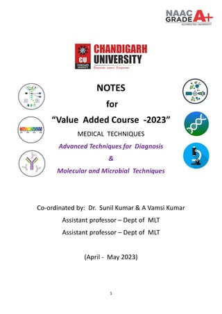
  • 1. 1 NOTES for “Value Added Course -2023” MEDICAL TECHNIQUES Advanced Techniques for Diagnosis & Molecular and Microbial Techniques Co-ordinated by: Dr. Sunil Kumar & A Vamsi Kumar Assistant professor – Dept of MLT Assistant professor – Dept of MLT (April - May 2023)
  • 2. 2 Table of Contents SYLLABUS................................................................................................................................................3 LECTURE PLAN ........................................................................................................................................4 1. FLOW CYTOMETRY .............................................................................................................................6 Flow cytometry Multiple Choice Questions with Answers...................................................................9 Flow cytometry Assignment Questions with Answers........................................................................12 FLOW CYTOMETRY CASE STUDIES .......................................................................................................15 2. HEMOCYTOMETRY: PRINCIPLES AND APPLICATIONS......................................................................17 Hemocytometry Multiple Choice Questions .......................................................................................20 Hemocytometry Assignment questions with answers........................................................................23 HEMOCYTOMETRY CASE STUDIES .......................................................................................................26 CHROMATOGRAPHY MULTIPLE CHOICE QUESTIONS ..........................................................................31 Chromatography Assignment questions with answers.......................................................................33 CHROMATOGRAPHY CASE STUDIES.....................................................................................................36 4. ELECTROPHORESIS: TYPES, PRINCIPLES AND APPLICATION...........................................................39 Electrophoreses Multiple choice questions.........................................................................................41 Electrophoreses Assignment questions with answers........................................................................44 ELECTROPHORESES CASE STUDIES.......................................................................................................47 5. PCR AND TRANSILLUMINATOR: THEORY AND ITS APPLICATIONS TO BIOMEDICAL FIELD.............49 PCR Multiple choice questions.............................................................................................................52 PCR ASSINGMENT QUESTIONS WITH ANSWERS................................................................................54 PCR CASE STUDIES................................................................................................................................57 Inoculation & Isolation of microbes Multiple choice questions ........................................................60 Inoculation & Isolation of microbes Assignment question with Answers..........................................62 INOCULATION & ISOLATION OF MICROBES CASE STUDIES.................................................................64
  • 3. 3 SYLLABUS Lecture No. Topic Reading Material / Reference No. of Hours Name of the Expert handling the Topic 1. Advance techniques for diagnosis 1. Flowcytometery: Principles and applications. 2. Hemocytometery: Principles and applications. 3. Chromatography: Types, Principles and application 4. Electrophoresis: Types, Principles and application 15 Sunil.e12102@ cumail.in 2. Molecular and microbial Techniques 1. PCR and transillumnator: Theory and its applications to biomedical field. 2. Inoculation and isolation of Microorganism from the different type of samples. 15 Attuluri.e13404 @cumail.in
  • 4. 4 LECTURE PLAN Lecture No. Topic Reading Material / Reference Date & Time 1 Introduction to Advanced Techniques for Diagnosis Basic introduction to diagnostic techniques 15-04-2023 12:00PM 2 Flow Cytometry: Principles Flow Cytometry: A Basic Introduction (Book) 15-04-2023 2:00PM 3 Flow Cytometry: Applications Flow Cytometry Applications in Cell and Molecular Biology (Review) 15-04-2023 2:00PM 4 Hemocytometry: Principles Hemocytometer: Protocol, Calculation, and Calibration (Article) 22-04-2023 12:00PM 5 Hemocytometry: Applications Hemocytometry: Methods and Applications (Review) 22-04-2023 2:00PM 6 Chromatography: Types Introduction to Chromatography (Book) 22-04-2023 3:00PM 7 Chromatography: Principles Chromatographic Separation Methods (Book) 23-04-2023 12:00PM 8 Chromatography: Applications Applications of Chromatography in Life Science (Review) 23-04-2023 2:00PM 9 Electrophoresis: Types Introduction to Gel Electrophoresis (Article) 23-04-2023 3:00PM 10 Electrophoresis: Principles Principles and Methods of Electrophoresis (Book) 29-04-2023 12:00 PM 11 Electrophoresis: Applications Applications of Electrophoresis in Biomedical Research (Review) 29-04-2023 02:00 PM 12 Introduction to Molecular and Microbial Techniques Molecular Biology Techniques: A Classroom Laboratory Manual (Book) 29-04-2023 03:00 PM 13 PCR: Theory PCR: Principles, Procedures, and Applications (Book) 30-04-2023 12:00 PM 14 PCR: Applications to Biomedical Field PCR Applications in Medical Diagnostic: An Overview (Review) 30-04-2023 02:00 PM 15 Transilluminator: Theory Gel Documentation System: An Introduction (Article) 30-04-2023 03:00 PM 16 Transilluminator: Applications Applications of Gel Documentation Systems in Molecular Biology (Review) 06-05-2023 12:00PM 17 Inoculation Techniques Inoculation Methods in Microbiology: An Overview (Article) 06-05-2023 02:00PM 18 Isolation of Microorganisms from Different Samples Microbial Isolation Techniques (Book) 06-05-2023 03:00PM 19 Practical Session: Flow Cytometry Virtual Hands-on lab session 07-05-2023 12:00PM
  • 5. 5 20 Practical Session: Hemocytometry Virtual Hands-on lab session 07-05-2023 2:00PM 21 Practical Session: Chromatography Virtual Hands-on lab session 07-05-2023 3:00PM 22 Practical Session: Electrophoresis Virtual Hands-on lab session 13-05-2023 12:00PM 23 Practical Session: PCR and Transilluminator Virtual Hands-on lab session 13-05-2023 2:00PM 24 Practical Session: Inoculation Techniques Virtual Hands-on lab session 13-05-2023 3:00PM 25 Practical Session: Isolation of Microorganisms Virtual Hands-on lab session 14-05-2023 12:00PM 26 Case Studies: Advanced Diagnostic Techniques Discussion of real-life case studies 14-05-2023 2:00PM 27 Case Studies: Molecular and Microbial Techniques Discussion of real-life case studies 14-05-2023 3:00PM 28 Emerging Diagnostic Techniques Emerging Technologies in Medical Diagnostics (Review) 20-05-2023 12:00PM 29 Ethical Considerations in Diagnostic Techniques Ethical Issues in Biomedical Research and Diagnostic Testing (Article) 20-05-2023 2:00PM 30 Recap and Final Discussion Review of key concepts and Q&A session 20-05-2023 3:00PM
  • 6. 6 1. FLOW CYTOMETRY Introduction • Flow cytometry is a powerful and versatile technique used in the analysis and sorting of cells and particles. • The technique enables researchers to rapidly analyze and quantify multiple physical and chemical characteristics of single cells or particles as they flow in a fluid stream through a beam of light. • The resulting data can be used for various applications, including cell counting, cell sorting, and biomarker detection. I. Principles of Flow Cytometry A. Fluidics system • • It consists of a sheath fluid and a sample fluid, which are combined to form a laminar flow. • Cells in the sample fluid are hydrodynamically focused, allowing them to pass individually through the laser interrogation point. B. Optics system • The optics system consists of lasers, lenses, and filters that direct and collect the light emitted from the cells or particles.
  • 7. 7 • As cells pass through the laser interrogation point, they scatter light and emit fluorescence. • Light scattering is categorized into forward scatter (FSC) and side scatter (SSC). • FSC: Provides information about the size of the cell. • SSC: Provides information about the internal complexity or granularity of the cell. C. Detection system • The detection system consists of photodetectors, such as photomultiplier tubes (PMTs) or avalanche photodiodes (APDs), which convert the emitted light into electrical signals. • The electrical signals are then amplified and processed by analog-to-digital converters (ADCs) and digital signal processors (DSPs). • The resulting digital data are plotted on a histogram or scatter plot, allowing for visual analysis and quantification. II. Applications of Flow Cytometry A. Immunophenotyping • Immunophenotyping is the process of identifying and characterizing cells based on the expression of specific cell surface markers (antigens). • Flow cytometry enables the simultaneous analysis of multiple markers through the use of fluorochrome-conjugated antibodies. • Applications include the identification and quantification of immune cell subsets, cancer cells, and stem cells. B. Cell cycle analysis • Flow cytometry can be used to analyze the DNA content of cells, allowing for the determination of cell cycle distribution. • This can help researchers understand cell proliferation, cell cycle regulation, and the effects of various drugs on cell cycle progression. C. Apoptosis and cell viability • Flow cytometry can be used to assess cell viability and apoptosis by measuring various cellular parameters such as membrane permeability, mitochondrial potential, and activation of caspases. • Common viability and apoptosis assays include Annexin V/PI staining, 7-AAD staining, and TUNEL assay. D. Cell sorting • Fluorescence-activated cell sorting (FACS) is a specialized application of flow cytometry that enables the separation and collection of individual cells based on their characteristics.
  • 8. 8 • This technique is widely used in research and clinical settings for isolating specific cell populations or single cells for further analysis or therapeutic applications. Conclusion • Flow cytometry is a powerful and versatile technique in medical lab technology, enabling the rapid analysis and quantification of multiple cellular parameters. • Its applications range from basic research to clinical diagnostics, making it an essential tool for medical lab technology students to understand and master.
  • 9. 9 Flow cytometry Multiple Choice Questions with Answers 1. What is the primary purpose of flow cytometry? A. Measuring cell size B. Analyzing and quantifying multiple physical and chemical characteristics of single cells or particles C. Counting the number of cells in a sample D. Sorting cells based on their size Answer: B 2. Which two types of light scattering are primarily measured in flow cytometry? A. Forward scatter (FSC) and reverse scatter (RSC) B. Forward scatter (FSC) and side scatter (SSC) C. Side scatter (SSC) and backscatter (BSC) D. Forward scatter (FSC) and total scatter (TSC) Answer: B 3.In the fluidics system of flow cytometry, what is the purpose of hydrodynamic focusing? A. To separate cells based on size B. To align cells in a single-file manner C. To increase the speed of the sample fluid D. To mix the sample fluid with the sheath fluid Answer: B 4. What is the function of photodetectors, such as photomultiplier tubes (PMTs) or avalanche photodiodes (APDs), in flow cytometry? A. Emitting light for cells to pass through B. Converting emitted light into electrical signals C. Aligning cells in a single-file manner D. Focusing the laser beam onto cells Answer: B 5.Which of the following is NOT an application of flow cytometry? A. Immunophenotyping B. Cell cycle analysis C. Microscopy imaging D. Apoptosis and cell viability Answer: C 6. Fluorescence-activated cell sorting (FACS) is a specialized application of flow cytometry used for: A. Cell counting
  • 10. 10 B. Cell sorting and collection C. Cell staining D. Cell cycle analysis Answer: B 7. Which flow cytometry assay is commonly used for assessing apoptosis? A. Annexin V/PI staining B. 7-AAD staining C. BrdU incorporation D. DAPI staining Answer: A 8. In flow cytometry, forward scatter (FSC) provides information about: A. Cell size B. Internal complexity of the cell C. Expression of cell surface markers D. DNA content of the cell Answer: A 9. Which component of a flow cytometer is responsible for transporting and aligning the cells in a single-file manner? A. Fluidics system B. Optics system C. Detection system D. Processing system Answer: A 10. In flow cytometry, what is the role of fluorochrome-conjugated antibodies? A. Focusing the laser beam onto cells B. Converting emitted light into electrical signals C. Identifying and characterizing cells based on specific cell surface markers D. Separating and collecting individual cells Answer: C 11.What does the side scatter (SSC) in flow cytometry provide information about? A. Cell size B. Internal complexity or granularity of the cell C. Expression of cell surface markers D. DNA content of the cell Answer: B
  • 11. 11 12. Which of the following is NOT a component of the optics system in flow cytometry? A. Lasers B. Lenses C. Filters D. Photodetectors Answer: D 13. In flow cytometry, which component is responsible for converting electrical signals into digital data? A. Analog-to-digital converters (ADCs) B. Photomultiplier tubes (PMTs) C. Digital signal processors (DSPs) D. Avalanche photodiodes (APDs) Answer: A 14. Which of the following assays is commonly used for cell cycle analysis in flow cytometry? A. Annexin V/PI staining B. 7-AAD staining C. BrdU incorporation D. TUNEL assay Answer: C 15. What is the primary difference between flow cytometry and fluorescence-activated cell sorting (FACS)? A. Flow cytometry analyzes cells, while FACS sorts and collects cells. B. Flow cytometry uses lasers, while FACS does not. C. FACS requires the use of fluorochrome-conjugated antibodies, while flow cytometry does not. D. Flow cytometry is used for clinical diagnostics, while FACS is used for research purposes only. Answer: A
  • 12. 12 Flow cytometry Assignment Questions with Answers Question 1: Explain the three main components of flow cytometry and their functions. Answer: The three main components of flow cytometry are the fluidics system, optics system, and detection system. The fluidics system is responsible for transporting and aligning the cells in a single-file manner, utilizing a sheath fluid and a sample fluid that are combined to form a laminar flow. The optics system consists of lasers, lenses, and filters that direct and collect the light emitted from the cells or particles as they pass through the laser interrogation point, scattering light and emitting fluorescence. The detection system comprises photodetectors such as photomultiplier tubes (PMTs) or avalanche photodiodes (APDs), which convert the emitted light into electrical signals. These electrical signals are then amplified and processed by analog-to-digital converters (ADCs) and digital signal processors (DSPs), resulting in digital data that can be plotted on a histogram or scatter plot for analysis and quantification. Question 2: What information do forward scatter (FSC) and side scatter (SSC) provide in flow cytometry? Answer: Forward scatter (FSC) provides information about the size of the cell. Larger cells scatter more light in the forward direction, resulting in a higher FSC signal. Side scatter (SSC) provides information about the internal complexity or granularity of the cell. Cells with more complex internal structures, such as granules, scatter more light in the side direction, resulting in a higher SSC signal. Question 3: How does flow cytometry facilitate immunophenotyping? Answer: Flow cytometry facilitates immunophenotyping by enabling the simultaneous analysis of multiple markers on cells through the use of fluorochrome-conjugated antibodies. These antibodies specifically bind to cell surface markers (antigens) and emit fluorescence when excited by a laser. By using different fluorochromes for different antibodies, researchers can analyze the expression of multiple cell surface markers simultaneously, identifying and characterizing distinct cell populations. Question 4: Describe how flow cytometry can be used for cell cycle analysis. Answer: Flow cytometry can be used for cell cycle analysis by measuring the DNA content of cells. Cells are stained with DNA-binding dyes, such as propidium iodide (PI) or DAPI, which emit fluorescence when bound to DNA. As the cells pass through the flow cytometer's laser, the
  • 13. 13 emitted fluorescence is proportional to their DNA content. This information allows researchers to determine the cell cycle distribution by distinguishing cells in different phases of the cell cycle (G0/G1, S, and G2/M), providing insights into cell proliferation and the effects of drugs on cell cycle progression. Question 5: Explain two common assays used for assessing apoptosis and cell viability using flow cytometry. Answer: Annexin V/PI staining: Annexin V is a protein that binds to phosphatidylserine, which is externalized on the outer leaflet of the plasma membrane during early apoptosis. Propidium iodide (PI) is a DNA-binding dye that can only enter cells with compromised membranes, indicative of late apoptosis or necrosis. By staining cells with Annexin V and PI, researchers can distinguish viable cells (Annexin V-negative, PI-negative), early apoptotic cells (Annexin V-positive, PI-negative), and late apoptotic or necrotic cells (Annexin V-positive, PI-positive). Question 6: What is the primary difference between flow cytometry and fluorescence- activated cell sorting (FACS)? Answer: The primary difference between flow cytometry and fluorescence-activated cell sorting (FACS) is their purpose. Flow cytometry is primarily used to analyze and quantify multiple physical and chemical characteristics of single cells or particles. In contrast, FACS is a specialized application of flow cytometry that enables the separation and collection of individual cells based on their specific characteristics, such as size, granularity, or marker expression. Question 7: Design a simple experiment using flow cytometry to study the effects of a drug on immune cell populations. Provide an overview of the experimental design, including sample preparation, staining, data acquisition, and data analysis. Answer: Overview of the experiment: 1. Sample preparation: Obtain peripheral blood mononuclear cells (PBMCs) from healthy donors and culture them in appropriate media. Treat one group of cells with the drug of interest at different concentrations and durations, while leaving another group untreated as a control. 2. Staining: Use fluorochrome-conjugated antibodies specific for immune cell markers (e.g., CD3 for T cells, CD19 for B cells, and CD14 for monocytes) to stain the cells according to the manufacturer's protocol. 3. Data acquisition: Run the stained samples through a flow cytometer to measure fluorescence intensity and light scattering properties (FSC and SSC) for each cell. Acquire a minimum of 10,000 events per sample. 4. Data analysis: Use flow cytometry software to analyze the data by gating on specific immune cell populations based on their marker expression. Calculate the percentage of each immune cell subset in the treated and control groups and compare the results to assess the drug's effect on immune cell populations.
  • 14. 14 Question 8: What are some potential limitations and challenges associated with flow cytometry, and how can these be addressed or mitigated to ensure accurate and reliable results? Answer: Some potential limitations and challenges associated with flow cytometry include: 1. Autofluorescence: Cells may have intrinsic fluorescence, which can interfere with the detection of specific fluorochromes. This can be addressed by selecting fluorochromes with minimal overlap in their emission spectra and using appropriate compensation controls. 2. Spectral overlap: Emission spectra of different fluorochromes can overlap, making it difficult to distinguish between them. To mitigate this issue, use proper compensation controls and carefully select fluorochromes with minimal spectral overlap. 3. Non-specific antibody binding: Antibodies may bind non-specifically to cells, causing false-positive signals. To minimize non-specific binding, use appropriate isotype controls, Fc receptor blocking reagents, and optimize antibody concentrations. 4. Sample variability: Variability in sample preparation, staining, and instrument settings can lead to inconsistent results. Standardize protocols, use appropriate controls, and regularly perform instrument quality control checks to ensure consistent and accurate data. By addressing these challenges and implementing appropriate controls and optimization strategies, researchers can obtain accurate and reliable results from flow cytometry experiments.
  • 15. 15 FLOW CYTOMETRY CASE STUDIES Case Study 1: Immunophenotyping of Leukemia Patients Background: Leukemia is a heterogeneous group of hematological malignancies characterized by the uncontrolled proliferation of abnormal white blood cells. Flow cytometry plays a critical role in the diagnosis, classification, and monitoring of leukemia by immunophenotyping leukemic cells based on their specific cell surface markers. Objective: Use flow cytometry to distinguish between different types of leukemia in patient samples based on their immunophenotypic profiles. Methods: Collect bone marrow or peripheral blood samples from patients with suspected leukemia. Prepare single-cell suspensions, and stain them with a panel of fluorochrome-conjugated antibodies specific for various cell surface markers associated with different types of leukemia (e.g., CD45, CD19, CD34, CD33, etc.). Run the stained samples through a flow cytometer and analyze the data using appropriate gating strategies and software tools. Results and Interpretation: By analyzing the expression of specific cell surface markers, different types of leukemia can be identified: Acute Lymphoblastic Leukemia (ALL): High expression of CD19 and CD10, often accompanied by CD34 and TdT. Acute Myeloid Leukemia (AML): High expression of CD33, CD13, and CD117, often accompanied by CD34. Chronic Lymphocytic Leukemia (CLL): High expression of CD19, CD5, and CD23, with low expression of CD20. Chronic Myeloid Leukemia (CML): High expression of CD33, CD13, and CD34, often accompanied by CD38. By comparing the immunophenotypic profiles of patient samples to these reference profiles, clinicians can accurately diagnose and classify leukemia cases, guiding appropriate treatment and monitoring strategies. Case Study 2: Assessing the Effects of a Chemotherapeutic Drug on Apoptosis Background: Apoptosis, or programmed cell death, is an essential process in the maintenance of tissue homeostasis. Many chemotherapeutic drugs exert their anticancer effects by inducing apoptosis in malignant cells. Flow cytometry can be used to assess the apoptotic effects of these drugs on cancer cells. Objective: Evaluate the pro-apoptotic effects of a chemotherapeutic drug on a cancer cell line using flow cytometry. Methods:
  • 16. 16 Culture a cancer cell line and treat with the chemotherapeutic drug at various concentrations and durations. Harvest the cells and stain them with Annexin V-FITC and propidium iodide (PI) according to the manufacturer's protocol. Run the stained samples through a flow cytometer and analyze the data using appropriate gating strategies and software tools. Results and Interpretation: By analyzing the Annexin V/PI staining, the percentage of viable, early apoptotic, and late apoptotic/necrotic cells can be determined: Viable cells: Annexin V-negative, PI-negative Early apoptotic cells: Annexin V-positive, PI-negative Late apoptotic/necrotic cells: Annexin V-positive, PI-positive By comparing the percentages of apoptotic cells in treated and untreated samples, researchers can evaluate the pro-apoptotic effects of the chemotherapeutic drug on the cancer cell line. These findings can inform the selection and optimization of drug concentrations and treatment durations for effective cancer therapy.
  • 17. 17 2. HEMOCYTOMETRY: PRINCIPLES AND APPLICATIONS I. Introduction A. Definition of hemocytometry 1. Hemocytometry refers to the quantitative measurement of cells, particularly blood cells, in a given volume of a liquid sample. B. Importance of hemocytometry 1. Crucial in clinical diagnostics, research, and treatment monitoring 2. Determines cell concentration and viability 3. Supports diagnosis of various blood disorders and infections II. Principles of Hemocytometry A. Hemocytometer 1. Specialized counting chamber used for hemocytometry 2. Composed of a thick glass microscope slide with a grid of etched lines 3. Grid subdivided into various squares of known dimensions to facilitate counting B. Sample preparation 1. Dilution of the blood sample 2. Staining (optional) to differentiate between cell types or identify dead cells C. Counting method 1. Manual counting using a light microscope 2. Automated cell counters using electrical impedance, flow cytometry, or image analysis techniques III. Applications in Medical Lab Technology A. Complete Blood Count (CBC) 1. Measures the concentration of red blood cells (RBCs), white blood cells (WBCs), and platelets
  • 18. 18 2. Provides information on hemoglobin, hematocrit, and mean cell volume B. Differential leukocyte count 1. Determines the relative percentage of each type of WBC (neutrophils, lymphocytes, monocytes, eosinophils, and basophils) 2. Aids in the diagnosis of infections, inflammatory disorders, and malignancies C. Reticulocyte count 1. Measures the number of immature RBCs (reticulocytes) 2. Assesses bone marrow function and response to anemia treatment D. Cell viability assays 1. Evaluates the effectiveness of drug treatments, radiation, or other therapies on cell survival 2. Guides treatment decisions in cancer and other disorders IV. Limitations and Challenges A. Manual counting 1. Time-consuming and labor-intensive 2. Inherent variability due to human error 3. Requires skilled and experienced personnel B. Automated counters 1. Expensive initial investment 2. Maintenance and calibration requirements 3. Potential for inaccurate results due to instrument limitations or sample quality issues VI. Total White Blood Cell (TWBC) and Total Red Blood Cell (TRBC) Counts by Visual Method A. Total White Blood Cell (TWBC) Count by Visual Method 1. Objective: To determine the number of white blood cells per microliter (µL) of blood 2. Sample preparation a. Blood sample mixed with a diluent (e.g., Turk's solution) to lyse RBCs and enhance WBC visibility b. The diluted sample is loaded onto the hemocytometer 3. Counting procedure a. Using a light microscope, focus on the grid of the hemocytometer b. Count the WBCs within specified grid squares c.Apply the appropriate calculation to determine the TWBC concentration in the original blood sample B. Total Red Blood Cell (TRBC) Count by Visual Method 1. Objective: To determine the number of red blood cells per microliter (µL) of blood 2. Sample preparation a. Blood sample mixed with a diluent (e.g., Hayem's solution) to prevent RBC clumping b. The diluted sample is loaded onto the hemocytometer 3. Counting procedure a. Using a light microscope, focus on the grid of the hemocytometer b. Count the RBCs within specified grid squares
  • 19. 19 c. Apply the appropriate calculation to determine the TRBC concentration in the original blood sample C. Calculations for TWBC and TRBC Counts 1. Formula: (Total number of cells counted / Number of squares counted) × Dilution factor × 10^4 2. The result represents the cell concentration in cells per microliter (µL) of the original blood sample 3. Ensure correct dilution factors and grid square specifications are used for accurate results D. Importance of TWBC and TRBC Counts by Visual Method 1. TWBC and TRBC counts provide critical information for diagnosing and monitoring various medical conditions 2. The visual method is cost-effective and accessible in resource-limited settings 3. Visual counting serves as a useful technique for cross-checking automated cell counter results, ensuring accuracy and reliability VII. Conclusion A. Hemocytometry is a fundamental technique in medical lab technology with broad applications in diagnostics, research, and treatment monitoring. B. Understanding the principles and applications of hemocytometry is essential for students pursuing careers in medical lab technology.
  • 20. 20 Hemocytometry Multiple Choice Questions 1. What is the main purpose of hemocytometry? A. To measure blood pressure B. To quantify cells in a liquid sample C. To identify specific blood proteins D. To measure blood glucose levels Answer: B. To quantify cells in a liquid sample 2. What is a hemocytometer? A. A type of blood cell B. A specialized counting chamber C. A laboratory instrument for measuring blood pressure D. A device for separating blood components Answer: B. A specialized counting chamber 3. Which of the following is NOT an application of hemocytometry in medical lab technology? A. Complete Blood Count (CBC) B. Differential leukocyte count C. Blood glucose measurement D. Reticulocyte count Answer: C. Blood glucose measurement 4. Which diluent is commonly used for Total White Blood Cell (TWBC) count by the visual method? A. Turk's solution B. Hayem's solution C. Wright's stain D. Giemsa stain Answer: A. Turk's solution 5. In the visual method of Total Red Blood Cell (TRBC) count, what is the main purpose of using a diluent like Hayem's solution? A. To lyse red blood cells B. To prevent red blood cell clumping C. To stain red blood cells for easier identification D. To promote red blood cell agglutination Answer: B. To prevent red blood cell clumping 6. When manually counting cells using a hemocytometer, which factor must be considered to calculate the concentration of cells in the original blood sample? A. The number of cells counted B. The dilution factor C. The number of squares counted D. All of the above Answer: D. All of the above
  • 21. 21 7. Which of the following blood components is NOT a part of a Complete Blood Count (CBC)? A. Red blood cells B. White blood cells C. Platelets D. Blood glucose Answer: D. Blood glucose 8. In the context of hemocytometry, what is the main advantage of using automated cell counters over manual counting methods? A. They require less skill and experience B. They are less expensive C. They have a higher risk of inaccuracies due to instrument limitations D. They are more time-consuming Answer: A. They require less skill and experience 9. Which of the following is NOT a limitation of manual cell counting using a hemocytometer? A. Time-consuming and labor-intensive B. Inherent variability due to human error C. Requires skilled and experienced personnel D. Expensive initial investment Answer: D. Expensive initial investment 10. Which type of blood cell count is used to assess bone marrow function and response to anemia treatment? A. Red blood cell count B. White blood cell count C. Reticulocyte count D. Platelet count Answer: C. Reticulocyte count 11. Which of the following white blood cell types is NOT a part of the differential leukocyte count? A. Neutrophils B. Erythrocytes C. Monocytes D. Basophils Answer: B. Erythrocytes 12. In hemocytometry, the mean cell volume (MCV) provides information about: A. The average size of red blood cells B. The average size of white blood cells C. The total number of red blood cells D. The total number of white blood cells Answer: A. The average size of red blood cells 13. What is the primary purpose of staining in the context of hemocytometry?
  • 22. 22 A. To lyse red blood cells B. To differentiate between cell types or identify dead cells C. To prevent cell clumping D. To enhance cell visibility under a microscope Answer: B. To differentiate between cell types or identify dead cells 14. When using a hemocytometer, what is the purpose of the etched grid lines? A. To magnify the cells B. To facilitate counting by providing a defined area C. To stain the cells D. To separate the cells into different types Answer: B. To facilitate counting by providing a defined area 15. Which of the following is NOT an example of an automated cell counting method? A. Electrical impedance B. Flow cytometry C. Light microscopy D. Image analysis Answer: C. Light microscopy
  • 23. 23 Hemocytometry Assignment questions with answers Assignment Question 1: Describe the procedure for manually counting Total White Blood Cell (TWBC) and Total Red Blood Cell (TRBC) counts using a hemocytometer. Include information about sample preparation, counting, and calculation. Answer: Sample Preparation: a. TWBC: Mix the blood sample with a diluent such as Turk's solution, which lyses RBCs and enhances WBC visibility. For TRBC, mix the blood sample with a diluent like Hayem's solution, which prevents RBC clumping. b. Load the appropriately diluted sample onto the hemocytometer. Counting: a. Using a light microscope, focus on the grid of the hemocytometer. b. For TWBC, count the WBCs within specified grid squares. For TRBC, count the RBCs within specified grid squares. Calculation: a. Use the formula: (Total number of cells counted / Number of squares counted) × Dilution factor × 10^4 b. The result represents the cell concentration in cells per microliter (µL) of the original blood sample. Assignment Question 2: Explain the advantages and limitations of manual cell counting and automated cell counting in hemocytometry. Answer: Manual cell counting: Advantages: 1. Cost-effective and accessible in resource-limited settings. 2. Can serve as a cross-checking technique for automated cell counter results. Limitations: 1. Time-consuming and labor-intensive. 2. Inherent variability due to human error. 3. Requires skilled and experienced personnel. Automated cell counting: Advantages: 1. Faster and more efficient than manual counting. 2. Requires less skill and experience. 3. Reduces the risk of human error.
  • 24. 24 Limitations: 1. Expensive initial investment. 2. Maintenance and calibration requirements. 3. Potential for inaccurate results due to instrument limitations or sample quality issues. Assignment Question 3: Describe the importance of differential leukocyte count in clinical diagnostics and provide examples of medical conditions that it can help diagnose. Answer: The differential leukocyte count determines the relative percentage of each type of white blood cell (neutrophils, lymphocytes, monocytes, eosinophils, and basophils). This count aids in the diagnosis of infections, inflammatory disorders, and malignancies. Examples of medical conditions that can be diagnosed or monitored using differential leukocyte count include: Bacterial infections: Typically, an increase in neutrophils is observed. Viral infections: An increase in lymphocytes is commonly seen. Parasitic infections and allergic reactions: Eosinophil levels tend to rise. Chronic inflammatory disorders: An increase in monocytes may be observed. Leukemia: Abnormal or immature white blood cells may be present, and the overall WBC count may be altered. Assignment Question 4: Discuss the role of reticulocyte count in the context of anemia and explain how it is useful in assessing bone marrow function and response to treatment. Answer: Reticulocyte count measures the number of immature red blood cells (reticulocytes) in the blood. Reticulocytes are released from the bone marrow into the bloodstream as part of the normal red blood cell production process. In the context of anemia, reticulocyte count serves several important purposes: Assessing bone marrow function: A high reticulocyte count indicates that the bone marrow is actively producing red blood cells in response to anemia, whereas a low reticulocyte count suggests that the bone marrow is not producing an adequate number of red blood cells, which may indicate bone marrow dysfunction or suppression. Evaluating the cause of anemia: Reticulocyte count can help distinguish between different types of anemia. For instance, a high reticulocyte count may suggest hemolytic anemia or acute blood loss, while a low reticulocyte count may indicate iron deficiency anemia or aplastic anemia. Monitoring response to treatment: An increase in reticulocyte count following treatment for anemia (e.g., iron supplementation, erythropoietin administration, or blood transfusion)
  • 25. 25 indicates a positive response to the treatment, suggesting that the bone marrow is producing more red blood cells. Assignment Question 5: Explain the significance of hemocytometry in medical lab technology and list at least three applications of hemocytometry in clinical diagnostics, research, or treatment monitoring. Answer: Hemocytometry is a fundamental technique in medical lab technology that plays a crucial role in clinical diagnostics, research, and treatment monitoring. It is used to determine cell concentration and viability, which is essential for diagnosing and monitoring various blood disorders and infections. Three applications of hemocytometry in clinical diagnostics, research, or treatment monitoring include: Complete Blood Count (CBC): Measures the concentration of red blood cells, white blood cells, and platelets, providing information on hemoglobin, hematocrit, and mean cell volume. CBC is important for diagnosing various blood disorders, such as anemia, thrombocytopenia, and leukopenia. Differential leukocyte count: Determines the relative percentage of each type of white blood cell, aiding in the diagnosis of infections, inflammatory disorders, and malignancies. Cell viability assays: Evaluates the effectiveness of drug treatments, radiation, or other therapies on cell survival. This information can help guide treatment decisions in cancer and other disorders, as well as inform the development of new therapeutic strategies in research settings.
  • 26. 26 HEMOCYTOMETRY CASE STUDIES Case Study 1: A 30-year-old female patient presents to the clinic with fatigue, pallor, and shortness of breath. The physician orders a Complete Blood Count (CBC) and a reticulocyte count to determine the cause of her symptoms. CBC Results: Hemoglobin: 9 g/dL (Normal range: 12-15.5 g/dL) Hematocrit: 30% (Normal range: 36-46%) RBC count: 3.8 million/µL (Normal range: 4.2-5.4 million/µL) WBC count: 6,000/µL (Normal range: 4,000-11,000/µL) Platelet count: 250,000/µL (Normal range: 150,000-400,000/µL) Reticulocyte count: 1% (Normal range: 0.5-1.5%) Answer: Based on the results, the patient has anemia, as indicated by low hemoglobin, hematocrit, and RBC count. The normal reticulocyte count suggests that the bone marrow is not responding to the anemia, which could indicate iron deficiency anemia, vitamin B12 deficiency, or folic acid deficiency. Further diagnostic tests, such as serum iron, ferritin, and vitamin levels, would be necessary to pinpoint the specific cause of the patient's anemia and guide treatment. Case Study 2: A25-year-old male patient presents to the clinic with a high fever, sore throat, and swollen lymph nodes. The physician orders a CBC with differential leukocyte count to investigate the cause of his symptoms. CBC Results: Hemoglobin: 15 g/dL (Normal range: 13.5-17.5 g/dL) Hematocrit: 45% (Normal range: 41-53%) RBC count: 5 million/µL (Normal range: 4.5-5.9 million/µL) WBC count: 15,000/µL (Normal range: 4,000-11,000/µL) Platelet count: 300,000/µL (Normal range: 150,000-400,000/µL) Differential leukocyte count results: Neutrophils: 70% (Normal range: 40-60%) Lymphocytes: 20% (Normal range: 20-40%) Monocytes: 5% (Normal range: 2-10%) Eosinophils: 3% (Normal range: 1-4%) Basophils: 2% (Normal range: 0.5-1%) Answer: The patient's elevated WBC count and increased neutrophil percentage suggest a bacterial infection, consistent with the symptoms of fever, sore throat, and swollen lymph nodes. The physician might consider prescribing antibiotics to treat the suspected bacterial infection and recommend additional diagnostic tests, such as a throat swab culture, to confirm the diagnosis and guide appropriate antibiotic therapy.
  • 27. 27 Case Study 4: A 45-year-old female patient presents to the clinic with recurrent sinus infections, fatigue, and joint pain. The physician orders a CBC with differential leukocyte count to investigate the cause of her symptoms. CBC Results: Hemoglobin: 13 g/dL (Normal range: 12-15.5 g/dL) Hematocrit: 40% (Normal range: 36-46%) RBC count: 4.5 million/µL (Normal range: 4.2-5.4 million/µL) WBC count: 10,000/µL (Normal range: 4,000-11,000/µL) Platelet count: 275,000/µL (Normal range: 150,000-400,000/µL) Differential leukocyte count results: Neutrophils: 50% (Normal range: 40-60%) Lymphocytes: 35% (Normal range: 20-40%) Monocytes: 12% (Normal range: 2-10%) Eosinophils: 2% (Normal range: 1-4%) Basophils: 1% (Normal range: 0.5-1%) Answer: The patient's slightly elevated monocyte percentage may indicate a chronic inflammatory condition, such as an autoimmune disorder. The physician might consider ordering additional tests, such as antinuclear antibody (ANA) or rheumatoid factor (RF), to investigate the possibility of an autoimmune disorder, such as lupus or rheumatoid arthritis, which could explain the patient's fatigue, joint pain, and recurrent infections.
  • 28. 28 3. CHROMATOGRAPHY: TYPES, PRINCIPLES AND APPLICATION Chromatography is a laboratory technique used to separate and identify the components of a mixture. It is widely used in medical laboratory technology to analyze various types of biological samples, such as blood, urine, and cerebrospinal fluid. TYPES OF CHROMATOGRAPHY: Gas Chromatography (GC): It is used to separate and analyze volatile organic compounds. In GC, the sample is vaporized and passed through a column containing a stationary phase. The components in the mixture interact differently with the stationary phase and are separated based on their volatility. Liquid Chromatography (LC): It is used to separate and analyze non-volatile and semi- volatile compounds. In LC, the sample is dissolved in a liquid and passed through a
  • 29. 29 column containing a stationary phase. The components in the mixture interact differently with the stationary phase and are separated based on their solubility and polarity. High-Performance Liquid Chromatography (HPLC): It is a type of liquid chromatography that uses high pressure to increase the separation efficiency. It is commonly used in medical laboratories to analyze drugs, hormones, and other biomolecules. Thin Layer Chromatography (TLC): It is a type of chromatography where the stationary phase is coated on a thin layer of a solid support, such as a glass plate or a plastic sheet. The sample is applied as a spot on the stationary phase and is separated based on its interaction with the stationary phase. PRINCIPLES OF CHROMATOGRAPHY: The principle of chromatography is based on the differential interaction of the components of a mixture with the stationary and mobile phases. The stationary phase is a solid or liquid support, while the mobile phase is a gas or liquid that carries the sample through the stationary phase. The components in the mixture interact differently with the stationary phase, causing them to move at different rates through the column. The degree of separation depends on the interaction between the components and the stationary phase. APPLICATIONS OF CHROMATOGRAPHY: 1. Chromatography has a wide range of applications in medical laboratory technology. Some of the common applications are: 2. Drug analysis: Chromatography is used to analyze drugs and their metabolites in biological samples such as blood and urine. 3. Hormone analysis: Chromatography is used to analyze hormones such as insulin, estrogen, and testosterone. 4. Protein analysis: Chromatography is used to separate and purify proteins for various applications such as drug development, vaccine production, and biotechnology. 5. Environmental analysis: Chromatography is used to analyze pollutants and contaminants in environmental samples such as air, water, and soil. 6. Food analysis: Chromatography is used to analyze food additives, contaminants, and flavor compounds.
  • 30. 30
  • 31. 31 CHROMATOGRAPHY MULTIPLE CHOICE QUESTIONS 1. Which type of chromatography is used to analyze volatile organic compounds? A) Gas chromatography B) Liquid chromatography C) High-performance liquid chromatography D) Thin layer chromatography Answer: A) Gas chromatography 2. In chromatography, the stationary phase is: A) A gas B) A liquid C) A solid or liquid support D) A mobile phase Answer: C) A solid or liquid support 3. Which of the following is NOT a common application of chromatography in medical laboratory technology? A) Drug analysis B) Hormone analysis C) Protein analysis D) Weather analysis Answer: D) Weather analysis 4. What is the principle of chromatography based on? A) Differential interaction of the components with the stationary and mobile phases B) Interaction of the components with a gas phase C) Separation based on size D) Separation based on color Answer: A) Differential interaction of the components with the stationary and mobile phases 5. What is the purpose of high-performance liquid chromatography? A) To separate and analyze non-volatile and semi-volatile compounds B) To increase the separation efficiency of liquid chromatography C) To analyze volatile organic compounds D) To separate components based on their size Answer: B) To increase the separation efficiency of liquid chromatography 6. Which type of chromatography is commonly used to analyze contaminants in environmental samples? A) Gas chromatography B) Liquid chromatography C) High-performance liquid chromatography D) Thin layer chromatography Answer: B) Liquid chromatography 7. What is the mobile phase in chromatography?
  • 32. 32 A) A gas or liquid that carries the sample through the stationary phase B) A solid or liquid support C) A gas that interacts with the components of the mixture D) A liquid that dissolves the components of the mixture Answer: A) A gas or liquid that carries the sample through the stationary phase 8. What is the most common application of thin layer chromatography? A) Protein analysis B) Hormone analysis C) Separation of non-volatile compounds D) Separation of volatile compounds Answer: D) Separation of volatile compounds 9. What is the purpose of protein analysis using chromatography? A) To analyze drugs and their metabolites B) To separate and purify proteins for various applications C) To analyze hormones D) To analyze environmental pollutants Answer: B) To separate and purify proteins for various applications 10. Which type of chromatography is commonly used to analyze drugs and their metabolites in biological samples? A) Gas chromatography B) Liquid chromatography C) High-performance liquid chromatography D) Thin layer chromatography Answer: C) High-performance liquid chromatography
  • 33. 33 Chromatography Assignment questions with answers 1. Describe the principle of chromatography and how it works in separating the components of a mixture. Provide an example of a common application of chromatography in medical laboratory technology. Answer: Chromatography works based on the differential interaction of the components of a mixture with the stationary and mobile phases. The sample is passed through a column containing a stationary phase, and the components interact differently with the stationary phase, causing them to move at different rates and be separated from each other. An example of a common application of chromatography in medical laboratory technology is the analysis of hormones using liquid chromatography. 2. Compare and contrast gas chromatography and liquid chromatography. What are the differences in the stationary phase and mobile phase, and what types of compounds are commonly analyzed using each technique? Answer: Gas chromatography (GC) is used to analyze volatile organic compounds and has a stationary phase that is a solid support, while the mobile phase is a gas. Liquid chromatography (LC) is used to analyze non-volatile and semi-volatile compounds and has a stationary phase that is a liquid support, while the mobile phase is a liquid. GC is useful for analyzing compounds that can be vaporized, while LC is more versatile and can be used for a wider range of compounds. 3. Explain the purpose of high-performance liquid chromatography (HPLC) and how it differs from standard liquid chromatography. What are some common applications of HPLC in medical laboratory technology? Answer: High-performance liquid chromatography (HPLC) uses high pressure to increase the separation efficiency of liquid chromatography. It differs from standard liquid chromatography in that it requires specialized equipment, and is more efficient and accurate. HPLC is commonly used in medical laboratory technology to analyze drugs, hormones, and other biomolecules. 4. What are the advantages and limitations of thin layer chromatography (TLC) compared to other types of chromatography? Describe a scenario in which TLC might be the preferred method of analysis. Answer: Thin layer chromatography (TLC) is a simpler and less expensive form of chromatography than other types, and can be used to analyze small samples quickly. However, it is less efficient than other types of chromatography and may not provide enough separation for more complex mixtures. TLC might be the preferred method of analysis when analyzing small, simple mixtures or when speed and simplicity are more important than precision.
  • 34. 34 5. Analyze the role of chromatography in drug development and manufacturing. How is chromatography used to analyze the purity and concentration of drugs, and what are the implications of accurate drug analysis in clinical settings? Answers: Chromatography plays a critical role in drug development and manufacturing by ensuring the purity and concentration of drugs. Chromatography is used to analyze drugs and their metabolites in biological samples, as well as to separate and purify proteins for various applications. Accurate drug analysis is essential in clinical settings to ensure the safety and efficacy of treatments, and to minimize the risk of adverse drug reactions. 6. Analyze the role of gas chromatography in forensic toxicology. How is this technique used to detect and analyze drugs and other toxic substances in biological samples, and what are some common limitations and challenges associated with this analysis? Answer: Gas chromatography is a common technique used in forensic toxicology to analyze biological samples for the presence of drugs and other toxic substances. This technique is useful because it can analyze volatile compounds and is very sensitive, but there are also some limitations and challenges associated with this analysis. For example, GC cannot analyze non- volatile or polar compounds and requires specialized equipment and expertise to perform accurately. 7. Describe the use of liquid chromatography-mass spectrometry (LC-MS) in clinical laboratory settings. How does this technique improve upon traditional liquid chromatography, and what are some common applications of LC-MS in clinical diagnostics? Answer: Liquid chromatography-mass spectrometry (LC-MS) is a powerful technique used in clinical laboratory settings to analyze complex biological samples. This technique combines the separation power of liquid chromatography with the specificity and sensitivity of mass spectrometry to provide accurate and precise analyses of various biomolecules. LC-MS is commonly used in clinical diagnostics to analyze drugs, hormones, and other biomolecules in biological samples. 8. Discuss the importance of proper sample preparation in chromatography. What are some common methods used to prepare samples for analysis, and what factors must be considered when selecting a preparation method? Answer: Proper sample preparation is critical for accurate and reliable chromatography analyses. Sample preparation methods vary depending on the type of sample being analyzed and the type of chromatography being used. Common sample preparation methods include filtration, centrifugation, extraction, and derivatization. Factors to consider when selecting a sample preparation method include sample matrix, analyte stability, and detection limits.
  • 35. 35 9. Explain the use of chromatography in food safety and quality control. What types of compounds can be analyzed using this technique, and what are some common applications of chromatography in the food industry? Answer: Chromatography is commonly used in food safety and quality control to analyze a wide range of compounds, including additives, contaminants, and flavor compounds. This technique can be used to identify and quantify these compounds in various food products, such as meat, dairy, and produce. Some common applications of chromatography in the food industry include analysis of pesticide residues, detection of food fraud, and monitoring of food additives and preservatives. 10. Discuss the potential risks associated with using chromatography in a laboratory setting. What safety measures should be taken to minimize these risks, and how can laboratory personnel protect themselves from exposure to hazardous substances? Answer: Chromatography involves the use of hazardous chemicals and materials, which can pose health and safety risks if not handled properly. To minimize these risks, laboratory personnel should be trained in proper handling, storage, and disposal of hazardous substances. Safety measures should be taken, such as wearing appropriate personal protective equipment, using fume hoods, and following established protocols for handling and disposing of hazardous materials. Regular safety inspections and risk assessments can also help identify potential hazards and prevent accidents.
  • 36. 36 CHROMATOGRAPHY CASE STUDIES 1. Case study: A patient is admitted to the hospital with symptoms of a drug overdose. The medical team suspects that the patient has taken a combination of drugs, but they are not sure which ones. How can chromatography be used to analyze the patient's blood sample and determine which drugs are present? Answer: Chromatography can be used to analyze the patient's blood sample and determine which drugs are present. High-performance liquid chromatography (HPLC) is commonly used to analyze drugs and their metabolites in biological samples such as blood. The blood sample can be passed through a column containing a stationary phase, and the components can be separated based on their interaction with the stationary phase. The separated components can then be detected and identified using mass spectrometry or other techniques. By analyzing the blood sample using chromatography, the medical team can determine which drugs the patient has taken and tailor their treatment accordingly. 2. Case study: A laboratory is analyzing a sample of water from a nearby river and wants to determine the levels of contaminants present. How can chromatography be used to analyze the sample and identify the types and concentrations of pollutants? Answer: Chromatography can be used to analyze the water sample and determine the types and concentrations of pollutants present. Liquid chromatography (LC) is commonly used to analyze pollutants and contaminants in environmental samples such as water. The water sample can be passed through a column containing a stationary phase, and the components can be separated based on their interaction with the stationary phase. The separated components can then be detected and identified using techniques such as mass spectrometry or UV spectroscopy. By analyzing the water sample using chromatography, the laboratory can determine the types and concentrations of pollutants present and monitor the environmental impact of human activity. 3. Case study: A food manufacturer is testing a new batch of spices and wants to ensure that they are free of contaminants and meet quality standards. How can chromatography be used to analyze the spices and identify any impurities or adulterants? Answer: Chromatography can be used to analyze the spices and identify any impurities or adulterants present. Thin layer chromatography (TLC) is commonly used to analyze food additives, contaminants, and flavor compounds. The spices can be applied to a thin layer of a solid support, and the components can be separated based on their interaction with the stationary phase. The separated components can then be detected and identified using techniques such as UV spectroscopy or mass spectrometry. By analyzing the spices using chromatography, the food manufacturer can ensure that they are free of contaminants and meet quality standards. 4. Case study: A research team is analyzing a sample of proteins for use in drug development. How can chromatography be used to separate and purify the proteins and prepare them for further analysis?
  • 37. 37 Answer: Chromatography can be used to separate and purify the proteins and prepare them for further analysis. Column chromatography is commonly used to separate and purify proteins for various applications. The protein sample can be applied to a column containing a stationary phase, and the components can be separated based on their interaction with the stationary phase. The separated components can then be eluted from the column and collected for further analysis. By analyzing the proteins using chromatography, the research team can separate and purify the proteins and prepare them for use in drug development or other applications. 5. Case study: A company is developing a new drug and wants to ensure that it is safe and effective before seeking regulatory approval. How can chromatography be used to analyze the drug and its metabolites in biological samples and ensure its purity and concentration? Answer: Chromatography can be used to analyze the drug and its metabolites in biological samples and ensure its purity and concentration. High-performance liquid chromatography (HPLC) is commonly used to analyze drugs and their metabolites in biological samples such as blood or urine. The sample can be passed through a column containing a stationary phase, and the components can be separated based on their interaction with the stationary phase. The separated components can then be detected and quantified using techniques such as mass spectrometry. By analyzing the drug using chromatography, the company can ensure its purity and concentration and monitor its efficacy and safety in clinical trials. 6. Case study: A laboratory is analyzing a sample of soil from a contaminated site and wants to determine the levels of heavy metals present. How can chromatography be used to analyze the sample and identify the types and concentrations of heavy metals? Answer: Chromatography can be used to analyze the soil sample and determine the types and concentrations of heavy metals present. Ion chromatography (IC) is commonly used to analyze heavy metals in environmental samples such as soil. The soil sample can be dissolved in a liquid phase and passed through a column containing a stationary phase. The heavy metal ions can be separated based on their interaction with the stationary phase, and the separated components can then be detected and identified using techniques such as UV spectroscopy or mass spectrometry. By analyzing the soil sample using chromatography, the laboratory can determine the types and concentrations of heavy metals present and assess the environmental impact of human activity. 7. Case study: A pharmaceutical company is analyzing a sample of a new drug formulation and wants to ensure that it meets quality standards and regulatory requirements. How can chromatography be used to analyze the drug and determine its chemical properties and stability? Answer: Chromatography can be used to analyze the drug and determine its chemical properties and stability. Gas chromatography (GC) or liquid chromatography (LC) is commonly used to analyze drugs for purity, stability, and other quality control parameters. The drug sample can be passed through a column containing a stationary phase, and the components can be separated based on their interaction with the stationary phase. The separated components
  • 38. 38 can then be detected and identified using techniques such as mass spectrometry or UV spectroscopy. By analyzing the drug using chromatography, the pharmaceutical company can ensure that it meets quality standards and regulatory requirements and monitor its stability and chemical properties over time. 8. Case study: A laboratory is analyzing a sample of wine and wants to determine the types and concentrations of flavor compounds present. How can chromatography be used to analyze the sample and identify the flavor compounds? Answer: Chromatography can be used to analyze the wine sample and identify the flavor compounds present. Gas chromatography-mass spectrometry (GC-MS) is commonly used to analyze flavor compounds in food and beverages. The wine sample can be extracted using a liquid phase and injected into a GC-MS instrument. The components can be separated based on their interaction with the stationary phase, and the separated components can then be detected and identified using mass spectrometry. By analyzing the wine using chromatography, the laboratory can determine the types and concentrations of flavor compounds present and assess the quality and characteristics of the wine.
  • 39. 39 4. ELECTROPHORESIS: TYPES, PRINCIPLES AND APPLICATION Electrophoresis is a laboratory technique used to separate and analyze molecules based on their electrical charge and size. It is commonly used in medical laboratories for DNA analysis, protein separation, and clinical diagnosis. Types of Electrophoresis: Agarose Gel Electrophoresis: This method is used for the separation of large molecules such as DNA, RNA, and proteins. The molecules are placed in a gel matrix and an electric current is passed through it. The molecules move through the gel matrix based on their charge and size. The separated molecules can then be visualized by staining the gel. Polyacrylamide Gel Electrophoresis: This method is used for the separation of smaller molecules such as proteins and nucleic acids. The gel matrix used in this method is made up of polyacrylamide, which can create a more detailed separation of molecules based on their size. Principles of Electrophoresis: Electrophoresis is based on the principle that charged molecules will move in an electric field towards the oppositely charged electrode. The rate of movement is determined by the size, charge, and shape of the molecule, as well as the strength of the electric field. Application of Electrophoresis: DNA analysis: Electrophoresis is commonly used for the analysis of DNA. It can be used to separate DNA fragments of different sizes, which can then be used for genetic testing or forensic analysis. Protein separation: Electrophoresis is used to separate and identify proteins in a sample. It is used in clinical laboratories to detect abnormal proteins in diseases such as multiple myeloma.
  • 40. 40 Clinical diagnosis: Electrophoresis can be used for the diagnosis of certain diseases, such as sickle cell anemia. It can also be used to monitor disease progression and treatment efficacy. Isoelectric focusing: This method is used to separate proteins based on their isoelectric point, which is the pH at which a protein has no net electrical charge. Proteins are placed in a gel matrix with a pH gradient and an electric field is applied. Proteins will move towards the area of the gel where the pH matches their isoelectric point and will stop moving once they reach that point. This method is useful for separating proteins with similar molecular weights but different isoelectric points. Capillary electrophoresis: This method uses a narrow capillary tube filled with a buffer solution and an electric field is applied. Molecules move through the capillary based on their charge and size, and they can be detected as they pass a detector at the end of the capillary. This method is used for separating small molecules such as amino acids, peptides, and drugs. Two-dimensional electrophoresis: This method combines two different types of electrophoresis to create a more detailed separation of molecules. The first dimension separates molecules based on their charge, and the second dimension separates them based on their size. This method is commonly used for proteomic analysis to identify proteins and their post- translational modifications. Western blotting: This is a technique used to detect a specific protein in a sample using electrophoresis. Proteins are separated by electrophoresis and transferred to a membrane, which is then incubated with a specific antibody that binds to the protein of interest. The antibody can be detected using a secondary antibody that is linked to an enzyme or a fluorescent dye. Electrophoresis is a versatile technique that has many applications in medical laboratory science. It is important for students to understand the different types of electrophoresis, their principles, and their applications in order to effectively use this technique in their work. In conclusion, Electrophoresis is a powerful laboratory technique that can be used for a wide range of applications. It is essential for medical laboratory technology students to have a good understanding of the principles and types of electrophoresis, as it is commonly used in clinical diagnosis and research.
  • 41. 41 Electrophoreses Multiple choice questions 1. What is the principle of electrophoresis? A. Separation based on size B. Separation based on electrical charge C. Separation based on molecular weight D. Separation based on concentration Answer: B. Separation based on electrical charge. 2. Which of the following is used to separate large molecules such as DNA and RNA? A. Polyacrylamide gel electrophoresis B. Capillary electrophoresis C. Agarose gel electrophoresis D. Isoelectric focusing Answer: C. Agarose gel electrophoresis. 3. What is the purpose of isoelectric focusing? A. Separating molecules based on size B. Separating molecules based on charge C. Separating molecules based on isoelectric point D. Separating molecules based on concentration Answer: C. Separating molecules based on isoelectric point. 4. Which of the following is used to detect specific proteins in a sample? A. Two-dimensional electrophoresis B. Capillary electrophoresis C. Western blotting D. Agarose gel electrophoresis Answer: C. Western blotting. 5. What is the purpose of two-dimensional electrophoresis? A. Separating molecules based on size B. Separating molecules based on charge C. Separating molecules based on isoelectric point and size D. Separating molecules based on concentration Answer: C. Separating molecules based on isoelectric point and size. 6. Which of the following is used to separate proteins based on their molecular weight? A. Polyacrylamide gel electrophoresis B. Capillary electrophoresis C. Agarose gel electrophoresis D. Isoelectric focusing Answer: A. Polyacrylamide gel electrophoresis. 7. What is the purpose of capillary electrophoresis? A. Separating molecules based on size
  • 42. 42 B. Separating molecules based on charge C. Separating molecules based on isoelectric point D. Separating molecules based on concentration Answer: A. Separating molecules based on size. 8. Which of the following is an application of electrophoresis? A. DNA analysis B. Protein synthesis C. Bacterial culture D. Blood pressure measurement Answer: A. DNA analysis. 9. Which of the following is a type of electrophoresis used for separating proteins based on their isoelectric point? A. Polyacrylamide gel electrophoresis B. Capillary electrophoresis C. Western blotting D. Isoelectric focusing Answer: D. Isoelectric focusing. 10. Which of the following combines two different types of electrophoresis to create a more detailed separation of molecules? A. Two-dimensional electrophoresis B. Capillary electrophoresis C. Western blotting D. Agarose gel electrophoresis Answer: A. Two-dimensional electrophoresis. 11. What is the purpose of staining the gel in agarose gel electrophoresis? A. To visualize separated molecules B. To increase the resolution of the separation C. To make the gel more rigid D. To prevent the gel from melting Answer: A. To visualize separated molecules. 12. Which of the following is a common application of polyacrylamide gel electrophoresis? A. DNA analysis B. Protein synthesis C. Blood typing D. Glucose monitoring Answer: A. DNA analysis. 13. Which of the following molecules can be separated using electrophoresis? A. Proteins B. Nucleic acids C. Amino acids
  • 43. 43 D. All of the above Answer: D. All of the above. 14. Which of the following is a disadvantage of capillary electrophoresis compared to gel electrophoresis? A. Capillary electrophoresis requires more sample B. Capillary electrophoresis is slower C. Capillary electrophoresis is less sensitive D. Capillary electrophoresis is more expensive Answer: A. Capillary electrophoresis requires more sample. 15. Which of the following is a type of electrophoresis that separates proteins based on their size and charge in two dimensions? A. Isoelectric focusing B. Western blotting C. Two-dimensional electrophoresis D. Capillary electrophoresis Answer: C. Two-dimensional electrophoresis.
  • 44. 44 Electrophoreses Assignment questions with answers 1. Explain the principle of electrophoresis and the factors that affect the rate of molecule movement during electrophoresis. Answer: Electrophoresis is based on the principle that charged molecules will move in an electric field towards the oppositely charged electrode. The rate of movement is determined by the size, charge, and shape of the molecule, as well as the strength of the electric field. Small molecules will move faster than larger molecules, and molecules with a higher charge will move faster than molecules with a lower charge. The pH of the buffer solution can also affect the movement of molecules in electrophoresis. 2. Compare and contrast the methods of agarose gel electrophoresis and polyacrylamide gel electrophoresis. Provide examples of when each method would be used. Answer:Agarose gel electrophoresis is used for the separation of large molecules such as DNA, RNA, and proteins, while polyacrylamide gel electrophoresis is used for the separation of smaller molecules such as proteins and nucleic acids. Agarose gel electrophoresis is easier to use and less expensive than polyacrylamide gel electrophoresis, but it has a lower resolution. Polyacrylamide gel electrophoresis is more complex and expensive but has a higher resolution. Agarose gel electrophoresis would be used for separating DNA fragments in genetic testing, while polyacrylamide gel electrophoresis would be used for identifying and quantifying proteins in clinical diagnosis. 3. Describe the process of western blotting and its applications in medical laboratory science. Answer: Western blotting is a technique used to detect a specific protein in a sample using electrophoresis. Proteins are separated by electrophoresis and transferred to a membrane, which is then incubated with a specific antibody that binds to the protein of interest. The antibody can be detected using a secondary antibody that is linked to an enzyme or a fluorescent dye. Western blotting can be used for the detection of specific proteins in a sample, which can be helpful in the diagnosis and treatment of diseases such as cancer and autoimmune disorders. 4. Explain the purpose of isoelectric focusing and provide an example of its use in medical laboratory science. Answer: Isoelectric focusing is a method used to separate proteins based on their isoelectric point, which is the pH at which a protein has no net electrical charge. Proteins are placed in a gel matrix with a pH gradient, and an electric field is applied. Proteins will move towards the area of the gel where the pH matches their isoelectric point and will stop moving once they reach that point. Isoelectric focusing can be used to separate and identify different isoforms of a protein, which can be helpful in the diagnosis and treatment of diseases such as multiple sclerosis.
  • 45. 45 5. Discuss the advantages and disadvantages of capillary electrophoresis compared to gel electrophoresis. Answer: Capillary electrophoresis is faster, more sensitive, and more precise than gel electrophoresis. It can separate molecules based on size and charge, and it requires less sample volume than gel electrophoresis. However, capillary electrophoresis is more expensive than gel electrophoresis and requires more specialized equipment. Additionally, it can be more difficult to analyze the results of capillary electrophoresis than gel electrophoresis, as the molecules are not physically separated and must be detected using other methods. 6. Explain the process of two-dimensional gel electrophoresis and its applications in medical laboratory science. Answer: Two-dimensional gel electrophoresis combines two different types of electrophoresis to create a more detailed separation of molecules. The first dimension separates molecules based on their charge, and the second dimension separates them based on their size. This method is commonly used for proteomic analysis to identify proteins and their post- translational modifications. Two-dimensional gel electrophoresis can be used to compare protein expression levels in different samples, identify disease biomarkers, and study protein- protein interactions. 7. Describe the advantages and disadvantages of using polyacrylamide gel electrophoresis for protein separation. Answer: Polyacrylamide gel electrophoresis is a powerful tool for separating and identifying proteins in a sample. It has a high resolution and can separate proteins based on size and charge. However, it is more complex and expensive than other types of electrophoresis, and it requires specialized equipment and expertise. In addition, the process of staining and detecting proteins on a gel can be time-consuming and may require additional steps. Despite these limitations, polyacrylamide gel electrophoresis is still widely used in medical laboratory science for protein analysis. 8. Discuss the role of electrophoresis in DNA analysis and its applications in genetic testing. Answer: Electrophoresis plays a crucial role in DNA analysis, allowing scientists to separate and analyze DNA fragments based on their size. This technique is commonly used in genetic testing to identify genetic disorders, determine paternity, and identify DNA samples in forensic investigations. Gel electrophoresis is typically used for separating DNA fragments, while capillary electrophoresis is used for sequencing DNA. These techniques have revolutionized the field of genetic testing, making it possible to diagnose and treat genetic diseases more accurately and efficiently than ever before. 9. Explain the purpose of capillary electrophoresis for drug analysis and its advantages over other analytical techniques.
  • 46. 46 Answer: Capillary electrophoresis is a powerful analytical technique for drug analysis, allowing scientists to separate and identify small molecules such as drugs and metabolites. It has a high resolution and is faster than other analytical techniques such as high-performance liquid chromatography (HPLC). In addition, it requires less sample volume and has lower solvent consumption than HPLC. Capillary electrophoresis is used for drug discovery and development, pharmacokinetic studies, and forensic drug analysis. 10. Discuss the role of electrophoresis in clinical diagnosis and its applications in disease detection. Answer: Electrophoresis plays a critical role in clinical diagnosis, allowing medical professionals to detect and quantify abnormal proteins in patient samples. It is commonly used in the diagnosis and monitoring of diseases such as multiple myeloma, hemoglobinopathies, and liver disease. By separating and analyzing proteins in patient samples, electrophoresis can provide valuable information about disease progression and treatment efficacy. In addition, electrophoresis is used for the detection of infectious agents such as viruses and bacteria, making it an important tool for public health surveillance and disease control.
  • 47. 47 ELECTROPHORESES CASE STUDIES Case study 1: A patient with multiple myeloma is undergoing treatment to manage their condition. A serum protein electrophoresis (SPEP) test is ordered to monitor the levels of monoclonal immunoglobulins in the patient's blood. The results show a significant decrease in monoclonal immunoglobulin levels compared to the previous test. What does this indicate about the patient's condition? Answer: The decrease in monoclonal immunoglobulin levels indicates that the patient's treatment is effective in managing their multiple myeloma. Monoclonal immunoglobulins are produced by abnormal plasma cells in multiple myeloma, and their levels can be used to monitor disease progression and treatment efficacy. Case study 2: A newborn is screened for sickle cell anemia using electrophoresis. The results show a hemoglobin electrophoresis pattern consistent with sickle cell trait. What does this mean for the child's health? Answer: Sickle cell trait is a genetic condition that occurs when an individual inherits one copy of the sickle cell gene and one copy of a normal gene. While individuals with sickle cell trait usually have no symptoms, they are at risk for complications under certain conditions such as extreme physical exertion, dehydration, and high altitudes. It is important for individuals with sickle cell trait to be aware of their condition and take precautions to prevent complications. Case study 3: A patient presents with symptoms of liver disease, including jaundice and elevated liver enzymes. A serum protein electrophoresis (SPEP) test is ordered to investigate the cause of the patient's symptoms. The results show an abnormal protein band on the electrophoresis gel. What does this indicate about the patient's condition? Answer: The presence of an abnormal protein band on the electrophoresis gel indicates that the patient has an abnormal protein in their blood. This can be a sign of liver disease or other conditions that affect protein production or metabolism. Additional tests, such as liver function tests and imaging studies, may be necessary to diagnose the specific cause of the patient's symptoms. Case study 4: A patient with a history of autoimmune disorders presents with symptoms of joint pain and inflammation. A serum protein electrophoresis (SPEP) test is ordered to investigate the possibility of rheumatoid arthritis. The results show an elevated level of gamma globulins in the patient's blood. What does this indicate about the patient's condition? Answer: The elevated level of gamma globulins in the patient's blood indicates that the patient's immune system is producing antibodies in response to an autoimmune disorder. Gamma globulins are a type of antibody produced by plasma cells, and their levels can be used to
  • 48. 48 monitor the activity of the immune system in response to autoimmune disorders such as rheumatoid arthritis. Case study 5: A patient presents with symptoms of anemia, including fatigue and shortness of breath. A hemoglobin electrophoresis test is ordered to investigate the cause of the patient's anemia. The results show an abnormal hemoglobin electrophoresis pattern consistent with sickle cell disease. What does this mean for the patient's health? Answer: Sickle cell disease is a genetic condition that affects the shape of red blood cells, causing them to become stiff and sickle-shaped. This can lead to a variety of complications, including anemia, pain, and organ damage. Treatment for sickle cell disease focuses on managing symptoms and preventing complications, and may include blood transfusions, medications, and lifestyle modifications. Case study 6: A patient with a family history of breast cancer presents for genetic testing to determine their risk of developing the disease. A DNA electrophoresis test is ordered to detect mutations in the BRCA1 and BRCA2 genes. The results show a mutation in the BRCA1 gene. What does this mean for the patient's health? Answer: Mutations in the BRCA1 and BRCA2 genes are associated with an increased risk of developing breast and ovarian cancer. Women with a mutation in the BRCA1 gene have a lifetime risk of up to 72% of developing breast cancer, as well as an increased risk of ovarian and other types of cancer. Genetic counseling and close monitoring, including regular breast and ovarian cancer screening, can help to manage the patient's risk and prevent or detect cancer at an early stage.
  • 49. 49 5. PCR AND TRANSILLUMINATOR: THEORY AND ITS APPLICATIONS TO BIOMEDICAL FIELD. Polymerase Chain Reaction (PCR) is a powerful molecular biology technique used for amplifying a specific segment of DNA in vitro. It was first developed by Kary Mullis in the 1980s and has since become an essential tool in biomedical research, clinical diagnostics, and forensic analysis. The PCR process involves three main steps: Denaturation, Annealing, and Extension. During denaturation, the double-stranded DNA template is heated to a high temperature to separate the strands. Next, during annealing, the temperature is lowered to allow the primers to bind to the complementary sequences of the single-stranded DNA template. Finally, during extension, the temperature is raised again, and the polymerase enzyme binds to the primers and synthesizes a new DNAstrand using the template strand as a guide. The end result is a doubling of the number of DNA molecules in the sample, which can then be used for further analysis.
  • 50. 50 PCR has many applications in the biomedical field, including: Disease diagnosis: PCR can be used to detect the presence of pathogens such as viruses and bacteria in clinical samples, allowing for early detection and treatment of diseases. Genetic testing: PCR can be used to amplify specific regions of DNA for genetic testing, including carrier screening, prenatal diagnosis, and disease risk assessment. Forensic analysis: PCR can be used to amplify DNA samples from crime scenes, allowing for DNA profiling and identification of suspects. A transilluminator is a device used to visualize DNA fragments after they have been separated by gel electrophoresis. It emits UV light, which causes the DNA fragments to fluoresce and become visible. Transilluminators are commonly used in conjunction with PCR to analyze and visualize the amplified DNA fragments. PCR: There are different types of PCR, including reverse transcription PCR (RT-PCR) for amplifying RNA, and quantitative PCR (qPCR) for measuring the amount of DNA or RNA in a sample. PCR requires specific reagents, including a DNA template, primers, a DNA polymerase enzyme, and nucleotides (A, T, C, G) for building new DNA strands. PCR can be used for a wide range of applications, including cloning, mutagenesis, and DNA sequencing.
  • 51. 51 Some common PCR variations include nested PCR, multiplex PCR, and touchdown PCR. PCR is a sensitive technique, and precautions must be taken to prevent contamination and ensure accurate results. Transilluminators: Transilluminators come in different sizes and shapes, including handheld, benchtop, and overhead models. Transilluminators emit UV light in the range of 254-365 nm, which causes the DNA fragments to fluoresce. DNA fragments can be visualized on the transilluminator by staining the gel with ethidium bromide or other fluorescent dyes. Transilluminators are commonly used in molecular biology and genetics research, as well as in clinical and forensic laboratories. Care must be taken when working with transilluminators, as exposure to UV light can be harmful to the eyes and skin. Overall, PCR and transilluminators are versatile and powerful tools in the biomedical field, with many applications in research, clinical diagnostics, and forensic analysis. Understanding the principles and techniques involved in PCR and transilluminator use is crucial for medical lab technology students in order to perform accurate and reliable analyses in the lab. In conclusion, PCR and transilluminators are essential tools in the biomedical field for amplifying and visualizing DNA samples. As a medical lab technology student, it is important to have a thorough understanding of these techniques and their applications in order to perform accurate and reliable analyses in the lab.
  • 52. 52 PCR Multiple choice questions 1. Which of the following is NOT one of the steps in the PCR process? a) Denaturation b) Annealing c) Ligation d) Extension Answer: c) Ligation 2. What is the main application of PCR in the biomedical field? a) Cloning b) Protein analysis c) Disease diagnosis d) RNA sequencing Answer: c) Disease diagnosis 3. Which of the following is a variation of PCR that can be used to amplify multiple DNA fragments in a single reaction? a) Reverse transcription PCR b) Nested PCR c) Touchdown PCR d) Quantitative PCR Answer: b) Nested PCR 4. What is the function of a transilluminator in molecular biology? a) To amplify DNA b) To visualize DNA fragments c) To sequence DNA d) To clone DNA Answer: b) To visualize DNA fragments 5. What is the range of UV light emitted by transilluminators? a) 200-300 nm b) 254-365 nm c) 400-500 nm d) 600-700 nm Answer: b) 254-365 nm 6. What type of staining agent is commonly used to visualize DNA fragments on a transilluminator? a) Coomassie Blue b) Bromophenol Blue c) Ethidium Bromide d) Crystal Violet Answer: c) Ethidium Bromide
  • 53. 53 7. What is the primary concern when working with transilluminators? a) Contamination b) Temperature control c) UV exposure d) Electromagnetic interference Answer: c) UV exposure 8. Which of the following is a type of PCR used to measure the amount of DNA or RNA in a sample? a) Nested PCR b) Reverse transcription PCR c) Quantitative PCR d) Touchdown PCR Answer: c) Quantitative PCR 9. What type of sample can be used for PCR analysis? a) RNA b) Proteins c) Lipids d) DNA Answer: d) DNA 10. What is the main advantage of using PCR for disease diagnosis? a) It is faster than traditional methods b) It is less expensive than traditional methods c) It is more sensitive than traditional methods d) It is less invasive than traditional methods Answer: c) It is more sensitive than traditional methods 11. Which of the following is a technique used to separate DNA fragments based on size? a) PCR b) Transilluminator c) Gel electrophoresis d) DNA sequencing Answer: c) Gel electrophoresis 12. What is the role of primers in the PCR process? a) To separate the DNA strands b) To amplify the DNA template c) To bind to the complementary DNA sequence d) To synthesize new DNA strands Answer: c) To bind to the complementary DNA sequence
  • 54. 54 PCR ASSINGMENT QUESTIONS WITH ANSWERS 1. Explain the three main steps involved in the PCR process. What is the function of each step, and why is each step important? Answer: The three main steps of the PCR process are denaturation, annealing, and extension. During denaturation, the double-stranded DNA template is heated to a high temperature to separate the strands. During annealing, the temperature is lowered to allow the primers to bind to the complementary sequences of the single-stranded DNA template. Finally, during extension, the temperature is raised again, and the polymerase enzyme binds to the primers and synthesizes a new DNA strand using the template strand as a guide. Each step is important to ensure that the correct DNA sequence is amplified and that the amplification process is specific and efficient. 2. How is PCR used for disease diagnosis? Describe a specific example of how PCR has been used to detect a specific disease. Answer: PCR is used for disease diagnosis by amplifying specific DNA sequences from pathogens in clinical samples. For example, PCR has been used to detect the presence of the SARS-CoV-2 virus in patient samples during the COVID-19 pandemic. The PCR test uses specific primers to amplify a segment of the viral RNA genome, which can be detected using fluorescent probes. This allows for rapid and accurate diagnosis of the disease, even in asymptomatic individuals. 3. What is the purpose of a transilluminator in molecular biology? How does a transilluminator work, and what are the potential hazards of using a transilluminator? Answer: A transilluminator is used in molecular biology to visualize DNA fragments after gel electrophoresis. A transilluminator works by emitting UV light, which causes the DNA fragments to fluoresce and become visible. However, exposure to UV light can be harmful to the eyes and skin, so precautions must be taken when using a transilluminator. Protective goggles and gloves should be worn, and the device should be shielded to prevent UV exposure. 4. What are the advantages and disadvantages of PCR compared to traditional methods for disease diagnosis? Answer: The advantages of PCR compared to traditional methods for disease diagnosis include increased sensitivity, specificity, and speed. PCR can detect low levels of pathogens in clinical samples and can provide results within a few hours. Traditional methods, such as culture or serology, can take several days or even weeks to provide results. However, PCR requires specialized equipment and reagents, and can be more expensive than traditional methods. Additionally, PCR can produce false positives or false negatives if proper controls are not used. 5. What is the difference between PCR and qPCR? How is qPCR used in research and clinical settings? Answer: PCR and qPCR are both techniques used to amplify DNA, but qPCR is a variation of PCR that allows for quantification of the amplified DNA. QPCR uses fluorescent probes to measure the amount of DNA in real-time during the amplification process. QPCR can be used
  • 55. 55 in research and clinical settings to measure gene expression levels, quantify viral or bacterial loads, and monitor treatment response in patients. 6. Describe the different types of PCR variations and their applications. Provide an example of when each type of PCR variation might be used in research or clinical settings. Answer: The different types of PCR variations include reverse transcription PCR (RT-PCR), nested PCR, multiplex PCR, touchdown PCR, and digital PCR. RT-PCR is used to amplify RNA instead of DNA and can be used to measure gene expression levels. Nested PCR is used to amplify a specific DNA fragment that is nested within a larger DNA fragment, and can be used for detecting low levels of pathogens in clinical samples. Multiplex PCR is used to amplify multiple DNA fragments in a single reaction, and can be used for genotyping or detecting multiple pathogens in a single sample. Touchdown PCR is used to improve specificity by gradually lowering the annealing temperature during the reaction. Digital PCR is used to measure the absolute amount of DNA in a sample and can be used for quantifying rare mutations or monitoring disease progression. 7. What are some common sources of contamination in PCR reactions, and how can they be prevented? Answer: Common sources of contamination in PCR reactions include airborne DNA, contamination from reagents, and cross-contamination from other samples. To prevent contamination, good laboratory practices should be followed, including using separate areas for preparing and handling reagents and samples, wearing clean gloves and lab coats, and using filtered pipette tips. Negative controls, such as water or no-template controls, should also be included in each PCR reaction to detect contamination. 8. Explain the concept of gel electrophoresis and its role in molecular biology research. How does a transilluminator aid in gel electrophoresis? Answer: Gel electrophoresis is a technique used to separate DNA fragments based on their size and charge. A sample of DNA is loaded onto a gel matrix and an electrical current is applied, causing the DNA fragments to migrate towards the positive electrode. The DNA fragments are then visualized using a staining agent, such as ethidium bromide, and a transilluminator is used to visualize the DNA fragments under UV light. Gel electrophoresis is a powerful tool in molecular biology research for analyzing DNA fragments, determining the size and purity of PCR products, and detecting mutations. 9. What is the role of primers in PCR, and how are they designed? Describe the factors that need to be considered when designing primers for a PCR reaction. Answer: Primers are short DNA sequences that are designed to bind to a specific region of the DNA template during PCR. Primers are typically 20-30 nucleotides in length and are designed to be complementary to the DNA template sequence, with a melting temperature that is appropriate for the PCR reaction. Factors that need to be considered when designing primers include the length and specificity of the primers, the presence of secondary structures in the DNA template, and the potential for primer-dimer formation or off-target amplification.
  • 56. 56 10. What are some potential applications of PCR and transilluminators in forensic science? Describe how PCR and transilluminators might be used to analyze DNA evidence in a criminal investigation. Answer: PCR and transilluminators are commonly used in forensic science for analyzing DNA evidence in criminal investigations. PCR can be used to amplify specific DNA fragments from crime scene samples, such as blood or semen, allowing for DNA profiling and identification of suspects. Transilluminators are used to visualize the amplified DNA fragments after gel electrophoresis and can aid in the identification of DNA matches between crime scene samples and suspects. PCR and transilluminators can also be used to analyze degraded or mixed DNA samples, allowing for more accurate analysis of DNA evidence.
  • 57. 57 PCR CASE STUDIES Case study 1: A patient presents to the clinic with symptoms of COVID-19. A PCR test is ordered to confirm the diagnosis. Describe the steps involved in the PCR test, and explain how the results are interpreted. Answer: The PCR test for COVID-19 involves several steps. First, a sample is collected from the patient, typically from the nasopharynx or oropharynx. The sample is then processed to extract the RNA from the virus. The RNA is then used as a template for the PCR reaction, which involves adding specific primers, nucleotides, and a DNA polymerase enzyme. The reaction is then cycled through a series of temperature changes to denature the RNA, anneal the primers, and extend the new DNA strands. The amplified DNA fragments are then detected using fluorescent probes, and the results are interpreted based on the presence or absence of the target sequence. A positive result indicates the presence of the virus in the patient's sample, while a negative result indicates the absence of the virus or levels below the limit of detection. Case study 2: A researcher is studying the expression of a specific gene in cancer cells. They perform qPCR to quantify the amount of mRNA for the gene of interest. Describe the steps involved in qPCR, and explain how the results are analyzed. Answer: QPCR involves several steps, including reverse transcription to convert the mRNA into cDNA, amplification of the cDNA using specific primers and probes, and real-time monitoring of the amplification using fluorescent dyes. The data generated from the qPCR reaction can be analyzed using various methods, including relative quantification and absolute quantification. Relative quantification involves comparing the expression of the gene of interest to a reference gene or control sample, while absolute quantification involves determining the copy number or concentration of the gene of interest in the sample. The results of qPCR can be used to measure gene expression levels, detect mutations or polymorphisms, and monitor disease progression or treatment response. Case study 3: A forensic scientist is analyzing DNA evidence from a crime scene. They perform PCR to amplify specific DNA fragments and then visualize the fragments using gel electrophoresis and a transilluminator. Describe the steps involved in PCR and gel electrophoresis, and explain how the results are interpreted. Answer: PCR involves amplifying specific DNAfragments using specific primers, nucleotides, and a DNA polymerase enzyme. The amplified DNA fragments are then visualized using gel electrophoresis, which involves separating the fragments based on their size and charge using an electrical current applied to a gel matrix. The fragments are then stained with ethidium bromide and visualized using a transilluminator. The results of the PCR and gel electrophoresis are interpreted based on the size and intensity of the DNA fragments. The DNA fragments can
  • 58. 58 be compared to DNA from known individuals or a database to determine if there is a match, and the results can be used as evidence in a criminal investigation. Case study 4: A patient with a suspected genetic disorder is undergoing genetic testing. The lab technician performs PCR to amplify specific DNA fragments and then visualizes the fragments using gel electrophoresis and a transilluminator. Describe the steps involved in PCR and gel electrophoresis, and explain how the results are interpreted. Answer: PCR involves amplifying specific DNAfragments using specific primers, nucleotides, and a DNA polymerase enzyme. The amplified DNA fragments are then visualized using gel electrophoresis, which involves separating the fragments based on their size and charge using an electrical current applied to a gel matrix. The fragments are then stained with ethidium bromide and visualized using a transilluminator. The results of the PCR and gel electrophoresis are interpreted based on the size and intensity of the DNA fragments. In this case, the lab technician is looking for specific mutations or variations in the DNA that may be associated with the genetic disorder. The results can then be used to diagnose the patient and provide guidance for treatment and management of the disorder. Case study 5: A researcher is studying the population genetics of a species of bird. They perform multiplex PCR to amplify multiple DNA fragments in a single reaction. Describe the steps involved in multiplex PCR, and explain how the results are analyzed. Answer: Multiplex PCR involves amplifying multiple DNA fragments in a single reaction using multiple primer sets and fluorescent probes. The steps involved in multiplex PCR are similar to those in regular PCR, but with multiple primer sets and probes. After the PCR reaction, the amplified DNA fragments are separated using gel electrophoresis and visualized using a transilluminator. The results of the multiplex PCR can be analyzed using software that identifies the different DNA fragments based on their size and fluorescence, allowing for genotyping and identification of genetic variations in the population. The results can also be used for phylogenetic and evolutionary analyses of the species.