Approach to undifferentiated tumors
•Télécharger en tant que PPTX, PDF•
17 j'aime•4,140 vues

Approach to undifferentiated tumors

Recommandé
Recommandé
Contenu connexe
Tendances
Tendances (20)
Similaire à Approach to undifferentiated tumors
Similaire à Approach to undifferentiated tumors (20)
Plus de Dr. Varughese George
Plus de Dr. Varughese George (20)
Dernier
Dernier (20)
Approach to undifferentiated tumors
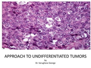
- 1. APPROACH TO UNDIFFERENTIATED TUMORS by Dr. Varughese George
- 2. Introduction “Undifferentiated” tumors • Tumors lacking evidence of lineage differentiation on the basis of routine light microscopic morphology. • Tumors mimicked by different types of tumors causing diagnostic challenge • Tumors that require an algorithmic diagnostic approach elucidating its true nature crucial for clinical management.
- 3. Diagnostic Algorithm for Workup of Undifferentiated Neoplasms
- 4. GENERAL APPROACH FOR UNDIFFERENTIATED TUMORS Categorize malignant tumors according to morphologic appearances. Undifferentiated tumors can be categorized according to morphologic appearances into 4 major groups:
- 5. Tumors according to morphologic appearances 1. Small round cell tumors Heterogeneous groups of tumors composed of relatively small, round to oval, closely packed undifferentiated cells, high nuclear to cytoplasmic ratio, scant cytoplasm, round nuclei Desmoplastic small round cell tumor The tumor cells appear monotonous and undifferentiated and are small to intermediate sized, round or polygonal, with small hyperchromatic nuclei, inconspicuous nucleoli and scant cytoplasm. Brisk mitotic activity and foci of necrosis are frequently found.
- 6. Small round cell tumors Differential Diagnosis Childhood • Lymphoblastic lymphoma • Rhabdomyosarcoma, solid alveolar type • Wilms tumor, blastema- predominant • Peripheral Neuroectodermal Tumor /Ewing sarcoma • Neuroblastoma • Medulloblastoma • Retinoblastoma • Small cell osteosarcoma Adulthood • Small cell carcinoma • Merkel cell carcinoma • Desmoplastic small round tumor • Mesenchymal chondrosarcoma
- 7. Tumors according to morphologic appearances 2. Epithelioid cell tumors Tumors have large round-oval to polygonal shape with a nesting/sheeting arrangement Photomicrograph of undifferentiated carcinomatous tumor composed of epithelioid cells with nesting arrangement and a desmoplastic stroma separating tumor cell nests (hematoxylin-eosin, original magnification 10).
- 8. Epithelioid cell tumors Differential Diagnosis • Rhabdomyoma • Granular cell tumor • Epithelioid sarcoma • Epithelioid variants of leiomyosarcoma • Epithelioid variant of malignant peripheral nerve sheath tumor • Angiosarcoma • Epithelioid hemangioendothelioma • Malignant extrarenal rhabdoid tumor • Pleomorphic rhabdomyosarcoma • Clear cell sarcoma • Alveolar soft part sarcoma • Metastatic tumors
- 9. Tumors according to morphologic appearances 3. Spindle cell tumor Tumors composed of spindle-shaped neoplastic cells with variable patterns of growth. Photomicrograph of undifferentiated sarcomatous tumor composed of spindled cells with a diffuse arrangement with no reactive stroma in between tumor cells. (hematoxylin-eosin, original magnification 20).
- 10. Spindle Cell Tumors Differential Diagnosis Muscle • Leiomyoma. • Leiomyosarcoma. • Rhabdomyosarcoma. Nerve sheath • Neurofibroma. • Malignant Peripheral Nerve Sheath Tumor. Vascular • Angiosarcoma • Kaposi’s sarcoma Myofibroblastic • Fibromatosis • Nodular Fascitis • Inflammatory Myofibroblastic Tumor
- 11. Spindle Cell Tumors Differential Diagnosis Fibrohistiocytic • Dermatofibroma • Dermatofibrosarcoma protuberans • Malignant Fibrous Histiocytoma Adipose • Dedifferentiated liposarcoma Non-sarcomas • Spindle cell carcinoma • Spindle cell melanoma Others • Gastrointestinal Stromal Tumors • Solitary fibrous tumor • Hemangiopericytoma • Synovial Sarcoma
- 12. Tumors according to morphologic appearances 4. Pleomorphic tumor Tumors with cellular component showing high-grade sarcoma-like features with marked cellular pleomorphism and marked nuclear atypia. Highly pleomorphic tumor cells, including multinucleated tumor giant cells haphazardly scattered in the stroma with no distinct areas of storiform growth pattern.
- 13. Pleomorphic tumors Differential Diagnosis Skin: • Benign fibrous histiocytoma • Carcinoma • Melanoma • Atypical fibroxanthoma Limbs: • Unclassified pleomorphic sarcomas (“Malignant Fibrous Histiocytoma”) • High-grade myxofibrosarcoma • Leiomyosarcoma • Liposarcoma (pleomorphic or dedifferentiated) • Rhabdomyosarcoma Extremities: • Giant cell tumor of tendon sheath Retroperitoneum • Dedifferentiated liposarcoma • Leiomyosarcoma
- 14. General approach for undifferentiated tumors Determine a main lineage of differentiation. • Morphologic clues such as a specific growth pattern and nuclear or cytoplasmic characteristics can guide to a diagnosis in some cases of undifferentiated tumors.
- 15. Lineages of tumor differentiation Main lineages of tumor differentiation are : - 1. Epithelial tumors (carcinomas) 2. Melanocytic tumors (malignant melanoma) 3. Hematopoietic and lymphoid tumors 4. Mesenchymal tumors (sarcomas)
- 16. Epithelial tumors • Poorly differentiated comprises ≈ 15% to 20% of carcinomas. • Cytokeratin stains are excellent marker of epithelial differentiation strongly & diffusely expressed in carcinomas. • Diagnosis of carcinoma must be seriously evaluated when an epithelioid tumor is overwhelmingly positive for pankeratin stains.
- 17. Melanocytic tumors • A diffuse strong staining with S100, CK (-) in undifferentiated tumor is good for a melanoma. • S100 is not specific for melanoma as it is also expressed by some carcinomas & sarcomas. • Unusual variants (de-differentiated liposarcoma/mesenchymal chondrosarcoma/malignant peripheral nerve sheath tumor) may pose a challenge to distinguish from melanoma. • Requires confirmation by additional melanoma markers such as HMB-45, Melan A, tyrosinase, or NKI/C3.
- 18. Hematopoietic and lymphoid tumors • If tumor is LCA (+++) and keratin (-),further workup is directed toward classifying the lymphoma using pan-B, pan T-cell (CD20, CD79a, and CD3) & others. • If LCA is ±; pan-B/pan-T cell markers are –ve with morphology still suggestive of lymphoid neoplasm, it could be a myeloid neoplasm. • Myeloperoxidase, chloracetate esterase, lysozyme stains & CD117 demonstrate myeloid lineage. • CD43 and CD68 stains are also positive in granulocytic sarcomas.
- 19. Mesenchymal tumors • Strong vimentin expression in a non-melanocytic, non- lymphoid neoplasm is generally an indication of being a sarcoma. • Sarcomas may have – small round blue-cell tumor morphology (Ewing’s sarcoma) – spindle and epithelioid cells (synovial sarcoma) – pure epithelioid cells (epithelioid sarcoma) • Not all sarcomas are negative for epithelial markers. • Epithelioid sarcoma & synovial sarcoma shows strong staining for epithelial markers.
- 20. General approach for undifferentiated tumors Specify a diagnosis. • Once the main lineage of tumor differentiation is determined, one can proceed to make a much more specific diagnosis. • In this step, clinical correlation as well as additional ancillary studies is needed.
- 21. Immunohistochemical Reagents • Intermediate Filaments Cytokeratins are constituents of the intermediate filaments (IFs) of epithelial cells. LMW CKs, including CK8, CK18, & CK19, recognized by antibodies CAM 5.2 or 35BH11 & a cocktail of keratins (pankeratin), recognized by the antibody AE1/AE3, are useful screening markers for the recognition of epithelial differentiation. • Vimentin is intermediate filament characteristic of mesenchymal cells present in virtually all sarcomas ,melanomas and lymphomas. • Neural IFs include neurofilament proteins and GFAP.
- 22. Immunohistochemical Reagents • CD45 [LCA]) is surface antigen expressed by virtually all hematolymphoid proliferations and monoclonal antibodies. • EMA represents complex membrane glycoprotein isolated from milk fat globules & used for detection of epithelial differentiation, especially in sarcomatoid carcinoma or those undifferentiated carcinomas that are negative or only focally positive for CKs • MOC-31 is a 41-kDa glycoprotein that is cell membrane- based & widely distributed in epithelial cells & tumors in many tissue sites. • PLAP is oncofetal antigen in germ cell tumors, some genitourinary, gastrointestinal & pulmonary carcinomas
- 23. Immunohistochemical Reagents • CEA is serologic indicator of the growth of colorectal cancer. • CD15 is hematopoietic differentiation antigen shared by granulocytes, monocytes, Reed-Sternberg cells, subsets of neoplastic B-cells & T-lymphocytes. • CD30 is relatively restricted in distribution to activated lymphoid cells, Reed-Sternberg cells, a subset of large-cell lymphomas & particular malignant germ cell tumors. • Calretinin is calcium-binding protein expressed by mesothelial cells & malignant mesotheliomas.
- 24. Immunohistochemical Reagents • S100 protein is calcium-flux determinant expressed by normal melanocytes in identification of malignant melanoma, clear-cell sarcoma, glioma, and malignant peripheral nerve sheath tumors. • Other Melanocyte Markers include HMB-45, antityrosinase, MART-1 (Melan-A), and PNL2 • HMB-45 is a quite specific marker for 90-100% of conventional primary melanomas. • Chromogranin A (CGA) is a protein indigenous to matrices of neurosecretory granules in neuroendocrine tissues. • Synaptophysin is integral membrane glycoprotein of presynaptic vesicles detectable in normal & neoplastic neuroendocrine cells.
- 25. Immunohistochemical Reagents • CD56 is neural-cell adhesion molecule, expressed on surfaces of neuroendocrine epithelia, Schwann cells , selected neuroepithelial elements & tumors. • CD99 is membranocytoplasmic protein expressed in virtually all primitive neuroectodermal tumors (PNETs) and Ewing's sarcomas. • FLI-1 is a protooncogene in ETS family of transcription factor loci involved in the regulation of cell growth & differentiation, especially in elements of the hematopoietic system & endothelia. • NB84 is monoclonal antibody raised against neuroblastomatous tumor tissue.
- 26. Immunohistochemical Reagents • Desmin is expressed both in smooth and striated muscle cells with variable expression in myofibroblasts. • Muscle-Specific Actin are confined to cells with muscular differentiation & aids in diagnosis of mesenchymal neoplasms. • Smooth muscle actin (SMA), aka α-actin, is a smooth muscle specific isoform of actin, absent in striated muscle.
- 27. Immunohistochemical Reagents • CD34 is a potential indicator of vascular differentiation highly sensitive for endothelial differentiation & recognizes >85% of angiosarcomas and Kaposi's sarcomas. • CD31 is a transmembrane glycoprotein shared by vascular lining cells, megakaryocytes, platelets & selected other hematopoietic elements; highly restricted to endothelial neoplasms like angiosarcoma.
- 28. Immunohistochemical Reagents • Thrombomodulin (CD141) is a membrano-cytoplasmic glycoprotein distributed among endothelial cells, mesothelial cells, osteoblasts, mononuclear phagocytic cells & selected epithelia. • CD117 is cell-membrane determinant expressed by gastrointestinal stromal tumors (GISTs), mast-cell neoplasms, seminomas, small-cell carcinomas, primitive neuroectodermal tumors, granulocytic sarcomas & melanomas. • Podoplanin is mucin-type glycoprotein localized to cell membranes of endothelia, mesothelium, osteocytes, glandular myoepithelial cells, ependyma, & reticulum- dendritic cells of lymphoid organ.
- 29. Immunohistochemistry • Epithelial origin – Cytokeratins • Melanoma – S100 – HMB45 • Germ Cell Tumour – AFP – bHCG – PLAP • Neuroendocrine – Chromogranin – Synaptophysin – CD56 • Lymphoma – CD45 – CD20 – CD10 – CD3 • Thyroid – Thyroglobulin – TTF1 • Prostate – PSA • Sarcoma – AML – CD31 – CD34
- 30. Why Gene Expression Profiling Can Classify Tumor Types Adrenal Gland Endometrium Pancreas Brain Breast Uterus Esophagus Gall Bladder Kidney Liver Lung Ovary Skin Bone Stomach Thyroid Head & Neck Prostate Germ Cell Soft Tissue Lymph Cervix Bladder GIST Colon Adrenal Gland Endometrium Pancreas Brain Breast Uterus Esophagus Gall Bladder Kidney Liver Lung Ovary Skin Bone Stomach Thyroid Head & Neck Prostate Germ Cell Soft Tissue Lymph Cervix Bladder GIST Colon Adrenal Gland Endometrium Pancreas Brain Breast Uterus Esophagus Gall Bladder Kidney Liver Lung Ovary Skin Bone Stomach Thyroid Head & Neck Prostate Germ Cell Soft Tissue Lymph Cervix Bladder GIST Colon • Tumors from different origins are derived from cells from different developmental processes • Gene expression is distinct between different developmentally- derived cell types
- 31. TO ISOLATE THE SITE CK7 AND CK 20 4 DIFFERENT COMBINATION Colon ca
- 33. Photomicrograph of urothelial carcinoma with positive staining for CK7 (original magnifications x20). Photomicrograph of urothelial carcinoma with positive staining for CK20 (original magnifications x20).
- 34. Photomicrograph of Merkel cell carcinoma showing positive staining for CK20 with a distinct paranuclear dot-like pattern (original magnification x40).
- 35. Expression of EMA in Carcinomas CK+ / EMA – • HCC (Cam5.2+, AE1/AE3–, CK903–) • Adrenocortical neoplasms (frequently negative for all CKs) • Most neuroendocrine neoplasms • Embryonal carcinoma, yolk sac tumor • Thyroid CK– / EMA+ • Meningioma • Perineurioma • Plasma cell neoplasms • Anaplastic large cell lymphoma • Popcorn or lymphocyte predominant (LP) cells [formerly L&H cells] in • Hodgkin lymphoma • RCC (sometimes)
- 36. Expression of Vimentin in Carcinomas Vimentin +ve carcinomas • RCC, clear cell type • Endometrium • Mesothelioma • Salivary gland • Thyroid • Sweat gland • Spindle cell carcinoma of any site Vimentin –ve carcinomas • RCC, chromophobe type • Endocervix (adenocarcinoma) • Lung carcinoma • Breast • Ovary • Prostate • Colorectum • HCC
- 37. Clear cell papillary renal cell carcinoma Note papillary architecture and clear cell cytoplasm (hematoxylin-eosin, 200x) CK7 is positive in tumor (CK7, 200x) Vimentin is positive in tumor
- 38. Photomicrograph of sarcomatoid carcinoma with cytokeratin expression in both sarcomatous and carcinomatous areas (original magnification x20).
- 39. Photomicrograph of rhabdomyosarcoma showing a diffuse strong nuclear staining for myogenin (original magnification x10).
- 40. Photomicrograph of lung adenocarcinoma with diffuse nuclear staining for thyroid transcription factor 1 (original magnification x10) Photomicrograph of epithelioid mesothelioma with nuclear and cytoplasmic staining for calretinin (original magnification x20)
- 41. Tumors showing Coexpression of Cytokeratin and Vimentin Carcinomas that frequently express both Cytokeratin and Vimentin • Anaplastic thyroid carcinoma • Endometrial carcinoma • Mesothelioma • Metaplastic breast carcinoma • Myoepithelial carcinoma • Renal cell carcinoma • Sarcomatoid carcinoma • Thyroid carcinomas Mesenchymal tumors that frequently express both Cytokeratin and Vimentin • Adamantinoma • Chordoma • DPSRCT • Epithelioid angiosarcoma • Epithelioid sarcoma • Leiomyosarcoma • Malignant rhabdoid tumor • Synovial sarcoma Carcinomas that rarely express both Cytokeratin and Vimentin •Breast carcinoma •GI carcinoma •Lung, non–small cell carcinoma •Ovarian carcinoma •Prostatic carcinoma •Small cell carcinoma
- 43. Algorithmic immunohistologic diagnosis of malignant small round cell tumors
- 44. Algorithmic immunohistochemical diagnosis of malignant epithelioid cell tumors
- 45. Algorithmic immunohistologic diagnosis of malignant spindle cell tumors of skin and soft tissues
- 46. Algorithmic immunohistologic diagnosis of malignant pleomorphic tumors
- 47. Photomicrograph of epithelioid sarcoma showing positive staining for vimentin (original magnification x20). Photomicrograph of epithelioid sarcoma showing positive staining for CD34 (original magnifications x20).
- 48. Photomicrograph of epithelioid angiosarcoma showing positive nuclear staining for FLI-1 protein (original magnification x20). Photomicrograph of seminoma, showing a diffuse strong nuclear staining for OCT3/4 (original magnification x20).
- 49. Photomicrograph of desmoplastic small round cell tumor (hematoxylin-eosin, original magnifications x10 [a] and x20 [b]), showing coexpression of cytokeratin (original magnification x20 [c]) and desmin (original magnification x20 [d]). a b c d
- 50. 11A through D, A metastatic prostatic adenocarcinoma in the pleura at initial presentation. Note the prostatic adenocarcinoma with clear cell features on hematoxylin-eosin stain (11A), nuclear staining for NKX3.1 (11B), cytoplasmic PSA staining (11C), and cytoplasmic PSAP staining (11D).
- 51. 7A through F, An epithelioid angiosarcoma/ hemangioendothelioma from the left femur of a 76- year-old woman. Note that the tumor cells are positive for cytokeratin (AE1/3 and CAM 5.2 [C]), ERG (D), CD31 (E), and CD34 (F). Abbreviations: CD, cluster of differentiation; ERG, ETS related gene (original magnification3400 [A through F]).
- 52. Conclusion • Classification of a tumor beyond its apparent undifferentiated morphology is possible in majority of cases. • Ancillary techniques must always be correlated with H&E LIGHT MICROSCOPIC FINDINGS. • Never use irrelevant non-specific markers. • Always progress from preliminary marker to most specific marker. • Ancillary techniques helps only in guiding the diagnosis but the overall diagnosis is based on morphology.
- 53. References • Rekhtman N, Bishop JA. Quick reference handbook for surgical pathologists. Berlin Heidelberg: Springer; 2011 Aug 27. • Dabbs, David J. Diagnostic Immunohistochemistry E-Book: Theranostic and Genomic Applications. Elsevier Health Sciences, 2017. • Wick MR. Immunohistochemical approaches to the diagnosis of undifferentiated malignant tumors. Annals of diagnostic pathology. 2008 Feb 1;12(1):72-84. • Lin F, Liu H. Immunohistochemistry in undifferentiated neoplasm/tumor of uncertain origin. Archives of pathology & laboratory medicine. 2014 Dec;138(12):1583-610. • Pradniwat K, Treetipsatit J. A practical approach to undifferentiated tumors: A review on helpful markers and common pitfalls. • Bahrami A, Truong LD, Ro JY. Undifferentiated tumor: true identity by immunohistochemistry. Archives of pathology & laboratory medicine. 2008 Mar;132(3):326-48.
Notes de l'éditeur
- Malignant fibrous histiocytoma (more accurately known as undifferentiated pleomorphic sarcoma) has been reported in the prostate. The image shows highly pleomorphic tumor cells, including multinucleated tumor giant cells haphazardly scattered in the stroma. There were no distinct areas of storiform growth pattern. An extensive panel of immunohistochemical stains yielded positivity only for vimentin.
- e.g., those of the anterior pituitary gland, thyroid C-cell system, parathyroid glands, paraganglion system, adrenal medulla, and pancreatic islets