Unit- IX Excretory System.pptx
•Télécharger en tant que PPTX, PDF•
2 j'aime•1,926 vues

The urinary system removes waste from the body through urine. It consists of the kidneys, ureters, urinary bladder, and urethra. The kidneys filter waste from the blood to form urine. The ureters then carry urine from the kidneys to the bladder. The bladder stores urine until urination, when urine passes through the urethra and out of the body. The kidneys also help regulate water balance and electrolyte levels in the blood through filtration, reabsorption, and secretion processes in the nephrons.

Recommandé
Contenu connexe
Tendances
Tendances (20)
Similaire à Unit- IX Excretory System.pptx
Similaire à Unit- IX Excretory System.pptx (20)
Plus de Vipin Chandran
Plus de Vipin Chandran (20)
Dernier
Dernier (20)
Unit- IX Excretory System.pptx
- 1. UNIT IX- Excretory System Prepared by, Mr. Vipin Chandran
- 2. INTRODUCTION: As a result of activities of cells, lot of metabolic wastes, i.e. unwanted by-products are formed. The excess of these substances must be removed from the body so as to maintain a proper homeostasis. Excretion is thus defined as a process by which metabolic wastes are removed from the body and an osmotic balance is maintained. The major waste products include: Nitrogenous wastes like ammonia, urea, oxalate, creatinine & uric acid.
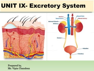
- 3. INTRODUCTION: The urinary system is one of the excretory systems of the body. It consists of the following structures 1) Two kidneys 2) Two ureters 3) One urinary bladder 4) One urethra Nephrology is the scientific study of anatomy, physiology & pathology of the kidneys. Branch of medicine that deals with male & female urinary systems & male reproductive system is called Urology.
- 4. Functions of Urinary system: 1) Kidneys regulate blood volume & composition; help regulate blood pressure, pH, glucose level, produce two hormone (calcitriol & erythropoietin) & excrete waste in urine. 2) Ureter transport urine from kidney to urinary bladder. 3) Urinary bladder stores urine and expels it into urethra. 4) Urethra discharge urine from body.
- 5. KIDNEYS: Gross Structure: 1) 2 kidney, each weight is 150 g in adults, located in retroperitoneally in the lumbar region of abdominal cavity on either side of vertebral column. 2) Kidney is bean-shaped organs, approx. 10cm long, 5cm wide & 2.5cm thick. The right kidney is slightly lower than the left because of liver. 3) Vertical section of kidney shows: Outer cortex- reddish in colour
- 6. KIDNEYS: Gross Structure: Inner medulla- pale in colour, it contains 10-15 renal pyramids which terminate medially in renal papillae. Papillae projects into calyces. Such 10-15 minor calyces join to form two major calyces which come out through pelvis of kidney to ureter. 4) The ureter exit from the hilus of the kidney & pass to the bladder. The blood vessels, lymphatics & nerves enter into or exit from the kidney via hilus.
- 8. KIDNEYS: Microscopic Structure: The basic functional unit of kidney is Nephron. There are approximately 1 to 1.3 million nephrons in each kidney which drain into renal pelvis. Total length of nephron ranges from 45 to 65 mm. Partsof nephronare: 1) Bowman’s capsule 2) Glomerulus 3) Proximal convoluted tubule (PCT) 4) Loop of Henle 5) Distal convoluted tubule (DCT) 6) Collecting tubules.
- 9. KIDNEYS: Microscopic Structure: Nephron consist of 2 parts: a) Renal corpuscle, where blood plasma is filtered. b) Renal tubule, where filtered fluid passes. 1) Bowman’s capsule is the initial dilated part of the nephron. 2) Glomerulus: It is formed by the invagination of a tuft of capillaries into the Bowman’s capsule. The capillaries are supplied by afferent arteriole & the blood leaves from the tuft by efferent arteriole. Bowman’s capsule & glomerulus together constitute Malpighian corpuscle.
- 10. Renal Corpuscle consist of Bowman’s Capsule & Glomerulus:
- 11. KIDNEYS: Microscopic Structure: Bowman’s capsule has two layers: Visceral cell layer is very closely applied to the loop capillaries so as to surround each loop on all sides. It is continuous at the site of entrance of afferent & efferent arterioles with parietal layer. Parietal cell layer is applied to Bowman’s capsule & forms the outer lining of the glomerulus. It is continuous with the PCT. A space is present between visceral and parietal layers of the bowman’s capsule called Bowman’s space.
- 12. KIDNEYS: Microscopic Structure: Glomerular membrane: Glomerular membrane is an extremely thin membrane, each glomerulus contains six lobules & each of these consists 3 to 6 capillary loops. Arrangement of afferent & efferent arterioles within glomerulus allows maintenance of higher pressure (about 45 mmHg). The major function of glomerular membrane is to produce an ultrafiltrate, i.e. the glomerular filtrate will contain all the constituents of plasma except proteins.
- 13. KIDNEYS: Microscopic Structure: 3) Proximal Convoluted Tubule: PCT lumen is continuous with that of Bowman’s capsule. It consists of singular layer of cells with curved outline & brush border formed by numerous microvilli which markedly increase the surface area for absorption. 4) Loop of Henle: It consist of descending limb which arises in continuity with the terminal part of PCT. Descending limb continues into the thick ascending limb which is formed by low cuboidal epithelium. 5) Distal Convoluted Tubule: Thick ascending limb of Henle is continued as DCT, this tubule come very close to its own glomerulus & establishes close proximity to the afferent & efferent arterioles of the glomerulus.
- 14. KIDNEYS: Microscopic Structure: 6) Collecting Tubules: DCT join to form collecting tubules. It passes through the renal cortex & medulla to empty into pelvis of the kidney. The whole kidney is enveloped by a thin but tough fibrous membrane called renal capsule. It limits the swelling, if the kidney becomes oedematous.
- 16. BLOOD SUPPLY OF KIDNEYS:
- 17. FUNCTIONS OF KIDNEY: The kidney perform the following main functions: a) Formation of urine b) Regulation of water balance c) Regulation of electrolyte balance d) Maintenance of acid-base balance e) Endocrine functions.
- 18. A. Formation of Urine: As blood passes through the kidneys, the nephrons clear the plasma of some substances e.g. urea, while simultaneously retaining other essential substances such as water. There are three linked mechanisms in the formation of urine: 1) Substance to be excreted are removed by glomerular filtrations & renal tubular secretion and passed into the urine. 2) Substances that the body needs, e.g. Na+, HCO3- are returned to the body by reabsorption processes.
- 19. Glomerular Filtration: It is the initial step in urine formation. The plasma that traverses the glomerular capillaries is filtered by highly permeable glomerular membrane & the resultant fluid, the glomerular filtrate, is passed into bowman’s capsule. Glomerular filtrate is called ultrafiltrate of plasma. It practically contains no protein & no cells. Glomerular Filtration Rate: It refers to the volume of glomerular filtrate formed each minute by all nephron in both the kidneys. Normal Value: 125 mL/ minute= 170-180 L per day.
- 20. Glomerular Filtration: Mechanism of Glomerular Filtration: For each nephron, Effective (Net) filtration pressure is given as: EFP = (Pcap- Pbow) - (COPcap-COPbow) Where, Pcap: Hydrostatic pressure in glomerular capillaries = 45 mmHG Pbow: Hydrostatic pressure in the Bowman’s capsule = 10 mmHG COPcap: Colloidal osmotic pressure of plasma in glomerular capillaries= 25 mmHG. COPbow: Colloidal osmotic pressure of filtrate in bowman’s capsule. Normally it is zero.
- 21. Reabsorption & Secretion in Renal Tubules: The terms renal tubular secretion & renal tubular reabsorption refer to the ‘direction of transport’. It is the process by which composition & volume of glomerular filtrate is changed during its passage through the tubules. i. Secretion refers to the transport of solutes from peritubular capillaries into tubular lumen, i.e. it is the addition of a substance to the filtrate. ii. Reabsorption denotes the active transport of solutes & the passive movement of water from tubular lumen into peritubular capillaries, i.e. it is the removal of a substance from the filtrate.
- 22. Reabsorption & Secretion in Renal Tubules: Renal tubular transport maximum (Tm): The renal tubular maximum refers to the maximum amount of a given solute that can be transported (reabsorbed or secreted) per minute by the renal tubules. Transport of individual substances in different segments of the renal tubule: i. The PCT reabsorbs 70% to 85% of the filtered Na+; Cl-; HCO3 - & water; & almost 100% of the filtered K+; HPO4 2- ; amino acids & glucose. ii. Reabsorption of water is passive, while reabsorption of solutes can be passive or active. Solute reabsorption generates an osmotic gradient which causes passive reabsorption of water (via osmosis).
- 23. Composition of Urine: 1) Colour: Pale yellow due to presence of pigments (Urochrome & Urobilin). 2) Salts: Inorganic (Na+, K+, Ca2+ & phosphate), Organic (Urea, uric acid & creatinine) 3) Volume: 1-2.5 L/day (average 1.5 L/day) 4) Specific gravity: 1.005-1.030 5) Reaction: pH ranges from 4.5 to 8 (average 6-6.5) 6) Microscopic examination (WBC, pus cells, hyaline casts).
- 24. B. Regulation of Water Balance: Since GFR is 125 mL/ minute, i.e. about 180 L/day of filtrate is formed, whereas normal urine volume is 1- 1.5 L/day. Thus, more than 99% of the filtrate (water) is normally reabsorbed. 1) Reabsorption from PCT: Passive reabsorption of 75- 80% of water occurs. 2) Reabsorption from Loop of Henle: In passing through the Loop of Henle, another 5-10% of the filtered water is removed. 3) Reabsorption in DCT & Collecting duct: In the early part of DCT approx. 5-8% of filtered water is removed passively, secondary to sodium reabsorption under the influence of aldosterone. In the terminal DCT & Collecting duct, ADH hormone facilitates another 10-12% of water reabsorbed.
- 25. B. Regulation of Water Balance: When ADH is present in high concentration, kidney excrete small volume of concentrated urine, but volume of urine cannot be less than 500 mL/ day because some solute require water for their excretion called obligatory volume of urine. This is the minimum urine volume required to excrete the solutes, otherwise it will result in accumulation of waste products in the body.
- 26. C. Regulation of Electrolyte Balance: 1) Kidney forms urine which varies widely in its solute concentration according to the need of the body, e.g. in overhydration, kidney can produce urine of 50 mosm/L (normal plasma osmol conc. Is 300 mosm/L) & during dehydration kidneys can produce urine of 1200 mosm/L. 2) Kidney regulate the concentration of water & electrolytes in blood. Thus it keeps the blood pressure under control.
- 27. Renin-Angiotensin-Aldosterone System (RAAS): 1) When blood volume & blood pressure decreases, walls of afferent arterioles are stretched less & juxtaglomerular cells secrete enzyme renin into the blood. 2) Renin clip off a 10- amino acid peptide called angiotensin I from angiotensinogen, which is synthesized by hepatocytes. 3) By clipping off two more amino acids, ACE converts angiotensin I to angiotensin II, which is the active form of the hormone. Angiotensin II affects renal physiology in 3 ways:
- 28. 1) It decreases GFR causing vasoconstriction of afferent arterioles. 2) It enhances reabsorption of Na+, Cl- & water in the PCT. 3) It stimulates the adrenal cortex to release aldosterone, it stimulates cells in collecting duct to reabsorb more Na+, Cl, which causes increase in blood volume & blood pressure. Renin-Angiotensin-Aldosterone System (RAAS):
- 29. D. Regulation of Acid- Base Balance: 1) Kidneys are responsible for clearing the body metabolically produced H+. 2) For all electrical neutrality of body fluids excretion of excess anion in urine is very important, this situation is prevented due to manufacture of two important cations, H+ & NH4+. 3) Major sites of urine acidification are DCT & Collecting tubules. 4) Three important reactions (buffer systems) in tubular fluid that remove free H+: Bicarbonate system Dibasic phosphate system Ammonia system
- 30. E. Endocrine Function: Kidneys are endocrine organs which secrete: 1) Renin: Renin is a major component of the renin- angiotensin-aldosterone mechanism & is useful to regulate blood pressure. 2) Renal erythropoietic factor (REF, Erythropoietin) it increases the number of circulating erythrocytes. 3) 1,25 DHCC (Dihydrocholecalciferol).
- 31. URETERS: Urinary passages include renal calyces & pelvis, ureter, urinary bladder & urethra, the urine produced by the nephrons continuously trickles from the renal papillae into the calyces & the papillae which extends through the hilum to continue as the ureter. 1) Ureters are 25 to 30 cm long tubes with a diameter of 3 mm, extend from hilum to urinary bladder. 2) They are located retroperitoneally but at the level of sacral bone, they pass obliquely through the posterior wall of urinary bladder to open up into its bottom. 3) Function: ureters convey urine from the kidneys to the urinary bladder by peristaltic contractions of the smooth muscular wall.
- 32. URINARY BLADDER: 1) Urinary bladder is mainly a smooth muscle hollow vesicle & is composed of following: i. The body comprises detrusor muscle. ii. The trigone, a small triangular area near the mouth of urinary bladder, through which both ureters & urethra pass. iii. Internal sphincter: The trigone muscle fibres get interlaced around the opening of urethra forming internal sphincter. Its main function is to maintain tonic closure of the urethral opening. iv. External sphincter: It is a voluntary skeletal muscle. Normally this sphincter remains tonically contracted which prevents constant dribbling of urine.
- 33. URINARY BLADDER: v. Urinary bladder wall is made of many layers, including: Urothelium or transitional epithelium Lamina propria, its connective tissue Detrusor muscle, present outside the lamina propria Fatty connective tissue, it present outside of bladder & separates it from other organs.
- 34. URINARY BLADDER: 2) Physiological capacity of urinary bladder varies with age: At birth – 20-50 ml At 1 year – 200 ml Adults – 600 ml 3) Urine stored in the bladder remains unchanged in the chemical composition. 4) Urinary bladder is innervated by efferent & afferent nerves, it indicate degree of distention & convey pain sensibility. 5) Micturition reflex: As the urinary bladder fills with urine, the wall stretches, impulses are initiated by stretch receptors in the bladder wall causing sensory signal through parasympathetic & sympathetic nerve, this whole constitute the micturition reflex.
- 35. URINARY BLADDER: 6) Mechanism of voluntary micturition: i. In response to desire to micturate, urinary bladder can be voluntarily emptied. Impulses from cerebral cortex motor area pass down to sacral segment causing stimulation of parasympathetic fibres. Then impulse pass to cause contraction of body of urinary bladder wall & relaxation of trigone & internal sphincter, this finally result in emptying of bladder. ii. First urge to pass urine is felt at urinary bladder volume of approx. 150 ml. iii. A marked sense of fullness or discomfort is felt at about 400 ml, which normally results in initiation of micturition reflex.
- 36. URINARY BLADDER: iv. If inconvenient to micturate or desire to hold urine, impulses from cerebral cortex relaxes the detrusor muscle. v. By constant practice urinary bladder can be trained to accommodate very large volume of urine before uncontrollable & unbearable rise of intra- vesicle pressure occurs.