
Recommandé
Contenu connexe
Tendances
Tendances (20)
En vedette
En vedette (20)
Similaire à LIVER FUNCTION TEST
Similaire à LIVER FUNCTION TEST (20)
Dernier
Dernier (20)
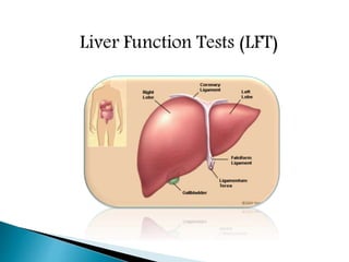
LIVER FUNCTION TEST
- 2. Synthetic functions : synthesis of plasma proteins, cholesterol, triacyl glycerol, lipoprotein Metabolic function: protein metabolism, ketogenesis, TCA cycle, production of ATP Detoxification & excretion: ammonia to urea, bilirubin, cholesterol, drug metabolites. Homeostasis : blood glucose regulation Storage function: Vitamin A,D,K,B12 Production of bile salts
- 3. Screening: They are non invasive yet sensitive modality for liver dysfunction. Pattern of disease: They are helpful to recongnize pattern of various diseases. Like being helpful in differentiating between acute viral hapatitis and various cholestatic disorders and chronic liver disease.(CLD) Assess severity : They are heplful to assess the severity and predict the outcome of certain diseases like primary biliary cirrhosis. Follow up: They are helpful in the follow up of certain liver diseases and also helpful in evaluating response to therapy like autoimmune hepatitis
- 4. 1. Tests of the liver’s capacity to transport organic anions & to metabolise drugs-serum bilirubin, urine bilirubin, urobilinogen etc. 2. Tests that detect injury to hepatocytes (serum enzyme tests)- Aminotransferases, Alkaline phosphatase, glutamyl transpeptidase,5nucleotidase,leucine aminopeptidase etc. 3. Tests of the liver’s biosynthetic capacity-serum proteins, albumin, ceruloplasmin, prothrombin time etc.
- 5. Jaundice Suspected liver metastasis Alcoholic liver disease Any undiagnosed chronic illness Annual checkup of diabetic patients Coagulation disorders Therapy with statins to check hepatotoxicity
- 7. .Bilirubin is an endogenous anion derived from catabolism of heme. . It is conjugated by liver to form bilirubin levels ,more than 17umol/L suggests liver disease. .Classification of bilirubin into direct & indirect bilirubin is based on original van den bergh method of measuring bilirubin. .Van den Bergh is a chemical reaction used to measure bilirubin level in blood. The reaction is highly useful in understanding the nature of jaundice.
- 8. .Bilirubin is the excretory product formed by the catabolism of heme part of haemoglobin. .Porphyrin part of heme are converted to bilirubin in reticuloendothelial cells of liver, spleen, bone marrow. .Unconjugated bilirubin is bound to serum albumin & transferred to liver where it is conjugated to glucuronate by UDP GLUCURONYL TRANSFERASE. .Conjugated bilirubin is excreted into bile. A fraction of bilirubin from stool is reabsorbed into blood via portal circulation (enterohepatic circulation).
- 9. PRODUCTION & EXCRETION OF URINE
- 10. PRINCIPLE: When diazotised sulfanilic acid reacts with bilirubin, it forms ‘azobilirubin’ which is a purple colour product. .Increased conjugated bilirubin – gives colour immediately i.e direct positive. .Increased unconjugated bilirubin -gives colour only after addition of methanol i.e indirect positive
- 11. Hyperbilirubinemia : when the level of bilirubin exceeds 1mg/dl in serum. Based on the cause it is classified into congenital and acquired. Congenital hyperbilirubinemia: They result from abnormal uptake, conjugation or excretion of bilirubin due to inherited defects CRIGLER-NAJJAR SYNDROME: Here the defect is in conjugation due to severe deficiency of enzyme UDP-GLUCURONYL TRANSFERASE. Unconjugated bilirubin level increases to more than 20mg/dl and hence kernicterus occurs.
- 12. GILBERT SYNDROME: it is inherited as an autosomal dominant trait. The defect is in the uptake of bilirubin by the liver . Bilirubin level goes around 3mg/dl. DUBIN-JOHNSON SYNDROME: It is an autosomal recessive trait leading to defective excretion of conjugated bilirubin, so conjugated bilirubin in bold increases. There is defect in transport of conjugated bilirubin into bile. Bilirubin gets deposited in the liver and liver appears black. This condition is known as black liver jaundice. ROTOR SYNDROME: It is an autosomal recessive trait. In this bilirubin exrection is defective.
- 13. PHYSIOLOGICAL JAUNDICE: Also known as neonatal jaundice. This transient hyperbilirubinemia is due to an accelerated rate of destruction of RBCs & also because of immature hepatic system of conjugation of bilirubin. In all new born infants after the 2nd day of life, mild jaundice appears. In such cases bilirubin does not increase above 5mg/dl. BREAST MILK JAUNDICE: In some breast-fed infants, prolongation of jaundice has attributed to high level of estrogen derivative in maternal blood, which is excreted through the milk. This would inhibit the glucuronyl transferase system .Sulfa and other drugs may release bilirubin from albumin and may cause jaundice in new born.
- 14. JAUNDICE :- Jaundice is the yellowish discolouration of sclera, skin, mucous membrane. It is characteristic of liver disease but will occur when rate of hemolysis is increased leading to the elevation of serum albumin.
- 15. Hemolytic disease of newborn: Rh incompatibilty – Erythroblastosis fetalis Hemolytic diseases of Adults: condition is seen in increased rate of hemolysis. It usually occurs in adults. There is increase in unconjugated bilirubin in blood, absence of bilirubinuria & excessive excretion of UBG in urine & SBG in feces.
- 16. .The most common cause is viral hepatitis, caused by hepatitis viruses A,B,C,D or E. .In pure hepatocellular disease, conjugation in liver is decreased leading to increase in free bilirubin in circulation. .The UBG level in urine may be normal or decreased in hepatocellular jaundice. .In this, a biphasic reaction is observed, because both conj. & unconj. bilirubins are increased
- 17. .Conjugated bilirubin is increased in blood & it is excreted in urine. If there is complete obstruction, UBG will be decreased in urine or even absent. .In total obstruction of biliary tree, the bile doesn’t enter the intestine. Since no pigments are entering the gut, the feces become clay coloured. .Van den Bergh test is direct positive as conj. bilirubin is elevated.
- 18. Types of bilirubin Class of jaundice Causes Unconjugated Prehepatic or hemolytic Abnormal red cells; antibodies; drugs & toxins; thalassemia; hemoglobinopathies. Gilbert syndrome; Crigler-Najjar syndrome, GPD deficiency. Unconjugated & conjugated Hepatic or hepatocellular Viral hepatitis; toxic hepatitis; intrahepatic cholestasis Conjugated or obstructive Posthepatic .Intrahepatic cholestatis- chronic active hepatitis, obstructive stage of viral hepatitis, Dubin- johnson syndrome .Extra hepatic obstruction-stones in the gallbladder or biliary tract, carcinoma of head of pancreas, enlarged
- 19. Hemolytic jaundice Hepatocellular jaundice Obstructive jaundice Blood, free bilirubin Increased Increased Normal Blood, conj. Bilirubin Normal Increased Increased Blood, ALP Normal Increased Very high Urine, bile salts Nil Nil Present Urine, conj. Bilirubin Nil Present Present Urine bilinogens Increased Present Nil Fecal urobilinogen increased Decreased Absent
- 20. . The presence of urine bilirubin indicates hepatobiliary disease. Unconjugated bilirubin is tightly bound to albumin and not filtered by the glomerulus and thus not present in urine. Measurable amount of conjugated bilirubin in serum are found only in hepatobiliary disease. .Because the renal threshold for conjugated bilirubin is low and the laboratory methods can detect low levels of bilirubin in urine so conjugated bilirubin may be found in urine when the serum bilirubin levels are normal. This is the case in early acute viral hepatitis.
- 21. . An increase in the urobilinogen in urine is a sensitive indicator of hepatocellular dysfunction. It is a good indicator of alcoholic liver damage, well compensated cirrhosis or malignant disease of liver. In viral hepatitis it appears early in urine. It is markedly increased in hemolysis. . In cholestatic jaundice urobilinogen disappears from urine. It is intermittently present in case of gallstones. .Urobilinogen gives a purple reaction to Ehrlich’s aldehyde reagent.
- 22. BY AALOK KUMAR
- 23. A large number of enzyme estimations are available which are used to ascertain liver function. They are be divided into two groups: A). Enzymes indicating hepatocellular damage. B). Enzymes indicating cholestasis (obstruction). In liver cells injury , damage to the membrane of cells & organelles allows intracellular enzymes to leak into the blood
- 24. ENZYMES THAT DETECT HEPATOCELLUAR NECROSIS • ALT • AST ENZYMES THAT DETECT CHOLESTASIS • ALKALINE PHOSPHATASE • 5 NUCLEOTIDASE • LEUCINE AMINOPEPTIDASE
- 25. ALKALINE PHOSPHATASE SERUM TRANSFRASE ALT AST GGT 5’NT
- 27. AST (Aspartate transaminase)/SGOT (serum glutamate oxaloacetate transaminase) ALT (Alanine transaminase)/SGPT (serum glutamate pyruvate transaminase) SGOT SGPT
- 28. Normal range: 10-45 U/L. AST is found in both cytoplasm & mitochondria AST/GOT also reflects damage to the hepatic cells & is less specific for liver disease. It is a cardiac marker. AST help diagnose various heart, muscle or brain disorders, such as a myocardial infarct (heart attack).
- 29. Acute hemolytic anemia Cirrhosis of the liver Hepatitis Acute pancreatitis or inflammation of pancreas Acute renal failure or loss of kidney function. Heart attack Primary muscle disease Recent surgery
- 30. Normal Range: 5-40 U/L. ALT is a cytoplasmic enzyme. The activity of these enzymes is low in normal serum. ALT is specific for liver disease. Its elevations favor liver cell necrosis as a cholestasis.
- 31. Alcoholic liver disease Cancer of liver Hepatitis or inflammation of the liver Noncancerous tumor of the liver Use of medicines or drugs toxic to the liver Cirrhosis or scarring of the liver Death of liver tissue.
- 32. • Normal ratio is 0.7 to 1.4 • Useful in Wilson disease, chronic liver disease and alcoholic liver disease • AST/ALT ratio of > 2:1 is suggestive of and >3:1 is highly suggestive of Alcoholic liver disease • AST in Alcoholic live disease is rarely >300 IU/L. • ALT is usually normal in alcoholic liver disease ; can be sometimes low due to an alcohol induced deficiency of pyridoxal phosphate • AST/ALT <1 is seen in NASH and viral hepatitis.
- 33. • mAST/total AST ratio – marker of chronic alcohol consumption • This distinguishes those who consume excess alcohol from normal subjects irrespective of the presence or absence of liver disease
- 36. ALP occurs in in all tissues, especially liver, bone, bile duct, kidney & the placenta. The ALP used to help diagnose certain liver diseases and bone disorders. Normal range: 30 - 95 IU/L (3-13 kings unit) ALP is a hydrolase enzyme responsible for removing phosphate groups from many types of molecules, including nucleotides & proteins. Levels are significantly higher in growing children. A rise in serum ALP (normal 3-13 KA units/dl), usually associated with elevated serum bilirubin is an indicator of biliary obstruction (obstructive/post hepatic jaundice). ALP is also elevated in cirrhosis of liver & hepatic tumors.
- 38. Normal range: 10-15 U/L This is a microsomal enzyme widely distributed in body tissues, including liver. Measurement of γ - glutamyl transpeptidase (GGT) activity provides a sensitive index to asses liver abnormality. Serum GGT is highly elevated in biliary obstruction & alcoholism. GGT elevation parallels than of ALP. Increased level of GGT are observed in chronic alcoholism ,pancreatic disease ,MI ,renal failure ,diabetes mellitus. Several drugs (e.g. phenytoin) induce (liver synthesis) & increase this enzyme in circulation.
- 39. Normal range: 2-15 U/L The serum activity of 5'-nucleotidase is elevated in hepatobiliary disease & this parallels ALP. It is highest in post-hepatic obstructive jaundice. The 5'-nucleotidase is normal in patients with bone disease where as serum ALP increased .
- 41. Function test Pre-hepatic Jaundice Hepatic Jaundice Post-hepatic Jaundice Total bilirubin Normal / Increased Increased Conjugated bilirubin Normal Increased Increased Unconjugated bilirubin Normal / Increased Increased Normal Urobilinogen Normal / Increased Increased Decreased / Negative Urine Color Normal Dark (urobilinogen + conjugated bilirubin) Dark (conjugated bilirubin) Stool Color Normal Normal/Pale Pale Alkaline phosphatase levels Normal Increased Alanine transferase and Aspartate transferase levels Increased Conjugated Bilirubin in Urine Not Present Present Splenomegaly Present Present Absent Table of diagnostic tests
- 43. Plasma proteins Prothrombin time TESTS BASED ON SYNTHETIC FUNCTION
- 44. PROTEIN •Liver is the sole site for synthesis of most plasma proteins except immunoglobulins (gamma globulins) •Serum albumin comprises 60% of all plasma proteins TESTS FOR PROTEINS INCLUDE- 1. Total serum proteins 2. Serum albumin 3. Albumin/globulin ratio 4. Serum protein electrophoresis 5. Prealbumin 6. Procollagen III peptide 7. Ceruloplasmin 8. Alpha fetoprotein 9. Alpha antitrypsin
- 45. ALBUMIN •Synthesized exclusively by liver •Its half life is 18-20 days •Due to its slow turn over – not a good indicator of acute or mild hepatic dysfunction • In hepatitis - <3g/dl of albumin – possibility of chronic liver disease •Non hepatic causes of Hypoalbuminemia - • Protein losing enteropathy • Nephrotic syndrome
- 46. PREALBUMIN /TRANSTHYRETIN • Levels fall in liver disease • Half life – 2 days • Sensitive indicator of any changes affecting its synthesis and catabolism • PAB is a negative acute phase reactant • Particularly useful in drug-induced hepatotoxicity
- 47. • Normal plasma levels - 0.2-0.4g/L • Acute phase protein • Decreased in multiple conditions – 1. Wilson’s disease 2. Menkes disease 3. Aceruloplasminemia 4. Copper deficiency CERULOPLASMIN • Increased in – 1. Copper toxicity 2. Pregnancy 3. OCPs
- 48. PROCOLLAGEN III PEPTIDE • Cleavage product of the type III procollagen molecule • Radioimmunoassay • Elevated Conc. Of PIIIP- the transformation of viable hepatic tissue into connective tissue/fibrosis
- 49. • AFP is a gp and MW – 70,000 daltons • Normally present in fetus • Liver, yolk sac and small intestine • AFP- ELISA HCC Non seminomatous testicular cancer Ataxia telangiectasia Hereditary tyrosinemia Neonatal hyperbilirubinemia Chronic active hepatitis α- FETO PROTEIN
- 50. COAGULATION FACTORS •With the exception of F-VIII , all other factors are synthesized in liver •Half life ranges from 6hrs for F-VII to 5 days for fibrinogen • So their measurement is the single best measure of hepatic synthetic function • Tests – Serum prothrombin time •Marked increase in PT >5secs above the control and not corrected by Vit K administration – is a poor prognostic sign in acute viral hepatitis and other acute and chronic liver disease
- 51. Prothrombin is synthesized by liver Half life – 6 hrs so it indicate – present function of liver PT :blood test, time taken by blood to clot Normal value : 10 to 15 sec. PT is Prolonged - liver losses 80% of reserve capacity Commonly PT is used for detecting liver coagulopathies Note : Vit K deficiency Prolongs PT time But In case of LIVER diseases : PT remains prolonged even after parenteral administration of Vit. K
- 52. Lack sensitivity: The LFT may be normal in certain disesliver disease like cirrhosis, non cirrhotic portal fibrosis ,cogenital hepatic fibrosis Lack specificity: They lack specificity and are not specific for any particular disease. Serum albumin may be decreased in chronic disease and also in nephrotic syndrome. Amino-transferase may be raised in cardiac diseases and hepatic diseases. Except for serum bile acids the LFT are not specific for liver diseases Thus, we see that LFT have certain advantages as well as limitation at the same time