Presentation1, musculoskeletal anatomy.
•Télécharger en tant que PPTX, PDF•
15 j'aime•1,959 vues

Health&Medicine

Recommandé
Recommandé
Contenu connexe
Tendances
Tendances (20)
Similaire à Presentation1, musculoskeletal anatomy.
Similaire à Presentation1, musculoskeletal anatomy. (20)
Plus de Abdellah Nazeer
Plus de Abdellah Nazeer (20)
Dernier
Dernier (20)
Presentation1, musculoskeletal anatomy.
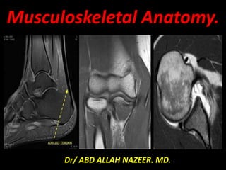
- 1. Musculoskeletal Anatomy. Dr/ ABD ALLAH NAZEER. MD.
- 2. MRI anatomy of the knee joint.
- 5. The sagittal plane is perpendicular to bicondylar plane, allowing a good study of anterior and posterior horn of the menisci and cruciate ligaments.
- 6. The frontal (coronal) plane parallel to bicondylar plan is systematic and can analyze the middle segment of the meniscus and collateral ligaments.
- 7. The axial plane provides a good analysis of periarticular tissues (muscles, tendons, nerves and vessels). They allow more precise study of patella-femoral cartilage and the popliteal fossa.
- 8. Exploring the meniscus requires two orthogonal planes: *The sagittal plane is the most appropriate for the exploration of the anterior and posterior horn of the menisci. *The frontal plane is exploring mainly the middle part of the menisci. *The axial sections have a poor intake due to the orientation of the menisci and partial volume effects with the articular surfaces.
- 10. In the frontal plane: • On the first anterior image: the anterior horn of medial meniscus appears, the anterior horn of lateral meniscus appears one or two cuts after. • On intermediate cuts: the middle segment of the two menisci, triangular in form, is similar in size and morphology.
- 11. In the axial plane: The menisci appear as a circular form closed for lateral meniscus “O” and open for the medial meniscus “C”
- 13. The medial meniscus, both horns are triangular in shape and have very sharp points. The posterior horn is always larger than the anterior horn. The semicircular medial meniscus has a wide posterior horn, narrows anteriorly and has open C shaped configuration.
- 14. On sagittal sections: The ACL is seen as a thin oblique band stretched between the femur and the tibia and has a moderate low signal in T1 and T2 weighted sequences (slightly more intense than the PCL). Its signal is rather heterogeneous and its contours may be poorly defined, due to its anatomical properties.
- 17. On coronal sections: The ACL has an oblique direction from tibial spines to the medial face of the lateral condyle.
- 18. On axial sections: The ACL has a triangular form and then an oval and oblique form with laterally direction from the lower to highest sections.
- 19. On sagittal sections: Due to its thickness, the PCL is most often seen on sagittal sections. When the knee is placed in slight external rotation, the PCL is usually fully visible on the same sagittal section.
- 20. On axial images: The PCL have: * Range form on proximal cuts * Triangular form on intermediate cuts * Oval form on distal cuts.
- 21. On coronal sections: It appears as a low signal oblique band top and medially. -The following criteria are needed to define a normal PCL: *Ligament with homogenous signal.mogeneous signal.
- 22. The MCL: have two bundles • The superficial bundle: is separated from the deep bundle by a layer of fat, and sometimes by a bursa. •The deep bundle: situated behind the previous and is formed of fibers that ranging from femur and tibia to articular fibro-cartilage. The MCL is then attached to the capsule and the medial meniscus.
- 23. The LCL is completely independent from the capsule and the lateral meniscus. This ligament inserts is the tuberosity of the lateral condyle of the femur and have a little obliquity down and back to attach to the upper of the fibula.
- 24. Patellar retinaculums: It appears as a low signal band extending from the patella to the femoral condyles on axial plane.
- 25. Patellar Ligament: • In sagittal sections: The patellar ligament appearing as a very thick band having a low signal.
- 27. The quadricipital tendon: The quadricipital tendon is constituted by the continuity of the muscle fascia of the four quadricipital muscles.
- 28. Muscles and neuro-vascular elements: Muscles appear with intermediate signal on PD weighted sequences and a low signal on T1weighted sequences and individualized by the interposition of the high signal of fatty tissue. Neuro-vascular elements are explored in axial sections. The popliteal artery and vein and the tibial nerve are seen in popliteal fossa runs between the twins muscles.
- 29. Adipose packages: There are many adipose packets in the knee joint. Each of which is located between the joint capsule in the lateral side and the articular cavity delimited by the synovial membrane in the medial side. They are intra-capsular but extra-synovial situation. The three anterior adipose packages include the anterior supra-patellar fat package, quadricipital package (QAP), the posterior supra-patellar fat, (PAP) and the Hoffa Adipose Packet (HAP).
- 30. The posterior bundle or “Wrisberg ligament” is the most constant and is found in 33% of MRI of the knee. It appears as a linear low signal band doubling the posterior PCL in the posterior cuts of coronal sections and as a nodular low signal structure located behind the PCL.
- 31. The anterior bundle or “Humphrey ligament”, less frequent and walks in front of the PCL, it appear as a nodular low signal structure located in front of the PCL on sagittal section.
- 32. Articular Cartilage: -In Spin echo T1weighted images, the cartilage is homogeneous and have an intermediate signal between that of the bone marrow and muscle. It differs from the low signal of meniscal and cortical signal. -In Spin echo T2, the presence of high signal intra-articular fluid is used to evaluate the thickness of the cartilage by arthrographic effect.
- 33. Femoro-tibial bone structures: -The Cortical bone appears as a linear structure of low signal intensity (black). -The medullar bone due to its fat content, rich in free protons, presents a very intense signal (white).
- 34. Ankle Joint.
- 39. (a) Coronal T1 weighted image of the ankle demonstrates the tibiotalar joint (blue arrow) and distal tibiofibular joint (red arrow). The osseous structures include the tibia (T), tibial plafond (Tib Plaf), medial malleolus (MM), lateral malleolus (LM), talus (Tal), superior faces (SF), lateral malleolar facet (LMF), medial malleolar facet (MMF), calcaneus (C), peroneal tubercle (PT), and sustentaculum tali (ST). (b) Sagittal T2 weighted image again demonstrates the tibiotalar joint (blue arrow), as well as the subtalar joint (yellow arrow). The talus to include the head (TH), neck (TN), and body (TB) are noted, as well as the calcaneus (C).
- 40. (a, b) Axial and coronal T1. The tendons of the ankle are divided into anterior (blue), posterior (green), medial (yellow), and lateral (red) compartments. The anterior compartment contains the tibialis anterior tendon, extensor hallucis longus tendon, extensor digitorum longus tendon, and peroneus tertius tendon. The posterior compartment contains the Achilles tendon. The medial compartment contains the tibialis posterior tendon, flexor digitorum longus tendon, and flexor hallucis longus tendon. The lateral compartment contains the peroneus longus and peroneus brevis tendons. Normal tendons are uniformly low in signal intensity throughout their course.
- 41. Anterior tendons. (a, b) Axial and coronal T1. From medial to lateral – TA (blue arrows), EHL (yellow arrows), EDL (red arrows), and PT (orange arrows). The asterisk (a) represents the extensor retinaculum.
- 42. Achilles tendon. (a) Axial T1. The Achilles tendon (red arrow) is concave anteriorly and convex along the posterior margin. (b) Sagittal PD FS. The normal Achilles tendon inserts onto the posterior surface of the calcaneus (blue arrow).
- 43. Medial tendons. (a) Axial T1. From lateral to medial – FHL (blue arrow), FDH (red arrow), TP (red arrow). The neurovascular bundle (purple ellipse) is located between the FHL and FDL. (b) Sagittal PD FS. The PT (yellow arrow) and FHL (red arrow) pass posterior to the medial malleolus (MM), with the FHL coursing posterolateral to the PT in the region of the tarsal tunnel. (c) Coronal T1. Superomedial to inferolateral – PT (yellow arrow), FDL (red arrow), FHL (blue arrow). The FHL passes under the sustentaculum tali (ST) of the calcaneus. (d) Sagittal PD FS. The FHL (blue arrow) is again visualized passing under the sustentaculum tali. (e) Sagittal PD FS. The PT (yellow arrow) is identified inserting onto the navicular (N). (f) Axial T2 FS. The FHL (blue arrow) is visualized crossing deep to the FDL (red arrow) within the plantar midfoot, an intersection known as Henry’s knot.
- 44. Lateral tendons. (a) Axial T1. The PB tendon (blue arrow) and PL tendon (red arrow) course posterior to the lateral malleolus (LM), with the PB tendon anteromedial to the PL tendon. The superior peroneal retinaculum overlies the tendons (orange arrow). (b) Coronal T1. The PB tendon (red arrow) is passes lateral to the PL tendon (blue arrow). (c) Sagittal PD FS. The PB tendon inserts onto the base of the 5th metatarsal (5th met). The PL tendon (red arrow) is partially visualized posterior to the PB tendon. (d, e) Axial and coronal T1. The PB tendon (blue arrow) passes anterosuperior and the PL tendon (blue arrow) courses posteroinferior to the peroneal tubercle of the calcaneus (PeT). (f) Sagittal PD FS. The PL tendon (red arrow) crosses the plantar aspect of the midfoot.
- 45. Deltoid ligament. (a) Coronal T2 FS. The tibionavicular ligament (blue arrow) is seen coursing along the medial aspect of the talus. (b) Coronal T2 FS. The tibiospring ligament (red arrow) arises from the tip of the medial malleolus (MM), and joins the spring ligament complex distally. The flexor retinaculum (yellow arrow) overlies the deltoid ligament. (c) Coronal T2 FS. The tibiocalcaneal ligament (green arrow) connects the medial malleolus to the sustentaculum tali. The PT (purple arrow) and FDL (pink arrow) course medial to the ligament. (d) Coronal T2 FS. Connecting the medial malleolus to the talar neck is the anterior tibiotalar ligament (orange arrow). (e) Coronal T2 FS. The posterior tibiotalar ligament (white arrow) attaches the medial malleolus to the medial talar body. (f) Axial T2 FS. The anterior (orange arrow) and posterior (white arrow) tibiotalar ligaments demonstrate normal inhomogeneous signal intensity.
- 46. Lateral ligaments. (a) Axial T1. The ATFL (blue arrow) extends from the distal lateral malleolus to the lateral talus. (b) Axial T2. The PTFL (orange arrow) is a broad band between the posterior tip of the LM and the lateral talus. (c) Coronal T2. The PTFL (orange arrow) is again seen extending from the distal LM to the talus. The posterior tibiofibular ligament (yellow arrow) lies superior to the PTFL, and attaches the LM to the tibial plafond (Tib Plaf). The normal inhomogeneous appearance of these ligaments is secondary to intervening fat. (d) Coronal T1. The CN ligament (red arrow) arises from the tip of the LM, and courses medial to the peroneal tendons (purple ellipse) to attach to the lateral calcaneus.
- 47. Syndesmotic ligaments. (a,b) Axial and coronal T1. The anterior tibiofibular ligament extends from the distal fibular to the distal tibia, just proximal to the tibiotalar joint. (c,d) Axial and coronal T1. The posterior tibiofibular ligament (yellow arrows) is an inhomogeneous band that joins the posterior distal tibia and fibula. (e,f) Axial and coronal T1. The interosseous ligament (red ellipse) is the distal continuation of the interosseous membrane, uniting the distal tibia and fibula.
- 48. Sinus tarsi. (a) Coronal T1. The normal fat is visualized within the sinus tarsi (yellow ellipse). (b) Sagittal PD FS. The fat within the tarsal sinus suppresses normally (blue arrow). (c) Axial T1. The talocalcaneal interosseous ligament (red arrow) courses within the sinus tarsi, and functions to maintain apposition of the talus and calcaneus.
- 49. Hip Joint.
- 71. Elbow Joint. Indication. -Pain. -Swelling. -Stiffness. -Deformity. -Instability. -Paraesthesias.
- 85. Shoulder Joint.
- 120. Thank You.