SlideShare une entreprise Scribd logo
1  sur  48
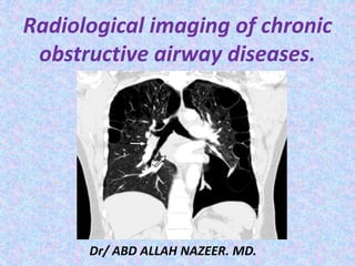
Radiological imaging of chronic
obstructive airway diseases.
Dr/ ABD ALLAH NAZEER. MD.
Chronic Bronchitis.
Definition: Persistent productive cough for at least 3 months in at least 2
consecutive years without any identifiable cause.
Damage to air ways caused mainly by chemicals.
Sources: Cigarette smoke, industrial gases, motor vehicle exhaust, etc,
Chronic asthmatic bronchitis- intermittent bronchospasm and wheezing.
Chest x-ray- This helps to show hyper-expansion of the lungs
associated with chronic bronchitis and COPD. The brochovascular
marking, cardiomeagly and may flatted out the diaphragm.
Chronic Bronchitis
with irregular
pattern of broncho-
vascular structures.
CT in chronic bronchitis, bronchial wall thickening may be seen in
addition to enlarged vessels. Repeated inflammation can lead to
scarring with brochovascular irregularity and fibrosis.
Chronic bronchitis and the lines that leave the right hilum
horizontally show irregular borders because of chronic inflammation
Emphysematous lung and chronic bronchitis.
COPD with chronic bronchitis.
Abnormal chest X-ray findings are usually not seen until COPD is
severe. In this case, the X-ray may show:
Flattening of the diaphragm, the large muscle that separates the lungs and heart
from the abdominal cavity.
Increased size of the chest, as measured from front to back.
A long narrow heart.
Abnormal air collections within the lung (focal bullae).
On the lateral radiograph, a "barrel chest" with widened anterior-posterior
diameter may be visualized. The "saber-sheath trachea" sign refers to marked
coronal narrowing of the intrathoracic trachea (frontal view) with concomitant
sagittal widening (lateral view).
CT finding in emphysema is diagnosed by alveolar septal destruction and
airspace enlargement, which may occur in a variety of distributions.
Centrilobular emphysema is predominantly seen in the upper lobes with
panlobular emphysema predominating in the lower lobes. Paraseptal
emphysema tends to occur near lung fissures and pleura. Formation of giant
bullae may lead to compression of mediastinal structures, while rupture of
pleural blebs may produce spontaneous pneumothorax /
pneumomediastinum.
COPD with hyperinflation and flattened diaphragm.
COPD with hyperinflation and flattened diaphragm and widening of retro-sternal space
Emphysema with horizontal ribs.
Bullous disease of the lungs-conventional radiograph and CT.
Giant Bullous Emphysema
Congenital Lobar Emphysema.
CLE almost always involves one lobe, with rates of
occurrence as follows:
Left upper lobe - 41%
Right middle lobe - 34%
Right upper lobe - 21%
Congenital lobar emphysema has 2 forms:
Hypoalveolar (fewer than expected number of alveoli)
Polyalveolar (greater than expected number of alveoli)
X-Ray shows unilateral –translucency.
CT Scan shows hyperinflation of one or more lobes with
attenuated pulmonary vasculatures, compression
atelectasis of the adjacent lung and mediastinal shift.
Left upper lobe congenital emphysema.
Right middle lobe congenital emphysema.
Pulmonary interstitial emphysema.
Pulmonary interstitial emphysema (PIE) refers to the abnormal location of air
within the pulmonary interstitium and lymphatics. It typically results from rupture
of overdistended alveoli following barotrauma in infants who have respiratory
distress syndrome. Interstitial emphysema can also occasionally be incidentally
detected in adults.
Radiographic features
Plain film - chest radiograph shows bubbles (round) or streaky (linear)
radioculencies in the interstitium radiating from the hilum . Affected segment is
often hyperexpanded and static in volume on multiple radiographs. Patients may
have pneumothorax, pneumomediastinum, or pneumopericardium in supine
patients, pneumomediastinum is evident by the sharp mediastinum sign
CT chest
shows cystic radioculencies in affected segment may characteristically show a line
and dot pattern with pulmonary arterial branches surrounded by radiolucent air
may help differentiate persistent PIE from a hyperlucent mass such as congenital
lobar emphysema, congenital pulmonary airway malformation (CPAM)
allows better visualization of a pneumothorax or pneumomediastinum
if incidentally detected in adults, it may appear as perivascular lucent or low-
attenuating halos and small cysts.
Pulmonary interstitial emphysema.
Pulmonary Interstitial Emphysema (PIE)
Pneumothorax and pulmonary interstitial emphysema
Persistent diffuse pulmonary interstitial emphysema
Pulmonary hypertension.
Pulmonary hypertension (PH) is an increase of blood pressure in the
pulmonary artery, pulmonary vein, or pulmonary capillaries,
together known as the lung vasculature, leading to shortness of
breath, dizziness, fainting, leg swelling and other symptoms.
Pulmonary hypertension is usually occur secondary to emphysema.
Pulmonary artery dilatation secondary to hypertension.
Pulmonary Arterial Hypertension.
Thank You.

Contenu connexe

Tendances

Chest x ray basic interpretation
Chest x ray basic interpretationChest x ray basic interpretation
Chest x ray basic interpretationVikram Patil
 
Presentation1.pptx, radiological signs in thoracic radiology.
Presentation1.pptx, radiological signs in thoracic radiology.Presentation1.pptx, radiological signs in thoracic radiology.
Presentation1.pptx, radiological signs in thoracic radiology.Abdellah Nazeer
 
Interstitial Lung Disease
Interstitial Lung DiseaseInterstitial Lung Disease
Interstitial Lung DiseaseKamal Bharathi
 
Presentation1.pptx. interpretation of x ray chest.
Presentation1.pptx. interpretation of x ray chest.Presentation1.pptx. interpretation of x ray chest.
Presentation1.pptx. interpretation of x ray chest.Abdellah Nazeer
 
Imaging in pulmonary tuberculosis
Imaging in pulmonary tuberculosisImaging in pulmonary tuberculosis
Imaging in pulmonary tuberculosisNISHANT RAJ
 
Differential diagnosis of cavitary lung lesions
Differential diagnosis of cavitary lung lesionsDifferential diagnosis of cavitary lung lesions
Differential diagnosis of cavitary lung lesionsDr.Bijay Yadav
 
Paediatric chest imaging
Paediatric chest imagingPaediatric chest imaging
Paediatric chest imagingSidra Afzal
 
Collapse- RADIOLOGY
Collapse- RADIOLOGYCollapse- RADIOLOGY
Collapse- RADIOLOGYNavdeep Shah
 
Interstitial lung diseases radiology
Interstitial lung diseases radiologyInterstitial lung diseases radiology
Interstitial lung diseases radiologyShrikant Nagare
 
Collapse and consolidation Lung Radiology
Collapse and consolidation Lung RadiologyCollapse and consolidation Lung Radiology
Collapse and consolidation Lung RadiologyNeelam Ashar
 
Presentation1.pptx, radiological imaging of chronic obstructive airway disease.
Presentation1.pptx, radiological imaging of chronic obstructive airway disease.Presentation1.pptx, radiological imaging of chronic obstructive airway disease.
Presentation1.pptx, radiological imaging of chronic obstructive airway disease.Abdellah Nazeer
 
Pleural effusion(X-ray Findings)
Pleural effusion(X-ray Findings)Pleural effusion(X-ray Findings)
Pleural effusion(X-ray Findings)z2jeetendra
 
Radiological imaging of copd
Radiological imaging of copdRadiological imaging of copd
Radiological imaging of copdharibabupedamajji
 
Radiology of Tuberculosis
Radiology of TuberculosisRadiology of Tuberculosis
Radiology of TuberculosisMohit Goyal
 

Tendances (20)

Chest x ray basic interpretation
Chest x ray basic interpretationChest x ray basic interpretation
Chest x ray basic interpretation
 
Radiological features of pneumonia
Radiological features of pneumoniaRadiological features of pneumonia
Radiological features of pneumonia
 
Presentation1.pptx, radiological signs in thoracic radiology.
Presentation1.pptx, radiological signs in thoracic radiology.Presentation1.pptx, radiological signs in thoracic radiology.
Presentation1.pptx, radiological signs in thoracic radiology.
 
Interstitial Lung Disease
Interstitial Lung DiseaseInterstitial Lung Disease
Interstitial Lung Disease
 
Presentation1.pptx. interpretation of x ray chest.
Presentation1.pptx. interpretation of x ray chest.Presentation1.pptx. interpretation of x ray chest.
Presentation1.pptx. interpretation of x ray chest.
 
Imaging in pulmonary tuberculosis
Imaging in pulmonary tuberculosisImaging in pulmonary tuberculosis
Imaging in pulmonary tuberculosis
 
Chest x ray pathology
Chest x ray pathologyChest x ray pathology
Chest x ray pathology
 
Differential diagnosis of cavitary lung lesions
Differential diagnosis of cavitary lung lesionsDifferential diagnosis of cavitary lung lesions
Differential diagnosis of cavitary lung lesions
 
Pneumothorax PPT
Pneumothorax PPTPneumothorax PPT
Pneumothorax PPT
 
Paediatric chest imaging
Paediatric chest imagingPaediatric chest imaging
Paediatric chest imaging
 
Collapse- RADIOLOGY
Collapse- RADIOLOGYCollapse- RADIOLOGY
Collapse- RADIOLOGY
 
Interstitial lung diseases radiology
Interstitial lung diseases radiologyInterstitial lung diseases radiology
Interstitial lung diseases radiology
 
Basics of CT chest
Basics of CT chestBasics of CT chest
Basics of CT chest
 
Collapse and consolidation Lung Radiology
Collapse and consolidation Lung RadiologyCollapse and consolidation Lung Radiology
Collapse and consolidation Lung Radiology
 
Presentation1.pptx, radiological imaging of chronic obstructive airway disease.
Presentation1.pptx, radiological imaging of chronic obstructive airway disease.Presentation1.pptx, radiological imaging of chronic obstructive airway disease.
Presentation1.pptx, radiological imaging of chronic obstructive airway disease.
 
Pleural effusion(X-ray Findings)
Pleural effusion(X-ray Findings)Pleural effusion(X-ray Findings)
Pleural effusion(X-ray Findings)
 
Radiological imaging of copd
Radiological imaging of copdRadiological imaging of copd
Radiological imaging of copd
 
Pleural effusion (dr. mahesh)
Pleural effusion (dr. mahesh)Pleural effusion (dr. mahesh)
Pleural effusion (dr. mahesh)
 
Radiology of Tuberculosis
Radiology of TuberculosisRadiology of Tuberculosis
Radiology of Tuberculosis
 
Chest xray
Chest xray  Chest xray
Chest xray
 

En vedette

Presentation1, film readiing for barium studies.
Presentation1, film readiing for barium studies.Presentation1, film readiing for barium studies.
Presentation1, film readiing for barium studies.Abdellah Nazeer
 
Presentation1, mri quiz cases.
Presentation1, mri quiz cases.Presentation1, mri quiz cases.
Presentation1, mri quiz cases.Abdellah Nazeer
 
Presentation1.pptx, radiological imaging of the larngeal diseases.
Presentation1.pptx, radiological imaging of the larngeal diseases.Presentation1.pptx, radiological imaging of the larngeal diseases.
Presentation1.pptx, radiological imaging of the larngeal diseases.Abdellah Nazeer
 
Hrct in diagnosis of diffuse lung diseases
Hrct in diagnosis of  diffuse lung diseasesHrct in diagnosis of  diffuse lung diseases
Hrct in diagnosis of diffuse lung diseasesAhmed Bahnassy
 
Chronic obstructive pulmonary disease
Chronic obstructive pulmonary diseaseChronic obstructive pulmonary disease
Chronic obstructive pulmonary diseaseEdson Mutandwa
 
Presentation1, radiological imaging of barium studies.
Presentation1, radiological imaging of barium studies.Presentation1, radiological imaging of barium studies.
Presentation1, radiological imaging of barium studies.Abdellah Nazeer
 
Presentation1.pptx, radiological imaging of large bowel diseases
Presentation1.pptx, radiological imaging of large bowel diseasesPresentation1.pptx, radiological imaging of large bowel diseases
Presentation1.pptx, radiological imaging of large bowel diseasesAbdellah Nazeer
 
Larynx pathologies by dr avinash
Larynx pathologies  by dr avinashLarynx pathologies  by dr avinash
Larynx pathologies by dr avinashAnish Choudhary
 
Presentation2.pptx, radiological imaging of the rectal diseases.
Presentation2.pptx, radiological imaging of the rectal diseases.Presentation2.pptx, radiological imaging of the rectal diseases.
Presentation2.pptx, radiological imaging of the rectal diseases.Abdellah Nazeer
 
Larynx anatomy ct and mri
Larynx anatomy ct and mriLarynx anatomy ct and mri
Larynx anatomy ct and mriAnish Choudhary
 
Larynx Imaging 3rd part laryngeal neoplasm CT MRI Dr Ahmed Esawy
Larynx Imaging 3rd  part laryngeal neoplasm CT MRI Dr Ahmed EsawyLarynx Imaging 3rd  part laryngeal neoplasm CT MRI Dr Ahmed Esawy
Larynx Imaging 3rd part laryngeal neoplasm CT MRI Dr Ahmed EsawyAHMED ESAWY
 
Presentation1, radiological imaging of diabetic foor and charcot joint.
Presentation1, radiological imaging of diabetic foor and charcot joint.Presentation1, radiological imaging of diabetic foor and charcot joint.
Presentation1, radiological imaging of diabetic foor and charcot joint.Abdellah Nazeer
 
Presentation1, radiological imaging of adhesive capsulitis(frozen shoulder).
Presentation1, radiological imaging of adhesive capsulitis(frozen shoulder).Presentation1, radiological imaging of adhesive capsulitis(frozen shoulder).
Presentation1, radiological imaging of adhesive capsulitis(frozen shoulder).Abdellah Nazeer
 
COPD (Chronic obstructive Pulmonary Disease) PowerPoint Presentation -aslam
COPD  (Chronic obstructive Pulmonary Disease) PowerPoint Presentation -aslamCOPD  (Chronic obstructive Pulmonary Disease) PowerPoint Presentation -aslam
COPD (Chronic obstructive Pulmonary Disease) PowerPoint Presentation -aslamDr.Aslam calicut
 
How to Make Awesome SlideShares: Tips & Tricks
How to Make Awesome SlideShares: Tips & TricksHow to Make Awesome SlideShares: Tips & Tricks
How to Make Awesome SlideShares: Tips & TricksSlideShare
 

En vedette (19)

Presentation1, film readiing for barium studies.
Presentation1, film readiing for barium studies.Presentation1, film readiing for barium studies.
Presentation1, film readiing for barium studies.
 
Presentation1, mri quiz cases.
Presentation1, mri quiz cases.Presentation1, mri quiz cases.
Presentation1, mri quiz cases.
 
Presentation1.pptx, radiological imaging of the larngeal diseases.
Presentation1.pptx, radiological imaging of the larngeal diseases.Presentation1.pptx, radiological imaging of the larngeal diseases.
Presentation1.pptx, radiological imaging of the larngeal diseases.
 
Laryngeal Cancer
Laryngeal CancerLaryngeal Cancer
Laryngeal Cancer
 
Hrct in diagnosis of diffuse lung diseases
Hrct in diagnosis of  diffuse lung diseasesHrct in diagnosis of  diffuse lung diseases
Hrct in diagnosis of diffuse lung diseases
 
Chronic obstructive pulmonary disease
Chronic obstructive pulmonary diseaseChronic obstructive pulmonary disease
Chronic obstructive pulmonary disease
 
Presentation1, radiological imaging of barium studies.
Presentation1, radiological imaging of barium studies.Presentation1, radiological imaging of barium studies.
Presentation1, radiological imaging of barium studies.
 
Hepatic imaging
Hepatic imagingHepatic imaging
Hepatic imaging
 
Presentation1.pptx, radiological imaging of large bowel diseases
Presentation1.pptx, radiological imaging of large bowel diseasesPresentation1.pptx, radiological imaging of large bowel diseases
Presentation1.pptx, radiological imaging of large bowel diseases
 
Larynx pathologies by dr avinash
Larynx pathologies  by dr avinashLarynx pathologies  by dr avinash
Larynx pathologies by dr avinash
 
Presentation2.pptx, radiological imaging of the rectal diseases.
Presentation2.pptx, radiological imaging of the rectal diseases.Presentation2.pptx, radiological imaging of the rectal diseases.
Presentation2.pptx, radiological imaging of the rectal diseases.
 
Emphysema
EmphysemaEmphysema
Emphysema
 
Larynx anatomy ct and mri
Larynx anatomy ct and mriLarynx anatomy ct and mri
Larynx anatomy ct and mri
 
Larynx Imaging 3rd part laryngeal neoplasm CT MRI Dr Ahmed Esawy
Larynx Imaging 3rd  part laryngeal neoplasm CT MRI Dr Ahmed EsawyLarynx Imaging 3rd  part laryngeal neoplasm CT MRI Dr Ahmed Esawy
Larynx Imaging 3rd part laryngeal neoplasm CT MRI Dr Ahmed Esawy
 
Presentation1, radiological imaging of diabetic foor and charcot joint.
Presentation1, radiological imaging of diabetic foor and charcot joint.Presentation1, radiological imaging of diabetic foor and charcot joint.
Presentation1, radiological imaging of diabetic foor and charcot joint.
 
Chest imaging
Chest imagingChest imaging
Chest imaging
 
Presentation1, radiological imaging of adhesive capsulitis(frozen shoulder).
Presentation1, radiological imaging of adhesive capsulitis(frozen shoulder).Presentation1, radiological imaging of adhesive capsulitis(frozen shoulder).
Presentation1, radiological imaging of adhesive capsulitis(frozen shoulder).
 
COPD (Chronic obstructive Pulmonary Disease) PowerPoint Presentation -aslam
COPD  (Chronic obstructive Pulmonary Disease) PowerPoint Presentation -aslamCOPD  (Chronic obstructive Pulmonary Disease) PowerPoint Presentation -aslam
COPD (Chronic obstructive Pulmonary Disease) PowerPoint Presentation -aslam
 
How to Make Awesome SlideShares: Tips & Tricks
How to Make Awesome SlideShares: Tips & TricksHow to Make Awesome SlideShares: Tips & Tricks
How to Make Awesome SlideShares: Tips & Tricks
 

Similaire à Presentation1.pptx, radiological imaging of copd.

Radiological signs in chest medicine Part 1
Radiological signs in chest medicine Part 1Radiological signs in chest medicine Part 1
Radiological signs in chest medicine Part 1Gamal Agmy
 
Critical chest radiographs cant miss
Critical chest radiographs cant missCritical chest radiographs cant miss
Critical chest radiographs cant missMEEQAT HOSPITAL
 
Radiological signs of chest diseases
Radiological signs of chest diseasesRadiological signs of chest diseases
Radiological signs of chest diseasesGamal Agmy
 
Interpretation of the paediatric chest x ray 1
Interpretation of the paediatric chest x ray 1Interpretation of the paediatric chest x ray 1
Interpretation of the paediatric chest x ray 1Archita Goel
 
Radiological imaging of pleural diseases
Radiological imaging of pleural diseases Radiological imaging of pleural diseases
Radiological imaging of pleural diseases Pankaj Kaira
 
Interactive radiology case presentation
Interactive radiology case presentationInteractive radiology case presentation
Interactive radiology case presentationGamal Agmy
 
unilateral hyperlucent lung in children
unilateral hyperlucent lung in childrenunilateral hyperlucent lung in children
unilateral hyperlucent lung in childrenAnnie Agarwal
 
Chest x ray 3
Chest x ray 3Chest x ray 3
Chest x ray 3Double M
 
Complex clinical imaging of radiological system
Complex clinical imaging of radiological system Complex clinical imaging of radiological system
Complex clinical imaging of radiological system Sonia Iyobosa Omoregie
 
Radiological findings of pulmonary consolidation and collapse
Radiological findings of pulmonary consolidation and collapseRadiological findings of pulmonary consolidation and collapse
Radiological findings of pulmonary consolidation and collapsejyotish roy
 
Pulmonary thromboembolism
Pulmonary thromboembolismPulmonary thromboembolism
Pulmonary thromboembolismBajanagaraju
 
Bullous disease of lung.pptx
Bullous disease of lung.pptxBullous disease of lung.pptx
Bullous disease of lung.pptxTarunTiwari57
 
Radiological Presentation of Pulmonary Pathology
Radiological Presentation of Pulmonary PathologyRadiological Presentation of Pulmonary Pathology
Radiological Presentation of Pulmonary PathologyGamal Agmy
 
Chest X-Ray in Pediatrics.pptx
Chest X-Ray in Pediatrics.pptxChest X-Ray in Pediatrics.pptx
Chest X-Ray in Pediatrics.pptxHurshidaShia
 
Radiology& Imaging in rehabilitation.pdf
Radiology& Imaging in rehabilitation.pdfRadiology& Imaging in rehabilitation.pdf
Radiology& Imaging in rehabilitation.pdfAndreiCealan1
 
Radiology Pulmonary Infections_Two.pptx
Radiology Pulmonary Infections_Two.pptxRadiology Pulmonary Infections_Two.pptx
Radiology Pulmonary Infections_Two.pptxAnge
 
10.Pneumothorax(
10.Pneumothorax(10.Pneumothorax(
10.Pneumothorax(ghalan
 

Similaire à Presentation1.pptx, radiological imaging of copd. (20)

Radiological signs in chest medicine Part 1
Radiological signs in chest medicine Part 1Radiological signs in chest medicine Part 1
Radiological signs in chest medicine Part 1
 
Critical chest radiographs cant miss
Critical chest radiographs cant missCritical chest radiographs cant miss
Critical chest radiographs cant miss
 
Radiological signs of chest diseases
Radiological signs of chest diseasesRadiological signs of chest diseases
Radiological signs of chest diseases
 
congenital lung.pptx
congenital lung.pptxcongenital lung.pptx
congenital lung.pptx
 
Interpretation of the paediatric chest x ray 1
Interpretation of the paediatric chest x ray 1Interpretation of the paediatric chest x ray 1
Interpretation of the paediatric chest x ray 1
 
Radiological imaging of pleural diseases
Radiological imaging of pleural diseases Radiological imaging of pleural diseases
Radiological imaging of pleural diseases
 
Air way disease
Air way diseaseAir way disease
Air way disease
 
Interactive radiology case presentation
Interactive radiology case presentationInteractive radiology case presentation
Interactive radiology case presentation
 
unilateral hyperlucent lung in children
unilateral hyperlucent lung in childrenunilateral hyperlucent lung in children
unilateral hyperlucent lung in children
 
Chest x ray 3
Chest x ray 3Chest x ray 3
Chest x ray 3
 
Complex clinical imaging of radiological system
Complex clinical imaging of radiological system Complex clinical imaging of radiological system
Complex clinical imaging of radiological system
 
Radiological findings of pulmonary consolidation and collapse
Radiological findings of pulmonary consolidation and collapseRadiological findings of pulmonary consolidation and collapse
Radiological findings of pulmonary consolidation and collapse
 
Pulmonary thromboembolism
Pulmonary thromboembolismPulmonary thromboembolism
Pulmonary thromboembolism
 
Bullous disease of lung.pptx
Bullous disease of lung.pptxBullous disease of lung.pptx
Bullous disease of lung.pptx
 
Radiological Presentation of Pulmonary Pathology
Radiological Presentation of Pulmonary PathologyRadiological Presentation of Pulmonary Pathology
Radiological Presentation of Pulmonary Pathology
 
Chest X-Ray in Pediatrics.pptx
Chest X-Ray in Pediatrics.pptxChest X-Ray in Pediatrics.pptx
Chest X-Ray in Pediatrics.pptx
 
Radiology& Imaging in rehabilitation.pdf
Radiology& Imaging in rehabilitation.pdfRadiology& Imaging in rehabilitation.pdf
Radiology& Imaging in rehabilitation.pdf
 
Casos clinico-Radiológicos
Casos clinico-RadiológicosCasos clinico-Radiológicos
Casos clinico-Radiológicos
 
Radiology Pulmonary Infections_Two.pptx
Radiology Pulmonary Infections_Two.pptxRadiology Pulmonary Infections_Two.pptx
Radiology Pulmonary Infections_Two.pptx
 
10.Pneumothorax(
10.Pneumothorax(10.Pneumothorax(
10.Pneumothorax(
 

Plus de Abdellah Nazeer

Muculoskeletal Pediatic Imaging..pptx
Muculoskeletal Pediatic Imaging..pptxMuculoskeletal Pediatic Imaging..pptx
Muculoskeletal Pediatic Imaging..pptxAbdellah Nazeer
 
Presentation1, Ultrasound of the bowel loops and the lymph nodes..pptx
Presentation1, Ultrasound of the bowel loops and the lymph nodes..pptxPresentation1, Ultrasound of the bowel loops and the lymph nodes..pptx
Presentation1, Ultrasound of the bowel loops and the lymph nodes..pptxAbdellah Nazeer
 
Presentation1 Short cases MD..pptx
Presentation1 Short cases MD..pptxPresentation1 Short cases MD..pptx
Presentation1 Short cases MD..pptxAbdellah Nazeer
 
Presentation1, MD MCQ Cases..pptx
Presentation1, MD MCQ Cases..pptxPresentation1, MD MCQ Cases..pptx
Presentation1, MD MCQ Cases..pptxAbdellah Nazeer
 
Presentation1, Short Cases Quiz..pptx
Presentation1, Short Cases Quiz..pptxPresentation1, Short Cases Quiz..pptx
Presentation1, Short Cases Quiz..pptxAbdellah Nazeer
 
Presentation1, radiological imaging of lateral hindfoot impingement.
Presentation1, radiological imaging of lateral hindfoot impingement.Presentation1, radiological imaging of lateral hindfoot impingement.
Presentation1, radiological imaging of lateral hindfoot impingement.Abdellah Nazeer
 
Presentation2, radiological anatomy of the liver and spleen.
Presentation2, radiological anatomy of the liver and spleen.Presentation2, radiological anatomy of the liver and spleen.
Presentation2, radiological anatomy of the liver and spleen.Abdellah Nazeer
 
Presentation1, artifacts and pitfalls of the wrist and elbow joints.
Presentation1, artifacts and pitfalls of the wrist and elbow joints.Presentation1, artifacts and pitfalls of the wrist and elbow joints.
Presentation1, artifacts and pitfalls of the wrist and elbow joints.Abdellah Nazeer
 
Presentation1, artifact and pitfalls of the knee, hip and ankle joints.
Presentation1, artifact and pitfalls of the knee, hip and ankle joints.Presentation1, artifact and pitfalls of the knee, hip and ankle joints.
Presentation1, artifact and pitfalls of the knee, hip and ankle joints.Abdellah Nazeer
 
Presentation1, radiological imaging of artifact and pitfalls in shoulder join...
Presentation1, radiological imaging of artifact and pitfalls in shoulder join...Presentation1, radiological imaging of artifact and pitfalls in shoulder join...
Presentation1, radiological imaging of artifact and pitfalls in shoulder join...Abdellah Nazeer
 
Presentation1, radiological imaging of internal abdominal hernia.
Presentation1, radiological imaging of internal abdominal hernia.Presentation1, radiological imaging of internal abdominal hernia.
Presentation1, radiological imaging of internal abdominal hernia.Abdellah Nazeer
 
Presentation11, radiological imaging of ovarian torsion.
Presentation11, radiological imaging of ovarian torsion.Presentation11, radiological imaging of ovarian torsion.
Presentation11, radiological imaging of ovarian torsion.Abdellah Nazeer
 
Presentation1, musculoskeletal anatomy.
Presentation1, musculoskeletal anatomy.Presentation1, musculoskeletal anatomy.
Presentation1, musculoskeletal anatomy.Abdellah Nazeer
 
Presentation1, new mri techniques in the diagnosis and monitoring of multiple...
Presentation1, new mri techniques in the diagnosis and monitoring of multiple...Presentation1, new mri techniques in the diagnosis and monitoring of multiple...
Presentation1, new mri techniques in the diagnosis and monitoring of multiple...Abdellah Nazeer
 
Presentation1, radiological application of diffusion weighted mri in neck mas...
Presentation1, radiological application of diffusion weighted mri in neck mas...Presentation1, radiological application of diffusion weighted mri in neck mas...
Presentation1, radiological application of diffusion weighted mri in neck mas...Abdellah Nazeer
 
Presentation1, radiological application of diffusion weighted images in breas...
Presentation1, radiological application of diffusion weighted images in breas...Presentation1, radiological application of diffusion weighted images in breas...
Presentation1, radiological application of diffusion weighted images in breas...Abdellah Nazeer
 
Presentation1, radiological application of diffusion weighted images in abdom...
Presentation1, radiological application of diffusion weighted images in abdom...Presentation1, radiological application of diffusion weighted images in abdom...
Presentation1, radiological application of diffusion weighted images in abdom...Abdellah Nazeer
 
Presentation1, radiological application of diffusion weighted imges in neuror...
Presentation1, radiological application of diffusion weighted imges in neuror...Presentation1, radiological application of diffusion weighted imges in neuror...
Presentation1, radiological application of diffusion weighted imges in neuror...Abdellah Nazeer
 
Presentation1, mr physics.
Presentation1, mr physics.Presentation1, mr physics.
Presentation1, mr physics.Abdellah Nazeer
 
Presentation1. ct physics.
Presentation1. ct physics.Presentation1. ct physics.
Presentation1. ct physics.Abdellah Nazeer
 

Plus de Abdellah Nazeer (20)

Muculoskeletal Pediatic Imaging..pptx
Muculoskeletal Pediatic Imaging..pptxMuculoskeletal Pediatic Imaging..pptx
Muculoskeletal Pediatic Imaging..pptx
 
Presentation1, Ultrasound of the bowel loops and the lymph nodes..pptx
Presentation1, Ultrasound of the bowel loops and the lymph nodes..pptxPresentation1, Ultrasound of the bowel loops and the lymph nodes..pptx
Presentation1, Ultrasound of the bowel loops and the lymph nodes..pptx
 
Presentation1 Short cases MD..pptx
Presentation1 Short cases MD..pptxPresentation1 Short cases MD..pptx
Presentation1 Short cases MD..pptx
 
Presentation1, MD MCQ Cases..pptx
Presentation1, MD MCQ Cases..pptxPresentation1, MD MCQ Cases..pptx
Presentation1, MD MCQ Cases..pptx
 
Presentation1, Short Cases Quiz..pptx
Presentation1, Short Cases Quiz..pptxPresentation1, Short Cases Quiz..pptx
Presentation1, Short Cases Quiz..pptx
 
Presentation1, radiological imaging of lateral hindfoot impingement.
Presentation1, radiological imaging of lateral hindfoot impingement.Presentation1, radiological imaging of lateral hindfoot impingement.
Presentation1, radiological imaging of lateral hindfoot impingement.
 
Presentation2, radiological anatomy of the liver and spleen.
Presentation2, radiological anatomy of the liver and spleen.Presentation2, radiological anatomy of the liver and spleen.
Presentation2, radiological anatomy of the liver and spleen.
 
Presentation1, artifacts and pitfalls of the wrist and elbow joints.
Presentation1, artifacts and pitfalls of the wrist and elbow joints.Presentation1, artifacts and pitfalls of the wrist and elbow joints.
Presentation1, artifacts and pitfalls of the wrist and elbow joints.
 
Presentation1, artifact and pitfalls of the knee, hip and ankle joints.
Presentation1, artifact and pitfalls of the knee, hip and ankle joints.Presentation1, artifact and pitfalls of the knee, hip and ankle joints.
Presentation1, artifact and pitfalls of the knee, hip and ankle joints.
 
Presentation1, radiological imaging of artifact and pitfalls in shoulder join...
Presentation1, radiological imaging of artifact and pitfalls in shoulder join...Presentation1, radiological imaging of artifact and pitfalls in shoulder join...
Presentation1, radiological imaging of artifact and pitfalls in shoulder join...
 
Presentation1, radiological imaging of internal abdominal hernia.
Presentation1, radiological imaging of internal abdominal hernia.Presentation1, radiological imaging of internal abdominal hernia.
Presentation1, radiological imaging of internal abdominal hernia.
 
Presentation11, radiological imaging of ovarian torsion.
Presentation11, radiological imaging of ovarian torsion.Presentation11, radiological imaging of ovarian torsion.
Presentation11, radiological imaging of ovarian torsion.
 
Presentation1, musculoskeletal anatomy.
Presentation1, musculoskeletal anatomy.Presentation1, musculoskeletal anatomy.
Presentation1, musculoskeletal anatomy.
 
Presentation1, new mri techniques in the diagnosis and monitoring of multiple...
Presentation1, new mri techniques in the diagnosis and monitoring of multiple...Presentation1, new mri techniques in the diagnosis and monitoring of multiple...
Presentation1, new mri techniques in the diagnosis and monitoring of multiple...
 
Presentation1, radiological application of diffusion weighted mri in neck mas...
Presentation1, radiological application of diffusion weighted mri in neck mas...Presentation1, radiological application of diffusion weighted mri in neck mas...
Presentation1, radiological application of diffusion weighted mri in neck mas...
 
Presentation1, radiological application of diffusion weighted images in breas...
Presentation1, radiological application of diffusion weighted images in breas...Presentation1, radiological application of diffusion weighted images in breas...
Presentation1, radiological application of diffusion weighted images in breas...
 
Presentation1, radiological application of diffusion weighted images in abdom...
Presentation1, radiological application of diffusion weighted images in abdom...Presentation1, radiological application of diffusion weighted images in abdom...
Presentation1, radiological application of diffusion weighted images in abdom...
 
Presentation1, radiological application of diffusion weighted imges in neuror...
Presentation1, radiological application of diffusion weighted imges in neuror...Presentation1, radiological application of diffusion weighted imges in neuror...
Presentation1, radiological application of diffusion weighted imges in neuror...
 
Presentation1, mr physics.
Presentation1, mr physics.Presentation1, mr physics.
Presentation1, mr physics.
 
Presentation1. ct physics.
Presentation1. ct physics.Presentation1. ct physics.
Presentation1. ct physics.
 

Dernier

VIP Hyderabad Call Girls KPHB 7877925207 ₹5000 To 25K With AC Room 💚😋
VIP Hyderabad Call Girls KPHB 7877925207 ₹5000 To 25K With AC Room 💚😋VIP Hyderabad Call Girls KPHB 7877925207 ₹5000 To 25K With AC Room 💚😋
VIP Hyderabad Call Girls KPHB 7877925207 ₹5000 To 25K With AC Room 💚😋mahima pandey
 
Call Girls Mussoorie Just Call 8854095900 Top Class Call Girl Service Available
Call Girls Mussoorie Just Call 8854095900 Top Class Call Girl Service AvailableCall Girls Mussoorie Just Call 8854095900 Top Class Call Girl Service Available
Call Girls Mussoorie Just Call 8854095900 Top Class Call Girl Service AvailableJanvi Singh
 
🚺LEELA JOSHI WhatsApp Number +91-9930245274 ✔ Unsatisfied Bhabhi Call Girls T...
🚺LEELA JOSHI WhatsApp Number +91-9930245274 ✔ Unsatisfied Bhabhi Call Girls T...🚺LEELA JOSHI WhatsApp Number +91-9930245274 ✔ Unsatisfied Bhabhi Call Girls T...
🚺LEELA JOSHI WhatsApp Number +91-9930245274 ✔ Unsatisfied Bhabhi Call Girls T...soniya pandit
 
Gastric Cancer: Сlinical Implementation of Artificial Intelligence, Synergeti...
Gastric Cancer: Сlinical Implementation of Artificial Intelligence, Synergeti...Gastric Cancer: Сlinical Implementation of Artificial Intelligence, Synergeti...
Gastric Cancer: Сlinical Implementation of Artificial Intelligence, Synergeti...Oleg Kshivets
 
Circulatory Shock, types and stages, compensatory mechanisms
Circulatory Shock, types and stages, compensatory mechanismsCirculatory Shock, types and stages, compensatory mechanisms
Circulatory Shock, types and stages, compensatory mechanismsMedicoseAcademics
 
Cardiac Output, Venous Return, and Their Regulation
Cardiac Output, Venous Return, and Their RegulationCardiac Output, Venous Return, and Their Regulation
Cardiac Output, Venous Return, and Their RegulationMedicoseAcademics
 
❤️Amritsar Escorts Service☎️9815674956☎️ Call Girl service in Amritsar☎️ Amri...
❤️Amritsar Escorts Service☎️9815674956☎️ Call Girl service in Amritsar☎️ Amri...❤️Amritsar Escorts Service☎️9815674956☎️ Call Girl service in Amritsar☎️ Amri...
❤️Amritsar Escorts Service☎️9815674956☎️ Call Girl service in Amritsar☎️ Amri...Sheetaleventcompany
 
Race Course Road } Book Call Girls in Bangalore | Whatsapp No 6378878445 VIP ...
Race Course Road } Book Call Girls in Bangalore | Whatsapp No 6378878445 VIP ...Race Course Road } Book Call Girls in Bangalore | Whatsapp No 6378878445 VIP ...
Race Course Road } Book Call Girls in Bangalore | Whatsapp No 6378878445 VIP ...dishamehta3332
 
Pune Call Girl Service 📞9xx000xx09📞Just Call Divya📲 Call Girl In Pune No💰Adva...
Pune Call Girl Service 📞9xx000xx09📞Just Call Divya📲 Call Girl In Pune No💰Adva...Pune Call Girl Service 📞9xx000xx09📞Just Call Divya📲 Call Girl In Pune No💰Adva...
Pune Call Girl Service 📞9xx000xx09📞Just Call Divya📲 Call Girl In Pune No💰Adva...Sheetaleventcompany
 
Gorgeous Call Girls Dehradun {8854095900} ❤️VVIP ROCKY Call Girls in Dehradun...
Gorgeous Call Girls Dehradun {8854095900} ❤️VVIP ROCKY Call Girls in Dehradun...Gorgeous Call Girls Dehradun {8854095900} ❤️VVIP ROCKY Call Girls in Dehradun...
Gorgeous Call Girls Dehradun {8854095900} ❤️VVIP ROCKY Call Girls in Dehradun...Sheetaleventcompany
 
Dehradun Call Girls Service {8854095900} ❤️VVIP ROCKY Call Girl in Dehradun U...
Dehradun Call Girls Service {8854095900} ❤️VVIP ROCKY Call Girl in Dehradun U...Dehradun Call Girls Service {8854095900} ❤️VVIP ROCKY Call Girl in Dehradun U...
Dehradun Call Girls Service {8854095900} ❤️VVIP ROCKY Call Girl in Dehradun U...Sheetaleventcompany
 
💚Chandigarh Call Girls Service 💯Piya 📲🔝8868886958🔝Call Girls In Chandigarh No...
💚Chandigarh Call Girls Service 💯Piya 📲🔝8868886958🔝Call Girls In Chandigarh No...💚Chandigarh Call Girls Service 💯Piya 📲🔝8868886958🔝Call Girls In Chandigarh No...
💚Chandigarh Call Girls Service 💯Piya 📲🔝8868886958🔝Call Girls In Chandigarh No...Sheetaleventcompany
 
Call Girls Shahdol Just Call 8250077686 Top Class Call Girl Service Available
Call Girls Shahdol Just Call 8250077686 Top Class Call Girl Service AvailableCall Girls Shahdol Just Call 8250077686 Top Class Call Girl Service Available
Call Girls Shahdol Just Call 8250077686 Top Class Call Girl Service AvailableDipal Arora
 
Whitefield { Call Girl in Bangalore ₹7.5k Pick Up & Drop With Cash Payment 63...
Whitefield { Call Girl in Bangalore ₹7.5k Pick Up & Drop With Cash Payment 63...Whitefield { Call Girl in Bangalore ₹7.5k Pick Up & Drop With Cash Payment 63...
Whitefield { Call Girl in Bangalore ₹7.5k Pick Up & Drop With Cash Payment 63...dishamehta3332
 
Bandra East [ best call girls in Mumbai Get 50% Off On VIP Escorts Service 90...
Bandra East [ best call girls in Mumbai Get 50% Off On VIP Escorts Service 90...Bandra East [ best call girls in Mumbai Get 50% Off On VIP Escorts Service 90...
Bandra East [ best call girls in Mumbai Get 50% Off On VIP Escorts Service 90...Angel
 
tongue disease lecture Dr Assadawy legacy
tongue disease lecture Dr Assadawy legacytongue disease lecture Dr Assadawy legacy
tongue disease lecture Dr Assadawy legacyDrMohamed Assadawy
 
Kolkata Call Girls Shobhabazar 💯Call Us 🔝 8005736733 🔝 💃 Top Class Call Gir...
Kolkata Call Girls Shobhabazar  💯Call Us 🔝 8005736733 🔝 💃  Top Class Call Gir...Kolkata Call Girls Shobhabazar  💯Call Us 🔝 8005736733 🔝 💃  Top Class Call Gir...
Kolkata Call Girls Shobhabazar 💯Call Us 🔝 8005736733 🔝 💃 Top Class Call Gir...Namrata Singh
 
👉 Chennai Sexy Aunty’s WhatsApp Number 👉📞 7427069034 👉📞 Just📲 Call Ruhi Colle...
👉 Chennai Sexy Aunty’s WhatsApp Number 👉📞 7427069034 👉📞 Just📲 Call Ruhi Colle...👉 Chennai Sexy Aunty’s WhatsApp Number 👉📞 7427069034 👉📞 Just📲 Call Ruhi Colle...
👉 Chennai Sexy Aunty’s WhatsApp Number 👉📞 7427069034 👉📞 Just📲 Call Ruhi Colle...rajnisinghkjn
 
Goa Call Girl Service 📞9xx000xx09📞Just Call Divya📲 Call Girl In Goa No💰Advanc...
Goa Call Girl Service 📞9xx000xx09📞Just Call Divya📲 Call Girl In Goa No💰Advanc...Goa Call Girl Service 📞9xx000xx09📞Just Call Divya📲 Call Girl In Goa No💰Advanc...
Goa Call Girl Service 📞9xx000xx09📞Just Call Divya📲 Call Girl In Goa No💰Advanc...Sheetaleventcompany
 
👉Chandigarh Call Girl Service📲Niamh 8868886958 📲Book 24hours Now📲👉Sexy Call G...
👉Chandigarh Call Girl Service📲Niamh 8868886958 📲Book 24hours Now📲👉Sexy Call G...👉Chandigarh Call Girl Service📲Niamh 8868886958 📲Book 24hours Now📲👉Sexy Call G...
👉Chandigarh Call Girl Service📲Niamh 8868886958 📲Book 24hours Now📲👉Sexy Call G...Sheetaleventcompany
 

Dernier (20)

VIP Hyderabad Call Girls KPHB 7877925207 ₹5000 To 25K With AC Room 💚😋
VIP Hyderabad Call Girls KPHB 7877925207 ₹5000 To 25K With AC Room 💚😋VIP Hyderabad Call Girls KPHB 7877925207 ₹5000 To 25K With AC Room 💚😋
VIP Hyderabad Call Girls KPHB 7877925207 ₹5000 To 25K With AC Room 💚😋
 
Call Girls Mussoorie Just Call 8854095900 Top Class Call Girl Service Available
Call Girls Mussoorie Just Call 8854095900 Top Class Call Girl Service AvailableCall Girls Mussoorie Just Call 8854095900 Top Class Call Girl Service Available
Call Girls Mussoorie Just Call 8854095900 Top Class Call Girl Service Available
 
🚺LEELA JOSHI WhatsApp Number +91-9930245274 ✔ Unsatisfied Bhabhi Call Girls T...
🚺LEELA JOSHI WhatsApp Number +91-9930245274 ✔ Unsatisfied Bhabhi Call Girls T...🚺LEELA JOSHI WhatsApp Number +91-9930245274 ✔ Unsatisfied Bhabhi Call Girls T...
🚺LEELA JOSHI WhatsApp Number +91-9930245274 ✔ Unsatisfied Bhabhi Call Girls T...
 
Gastric Cancer: Сlinical Implementation of Artificial Intelligence, Synergeti...
Gastric Cancer: Сlinical Implementation of Artificial Intelligence, Synergeti...Gastric Cancer: Сlinical Implementation of Artificial Intelligence, Synergeti...
Gastric Cancer: Сlinical Implementation of Artificial Intelligence, Synergeti...
 
Circulatory Shock, types and stages, compensatory mechanisms
Circulatory Shock, types and stages, compensatory mechanismsCirculatory Shock, types and stages, compensatory mechanisms
Circulatory Shock, types and stages, compensatory mechanisms
 
Cardiac Output, Venous Return, and Their Regulation
Cardiac Output, Venous Return, and Their RegulationCardiac Output, Venous Return, and Their Regulation
Cardiac Output, Venous Return, and Their Regulation
 
❤️Amritsar Escorts Service☎️9815674956☎️ Call Girl service in Amritsar☎️ Amri...
❤️Amritsar Escorts Service☎️9815674956☎️ Call Girl service in Amritsar☎️ Amri...❤️Amritsar Escorts Service☎️9815674956☎️ Call Girl service in Amritsar☎️ Amri...
❤️Amritsar Escorts Service☎️9815674956☎️ Call Girl service in Amritsar☎️ Amri...
 
Race Course Road } Book Call Girls in Bangalore | Whatsapp No 6378878445 VIP ...
Race Course Road } Book Call Girls in Bangalore | Whatsapp No 6378878445 VIP ...Race Course Road } Book Call Girls in Bangalore | Whatsapp No 6378878445 VIP ...
Race Course Road } Book Call Girls in Bangalore | Whatsapp No 6378878445 VIP ...
 
Pune Call Girl Service 📞9xx000xx09📞Just Call Divya📲 Call Girl In Pune No💰Adva...
Pune Call Girl Service 📞9xx000xx09📞Just Call Divya📲 Call Girl In Pune No💰Adva...Pune Call Girl Service 📞9xx000xx09📞Just Call Divya📲 Call Girl In Pune No💰Adva...
Pune Call Girl Service 📞9xx000xx09📞Just Call Divya📲 Call Girl In Pune No💰Adva...
 
Gorgeous Call Girls Dehradun {8854095900} ❤️VVIP ROCKY Call Girls in Dehradun...
Gorgeous Call Girls Dehradun {8854095900} ❤️VVIP ROCKY Call Girls in Dehradun...Gorgeous Call Girls Dehradun {8854095900} ❤️VVIP ROCKY Call Girls in Dehradun...
Gorgeous Call Girls Dehradun {8854095900} ❤️VVIP ROCKY Call Girls in Dehradun...
 
Dehradun Call Girls Service {8854095900} ❤️VVIP ROCKY Call Girl in Dehradun U...
Dehradun Call Girls Service {8854095900} ❤️VVIP ROCKY Call Girl in Dehradun U...Dehradun Call Girls Service {8854095900} ❤️VVIP ROCKY Call Girl in Dehradun U...
Dehradun Call Girls Service {8854095900} ❤️VVIP ROCKY Call Girl in Dehradun U...
 
💚Chandigarh Call Girls Service 💯Piya 📲🔝8868886958🔝Call Girls In Chandigarh No...
💚Chandigarh Call Girls Service 💯Piya 📲🔝8868886958🔝Call Girls In Chandigarh No...💚Chandigarh Call Girls Service 💯Piya 📲🔝8868886958🔝Call Girls In Chandigarh No...
💚Chandigarh Call Girls Service 💯Piya 📲🔝8868886958🔝Call Girls In Chandigarh No...
 
Call Girls Shahdol Just Call 8250077686 Top Class Call Girl Service Available
Call Girls Shahdol Just Call 8250077686 Top Class Call Girl Service AvailableCall Girls Shahdol Just Call 8250077686 Top Class Call Girl Service Available
Call Girls Shahdol Just Call 8250077686 Top Class Call Girl Service Available
 
Whitefield { Call Girl in Bangalore ₹7.5k Pick Up & Drop With Cash Payment 63...
Whitefield { Call Girl in Bangalore ₹7.5k Pick Up & Drop With Cash Payment 63...Whitefield { Call Girl in Bangalore ₹7.5k Pick Up & Drop With Cash Payment 63...
Whitefield { Call Girl in Bangalore ₹7.5k Pick Up & Drop With Cash Payment 63...
 
Bandra East [ best call girls in Mumbai Get 50% Off On VIP Escorts Service 90...
Bandra East [ best call girls in Mumbai Get 50% Off On VIP Escorts Service 90...Bandra East [ best call girls in Mumbai Get 50% Off On VIP Escorts Service 90...
Bandra East [ best call girls in Mumbai Get 50% Off On VIP Escorts Service 90...
 
tongue disease lecture Dr Assadawy legacy
tongue disease lecture Dr Assadawy legacytongue disease lecture Dr Assadawy legacy
tongue disease lecture Dr Assadawy legacy
 
Kolkata Call Girls Shobhabazar 💯Call Us 🔝 8005736733 🔝 💃 Top Class Call Gir...
Kolkata Call Girls Shobhabazar  💯Call Us 🔝 8005736733 🔝 💃  Top Class Call Gir...Kolkata Call Girls Shobhabazar  💯Call Us 🔝 8005736733 🔝 💃  Top Class Call Gir...
Kolkata Call Girls Shobhabazar 💯Call Us 🔝 8005736733 🔝 💃 Top Class Call Gir...
 
👉 Chennai Sexy Aunty’s WhatsApp Number 👉📞 7427069034 👉📞 Just📲 Call Ruhi Colle...
👉 Chennai Sexy Aunty’s WhatsApp Number 👉📞 7427069034 👉📞 Just📲 Call Ruhi Colle...👉 Chennai Sexy Aunty’s WhatsApp Number 👉📞 7427069034 👉📞 Just📲 Call Ruhi Colle...
👉 Chennai Sexy Aunty’s WhatsApp Number 👉📞 7427069034 👉📞 Just📲 Call Ruhi Colle...
 
Goa Call Girl Service 📞9xx000xx09📞Just Call Divya📲 Call Girl In Goa No💰Advanc...
Goa Call Girl Service 📞9xx000xx09📞Just Call Divya📲 Call Girl In Goa No💰Advanc...Goa Call Girl Service 📞9xx000xx09📞Just Call Divya📲 Call Girl In Goa No💰Advanc...
Goa Call Girl Service 📞9xx000xx09📞Just Call Divya📲 Call Girl In Goa No💰Advanc...
 
👉Chandigarh Call Girl Service📲Niamh 8868886958 📲Book 24hours Now📲👉Sexy Call G...
👉Chandigarh Call Girl Service📲Niamh 8868886958 📲Book 24hours Now📲👉Sexy Call G...👉Chandigarh Call Girl Service📲Niamh 8868886958 📲Book 24hours Now📲👉Sexy Call G...
👉Chandigarh Call Girl Service📲Niamh 8868886958 📲Book 24hours Now📲👉Sexy Call G...
 

Presentation1.pptx, radiological imaging of copd.

  • 1. Radiological imaging of chronic obstructive airway diseases. Dr/ ABD ALLAH NAZEER. MD.
  • 2.
  • 3.
  • 4.
  • 5.
  • 6.
  • 7.
  • 8.
  • 9.
  • 10.
  • 11.
  • 12.
  • 13.
  • 14.
  • 15.
  • 16.
  • 17. Chronic Bronchitis. Definition: Persistent productive cough for at least 3 months in at least 2 consecutive years without any identifiable cause. Damage to air ways caused mainly by chemicals. Sources: Cigarette smoke, industrial gases, motor vehicle exhaust, etc, Chronic asthmatic bronchitis- intermittent bronchospasm and wheezing.
  • 18. Chest x-ray- This helps to show hyper-expansion of the lungs associated with chronic bronchitis and COPD. The brochovascular marking, cardiomeagly and may flatted out the diaphragm. Chronic Bronchitis with irregular pattern of broncho- vascular structures. CT in chronic bronchitis, bronchial wall thickening may be seen in addition to enlarged vessels. Repeated inflammation can lead to scarring with brochovascular irregularity and fibrosis.
  • 19. Chronic bronchitis and the lines that leave the right hilum horizontally show irregular borders because of chronic inflammation
  • 20. Emphysematous lung and chronic bronchitis.
  • 21. COPD with chronic bronchitis.
  • 22. Abnormal chest X-ray findings are usually not seen until COPD is severe. In this case, the X-ray may show: Flattening of the diaphragm, the large muscle that separates the lungs and heart from the abdominal cavity. Increased size of the chest, as measured from front to back. A long narrow heart. Abnormal air collections within the lung (focal bullae). On the lateral radiograph, a "barrel chest" with widened anterior-posterior diameter may be visualized. The "saber-sheath trachea" sign refers to marked coronal narrowing of the intrathoracic trachea (frontal view) with concomitant sagittal widening (lateral view). CT finding in emphysema is diagnosed by alveolar septal destruction and airspace enlargement, which may occur in a variety of distributions. Centrilobular emphysema is predominantly seen in the upper lobes with panlobular emphysema predominating in the lower lobes. Paraseptal emphysema tends to occur near lung fissures and pleura. Formation of giant bullae may lead to compression of mediastinal structures, while rupture of pleural blebs may produce spontaneous pneumothorax / pneumomediastinum.
  • 23.
  • 24. COPD with hyperinflation and flattened diaphragm.
  • 25. COPD with hyperinflation and flattened diaphragm and widening of retro-sternal space
  • 27. Bullous disease of the lungs-conventional radiograph and CT.
  • 29.
  • 30.
  • 31.
  • 32.
  • 33.
  • 34.
  • 35. Congenital Lobar Emphysema. CLE almost always involves one lobe, with rates of occurrence as follows: Left upper lobe - 41% Right middle lobe - 34% Right upper lobe - 21% Congenital lobar emphysema has 2 forms: Hypoalveolar (fewer than expected number of alveoli) Polyalveolar (greater than expected number of alveoli) X-Ray shows unilateral –translucency. CT Scan shows hyperinflation of one or more lobes with attenuated pulmonary vasculatures, compression atelectasis of the adjacent lung and mediastinal shift.
  • 36. Left upper lobe congenital emphysema.
  • 37. Right middle lobe congenital emphysema.
  • 38. Right middle lobe congenital emphysema.
  • 39. Pulmonary interstitial emphysema. Pulmonary interstitial emphysema (PIE) refers to the abnormal location of air within the pulmonary interstitium and lymphatics. It typically results from rupture of overdistended alveoli following barotrauma in infants who have respiratory distress syndrome. Interstitial emphysema can also occasionally be incidentally detected in adults. Radiographic features Plain film - chest radiograph shows bubbles (round) or streaky (linear) radioculencies in the interstitium radiating from the hilum . Affected segment is often hyperexpanded and static in volume on multiple radiographs. Patients may have pneumothorax, pneumomediastinum, or pneumopericardium in supine patients, pneumomediastinum is evident by the sharp mediastinum sign CT chest shows cystic radioculencies in affected segment may characteristically show a line and dot pattern with pulmonary arterial branches surrounded by radiolucent air may help differentiate persistent PIE from a hyperlucent mass such as congenital lobar emphysema, congenital pulmonary airway malformation (CPAM) allows better visualization of a pneumothorax or pneumomediastinum if incidentally detected in adults, it may appear as perivascular lucent or low- attenuating halos and small cysts.
  • 42. Pneumothorax and pulmonary interstitial emphysema
  • 43. Persistent diffuse pulmonary interstitial emphysema
  • 44. Pulmonary hypertension. Pulmonary hypertension (PH) is an increase of blood pressure in the pulmonary artery, pulmonary vein, or pulmonary capillaries, together known as the lung vasculature, leading to shortness of breath, dizziness, fainting, leg swelling and other symptoms. Pulmonary hypertension is usually occur secondary to emphysema.
  • 45. Pulmonary artery dilatation secondary to hypertension.
  • 46.