Strategies for Unlocking Knowledge Management in Microsoft 365 in the Copilot...
Multiplex Western Blot
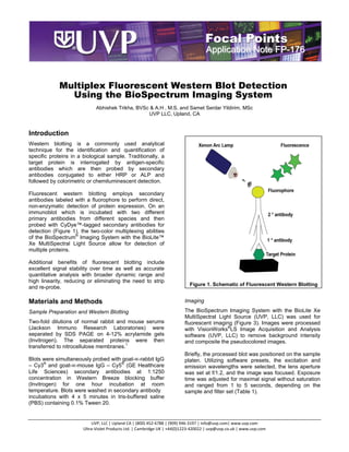
1. Application Note FP-176
Multiplex Fluorescent Western Blot Detection
Using the BioSpectrum Imaging System
Abhishek Trikha, BVSc & A.H , M.S. and Samet Serdar Yildirim, MSc
UVP LLC, Upland, CA
Introduction
Western blotting is a commonly used analytical
technique for the identification and quantification of
specific proteins in a biological sample. Traditionally, a
target protein is interrogated by antigen-specific
antibodies which are then probed by secondary
antibodies conjugated to either HRP or ALP and
followed by colorimetric or chemiluminescent detection.
Fluorescent western blotting employs secondary
antibodies labeled with a fluorophore to perform direct,
non-enzymatic detection of protein expression. On an
immunoblot which is incubated with two different
primary antibodies from different species and then
probed with CyDye™-tagged secondary antibodies for
detection (Figure 1), the two-color multiplexing abilities
of the BioSpectrum® Imaging System with the BioLite™
Xe MultiSpectral Light Source allow for detection of
multiple proteins.
Additional benefits of fluorescent blotting include
excellent signal stability over time as well as accurate
quantitative analysis with broader dynamic range and
high linearity, reducing or eliminating the need to strip
Figure 1. Schematic of Fluorescent Western Blotting
and re-probe.
Materials and Methods Imaging
Sample Preparation and Western Blotting The BioSpectrum Imaging System with the BioLite Xe
MultiSpectral Light Source (UVP, LLC) was used for
Two-fold dilutions of normal rabbit and mouse serums fluorescent imaging (Figure 3). Images were processed
(Jackson Immuno Research Laboratories) were with VisionWorks®LS Image Acquisition and Analysis
separated by SDS PAGE on 4-12% acrylamide gels software (UVP, LLC) to remove background intensity
(Invitrogen). The separated proteins were then and composite the pseudocolored images.
transferred to nitrocellulose membranes.1
Briefly, the processed blot was positioned on the sample
Blots were simultaneously probed with goat-∝-rabbit IgG platen. Utilizing software presets, the excitation and
– Cy3® and goat-∝-mouse IgG – Cy5® (GE Healthcare emission wavelengths were selected, the lens aperture
Life Sciences) secondary antibodies at 1:1250 was set at f/1.2, and the image was focused. Exposure
concentration in Western Breeze blocking buffer time was adjusted for maximal signal without saturation
(Invitrogen) for one hour incubation at room and ranged from 1 to 5 seconds, depending on the
temperature. Blots were washed in secondary antibody sample and filter set (Table 1).
incubations with 4 x 5 minutes in tris-buffered saline
(PBS) containing 0.1% Tween 20.
UVP, LLC | Upland CA | (800) 452-6788 | (909) 946-3197 | info@uvp.com| www.uvp.com
Ultra-Violet Products Ltd. | Cambridge UK | +44(0)1223-420022 | uvp@uvp.co.uk | www.uvp.com
2. Focal Points Application Note Page | 2
a) b)
Cy3 Cy5
c)
Multiplexed Cy3 and Cy5
®
Figure 2. Multiplex Fluorescent Western Blot Detection with CyDyes™ using BioSpectrum Imaging System
a) Rabbit IgG probed with Cy3–tagged goat anti-rabbit IgG b) Mouse IgG probed with Cy5-tagged goat anti-mouse IgG c)
Multiplex fluorescent detection of two fold serial dilution of mouse and rabbit serum proteins probed with Cy3–tagged goat
anti-rabbit IgG and Cy5-tagged goat anti-mouse IgG, respectively, on same immunoblot. CyDyes were imaged using a one
second exposure with the BioSpectrum Imaging System.
Once acquired, the original unaltered image was
archived and a copy was used for image analysis. The Fluorescent western blotting applications offer many
image was adjusted to globally remove background advantages over chemiluminescent or chromogenic
intensity and contrast, and was pseudocolored green visualization. Most significantly, fluorescent labels
and red using VisionWorksLS Software to indicate Cy3 permit multiplexing so that several proteins in a sample
and Cy5, respectively. can be detected and analyzed at the same time and on
a single protein blot. Fluorescent labels in particular
offer very low background and a high signal-to-noise
ratio for quantitative imaging.
Dye Excitation Emission
Cy3 525BP45 605BP50 The combination of the BioSpectrum Imaging System
Cy5 630BP30 730BP40 and the BioLite MultiSpectral Light Source provides not
only a full range of wavelengths for excitation light but
Table 1. Filters used for NIR blotting with 680 and 770
also rapid high resolution image capture through the use
to 800nm fluorescent tags.
of deeply cooled MegaCam 810 and OptiChemi™ 610
cameras and low light lenses.
Results and Discussion
Imaging Time
Figure 2 illustrates the multiplex imaging capabilities of
the BioSpectrum Imaging System, specifically Compared to laser scanning imaging systems where
separating out the signal of both Cy3 and Cy5 color imaging times range from 3 to 5 minutes, the
channels. BioSpectrum Imaging System provides a significant
advantage with imaging times ranging from 1 to 5
seconds for fluorescent Western blotting applications.
3. Focal Points Application Note Page | 3
Conclusion References
Fluorescent Western blot imaging with the BioSpectrum 1. Gallagher, S.R. and Wiley, E.A. Current
Imaging System and BioLite MultiSpectral Light Source Protocols: Essential Laboratory Techniques.
is a fast and efficient process for enabling researchers Wiley, 2012
to achieve simultaneous detection of multiple proteins
on the same immunoblot and generate high resolution,
publication-ready images.
Figure 3. BioSpectrum Connected to the BioLite
Together, the BioSpectrum and BioLite are a powerful
combination that are able to specifically excite and
illuminate at wavelengths from 365 to 765nm and read
emissions from 400 to 850nm. Use of up to eight
excitation and five emission wavelengths are possible
in a single experiment.
The BioSpectrum 810 and BioSpectrum 610 are
recommended for imaging multiplex fluorescent
Western blots.
4. Focal Points Application Note Page | 4
BioSpectrum and VisionWorks are registered trademarks of UVP, LLC. BioLite and OptiChemi are trademarks of UVP, LLC.
All other registered trademarks or trademarks are the property of their respective owners.