SlideShare une entreprise Scribd logo
1  sur  20
Acute Abdomen - Practical &
Diagnostic Approach
• Presented By

• DR Laith Fadhil Al-hialy
• MBchB PGSR
The 'acute abdomen' is a clinical condition characterized by
severe abdominal pain, requiring the clinician to make an
urgent therapeutic decision- surgical or none surgical

Major causes
















Acute appendicitis.
Acute Cholecystitis
Diverticulitis. or Meckel's diverticulitis
Acute pancreatitis.
Ectopic pregnancy.
Peptic ulcer disease, perforated DU ( 80% duodenum , pyloric antrum )
Salpingitis , PID , Expelled IUCCD
Intestinal obstruction, including paralytic ileus (a dynamic obstruction).
Gastroenteritis.
Acute intestinal ischemia/infarction or vasculitis.
Renal colic or renal tract pain , acute urinary retention.
Abdominal aortic aneurysm (AAA).
Gastrointestinal (GI) haemorrhage
Testicular torsion.
Nonsurgical disease, e.g. myocardial infarction, pericarditis, pneumonia, sickle
cell crisis, hepatitis, inflammatory bowel disease, opiate withdrawal, typhoid.
acute abdomen includes a wide spectrum of disorders, ranging
from life-threatening diseases to benign self-limiting conditions
Radiological strategy
 Before you perform an examination, obtain relevant information from the referring
clinician.
Don't let the clinician simply 'order' a plain X-ray film , sonogram or CT, but discuss
the patient's age and posture, laboratory results and the number one clinical
diagnosis and differential diagnosis.
Based on that information and your own degree of confidence with the modalities
decide for yourself whether to perform plan X-Ray film ,Sonography or CT.
Sonography has the advantage of close patient contact, enabling assessment of
the spot of maximum tenderness and the severity of illness without ionizing
radiation.
In general the diagnostic accuracy of CT is higher than Sonography.
We advocate the following two-step radiological approach of an acute abdomen.

1. Confirm or exclude the most common disease
2. Screen whole abdomen for general signs of pathology
Confirm or exclude most common disease
1st step focusing on most common causes

Location of pain determined strategy
•
•
•
•

RLQ : Appendicitis
RUQ : Cholecystitis
LLQ : Diverticulitis
LUQ : rare – mostly related to

gastric pathology

• 2nd step is
Screen the whole abdomen for the
pathological signs
•
•
•
•
•
•

Bowel wall thickening
Ileus ( dilated a tonic loop )
Air ( Pneumoperitoneum )
Fluid ( ascites )
Inflamed surrounding fat
Renal , uretric stone – pathology
A plain X-Ray
abdominal film
A normal film does not exclude an
ileus or other pathology and may
falsely reassure the clinician.
An ileus may not be appreciated on
a plain abdominal film if bowel
loops are filled with fluid only
without intraluminal air (figure).
Alternatively if a plain abdominal
film does indicate an ileus than
Sonography or CT are usually
needed to identify its cause.
Thus, a plain abdominal film is
seldomly useful, with the exception
of detection of kidney stones 70 % ,
GS 20 % , fecolith 10 % or a
Pneumoperitoneum.
For all other indications use
Sonography or CT.
LEFT: Plain abdominal film in a patient with an acute abdomen, showing no abnormalities.
RIGHT: Subsequent CT shows distended small bowel loops (arrowheads) that are not seen on
plain abdominal film because they are filled with fluid only and do not contain intraluminal air.
RLQ : Appendicitis
Pain in the RLQ, regardless of any
other symptom or laboratory results,
should be considered to be
appendicitis until proven otherwise.
If you are unable to find the appendix
you cannot rule out the diagnosis of
appendicitis unless a good alternative
diagnosis is found.
If you do not find the appendix and
there is no alternative diagnosis call
the results of the examination
indeterminate. Do not call it:' no
appendicitis'.

Inflamed Appendix
An inflamed appendix ( none
compressible tubal lesion ) has a
diameter larger than 6 mm, and is
usually surrounded by inflamed fat.
The presence of a fecolith or
hypervascularity on power Doppler
strongly supports inflammation.
DDX of RLQ disease
Mesenteric lymphadenitis.
Mesenteric lymphadenitis is a common
mimicker of appendicitis.
It is the second most common cause of right
lower quadrant pain after appendicitis.
It is defined as a benign self-limiting
inflammation of right-sided mesenteric
lymph nodes without an identifiable
underlying inflammatory process, occurring
more often in children than in adults..
This diagnosis can only be made confidently
when a normal appendix is found, because
adenopathy also frequently occurs with
appendicitis.
Key finding: Lymphadenopathy with a
normal appendix and normal mesenteric fat.

Bacterial ileocecitis
Infectious enter colitis may cause mild symptoms
resembling a common viral gastroenteritis, but it
may also clinically present with features
indistinguishable from appendicitis especially in
bacterial ileocecitis, caused by Yersinia,
Campylobacter, or Salmonella.
Key finding: ileocecal wall thickening without
inflamed fat, adenopathy, normal appendix
Right-sided diverticulitis
Right-sided colonic diverticulitis may
clinically mimic appendicitis or
Cholecystitis, though the patient's
history is generally more protracted.
In contrast to sigmoid diverticula,
right-sided colonic diverticula are
usually true diverticula, that is,
outpunching of the colonic wall
containing all layers of the wall.
This may possibly explain the
essentially benign self- limiting
character of right-sided diverticulitis

Salphingitis
Salphingitis is a common mimicker of
both of appendicitis and diverticulitis.
Transvaginal Sonography depicts an
inhomogeneous enlarged inflamed
ovary. ( adenixial mass lesion )
Epiploic appendagitis.
Epiploic appendages are small adipose
protrusions from the serosal surface of the
colon.
An epiploic appendage may undergo torsion
and secondary inflammation causing focal
abdominal pain that simulates appendicitis
when located in the right lower quadrant or
diverticulitis when located in the left lower
quadrant.
The characteristic ring-sign corresponds to
inflamed visceral peritoneal lining surrounding
an infracted fatty epiploic appendage. Epiploic

appendagitis has been reported in
approximately 1% of patients clinically
suspected of having appendicitis.
It is very important to make a positive
diagnosis of this characteristic entity since
epiploic appendagitis is a self-limiting
disease.
Both US and CT will depict an inflamed fatty
mass adjacent to the colon.
Key finding: inflamed fatty mass adjacent to
the colon with characteristic ring sign.
Urolithiasis
Urolithiasis often causes flank
pain, but an ureteral stone
(arrowhead) may occasionally
present with clinical signs
simulating appendicitis,
Cholecystitis or diverticulitis.
Appendicitis on the other hand
may cause hematuria, pyuria and
albuminuria in up to 25% of
patients because of direct
invaded ureteral inflammation
Note
Renal colic may associated by
paralytic ileus , sever pain air
swallowing commonly lead to
ileus
LLQ : Diverticulitis
If the pain is located in the LLQ your
main concern is sigmoid diverticulitis.
In diverticulitis Sonography and CT
show diverticulosis with segmental
colonic wall thickening and
inflammatory changes in the fat
surrounding a diverticulum.

LEFT: Sigmoid diverticulitis. Diverticulum
(arrow) is surrounded by hyper
attenuating fat. The sigmoid wall is
thickened.
RIGHT: Sigmoid carcinoma with limited fat
stranding.

Complications of
diverticulitis such
as abscess
formation or
perforation, can
best be excluded
with CT.
As DDX of LLQ lesion

Ruptured Aneurysm
Most abdominal aortic aneurysms rupture
into the left retro peritoneum (4).
Clinically this may simulate sigmoid
diverticulitis or renal colic due to
impingement of the hematoma on
adjacent structures.
However most patient will present with
the classic triad of hypotension, a
pulsating mass and back pain.
Continuous leakage will lead to rupture
into the peritoneal cavity and eventually
death.
Sonography is a quick and convenient
modality, but it is much less sensitive and
specific for the diagnosis of aneurysmal
rupture than CT.
The absence of sonographic evidence of
rupture does not rule out this entity if
clinical suspicion is high
RUQ : Cholecystitis
Cholecystitis occurs when a calculus
obstructs the cystic duct. The trapped
bile causes inflammation of the
gallbladder wall.
As gallstones are often occult on CT,
Sonography is the preferred imaging
method for the evaluation of
Cholecystitis, Some gallbladders happen
to be small and others are large. The

imaging appearance of Cholecystitis
consists of an enlarged hydropic
(meaning non-compressible)
gallbladder with a thickened wall in
the region of maximum tenderness
(the so-called 'Murphy sign‘)
Epigastric or upper
abdominal pain

Pancreatitis
CT depicts fat-stranding

(arrowheads) surrounding the
primary focus of the inflammation:
the pancreas
BY US focal edema on the pancreatic
fat , enlarge hypoechoic texture
pancreas
By X-ray , duodenal ileus , Lt plural
effusion , obliteration Lt poses shadow
Screen the whole abdomen for general signs of pathology
1.
2.
3.
4.
5.

Bowel wall thickening
Air ( Pneumoperitoneum )
Ileus ( dilated a tonic loop )
Fluid ( ascites )
Renal , uretric stone and
pathology
6. Inflamed surrounding fat

Bowel wall thickening
Thickening of bowel wall indicates inflammation or
tumor, and has an extensive differential diagnosis.
Thickening of small bowel loops usually indicates
regional inflammation, as small bowel tumors
(carcinoid, lymphoma, GIST) are relatively
infrequent.
In patients with local colonic wall thickening a
carcinoma is a prime concern.
Free air
The presence of free
intraperitoneal air is proof of
bowel perforation, and indicates
a surgical emergency.
A Pneumoperitoneum has only
two frequent causes:
- Perforation of a duodenal ,
gastric ulcer 80%
- Perforation of colonic
diverticulitis
Free air is usually not seen in
perforated appendicitis).
Always examine the images in
lung setting for better detection
of free intraabdominal air
(figure).
Ileus
Pathologic distention of bowel loops may be caused by obstruction or
paralysis.
Firstly determine which parts of the gut are affected: small bowel, large bowel, or
both. Look for normal non distended bowel loops, which, if present, strongly suggest
an obstructive cause for the ileus

Causes of paralytic Ileus
• post operative
• Peritonitis
• Inflammation ( appendicitis ,
cholecystis , pancreatitis )
• Trauma
• CHF , Uremia
• Hypokalaemia
• Pneumonia
•Drugs – morphine
•Renal colic

Causes of SBO
• adhesion 70-80 %
• Hernia
• volvulus
• intussusception
• GS Ileus
• tumor – lymphoma
• mesenteric ischemia

Causes of LBO
• Ca 60% - mostly sigmoid
• volvulus
• diverticulitis
• fecal impaction
• IBD - toxic mega colon
Distinction between small and Large bowel dilation
• No. of loops
• distribution of loops
• diameter
• Sold feces
• structure
• Radius of curvature

• Many
• Central
• 3-5 cm
• absent
• valvule
conniventes
• small

• Few
• peripheral
• 5 cm +
• present
• haustra
• large
Ascites
Asymptomatic person do not
have a detectable amount of
free intraperitoneal fluid, with
the exception of an incidental
drop of fluid in Douglas in
fertile women.
The presence of ascites is a
nonspecific sign of abdominal
pathology, indicating that
'something is wrong'.
You may want to perform a
US-guided diagnostic puncture
of the ascites, in order to
investigate whether it is

sterile reactive fluid, pus,
blood, urine, or bile.

Contenu connexe

Tendances

Volvulus in git
Volvulus in gitVolvulus in git
Volvulus in git
airwave12
 
Presentation2.pptx. radilogical imaging of intestinal obstruction.
Presentation2.pptx. radilogical imaging of intestinal obstruction.Presentation2.pptx. radilogical imaging of intestinal obstruction.
Presentation2.pptx. radilogical imaging of intestinal obstruction.
Abdellah Nazeer
 

Tendances (20)

CT Evaluation of the Acute Abdomen
CT Evaluation of the Acute AbdomenCT Evaluation of the Acute Abdomen
CT Evaluation of the Acute Abdomen
 
Acute cholecystitis/ RUQ Pain
Acute cholecystitis/ RUQ PainAcute cholecystitis/ RUQ Pain
Acute cholecystitis/ RUQ Pain
 
Acute abdomen
Acute abdomenAcute abdomen
Acute abdomen
 
Volvulus of colon
Volvulus of colonVolvulus of colon
Volvulus of colon
 
Aid of Imaging in diagnosis of Acute Abdomen
Aid of Imaging in diagnosis of Acute AbdomenAid of Imaging in diagnosis of Acute Abdomen
Aid of Imaging in diagnosis of Acute Abdomen
 
Acute Abdomen-Radiology
Acute Abdomen-RadiologyAcute Abdomen-Radiology
Acute Abdomen-Radiology
 
Acute Abdomen
Acute AbdomenAcute Abdomen
Acute Abdomen
 
Acute Abdomen by Dr KD DELE
Acute Abdomen by Dr KD DELEAcute Abdomen by Dr KD DELE
Acute Abdomen by Dr KD DELE
 
Haemorrhoids and perianal diseases
Haemorrhoids and perianal diseasesHaemorrhoids and perianal diseases
Haemorrhoids and perianal diseases
 
Acute abdomen
Acute abdomenAcute abdomen
Acute abdomen
 
Acute abdomen
Acute abdomenAcute abdomen
Acute abdomen
 
Xrays
XraysXrays
Xrays
 
Volvulus in git
Volvulus in gitVolvulus in git
Volvulus in git
 
Acute abdomen
Acute abdomenAcute abdomen
Acute abdomen
 
Abdominal pain- all quadrants- case based learning
Abdominal pain-  all quadrants- case based learningAbdominal pain-  all quadrants- case based learning
Abdominal pain- all quadrants- case based learning
 
Acute abdomen
Acute abdomenAcute abdomen
Acute abdomen
 
Abdominal X ray
Abdominal X rayAbdominal X ray
Abdominal X ray
 
Presentation2.pptx. radilogical imaging of intestinal obstruction.
Presentation2.pptx. radilogical imaging of intestinal obstruction.Presentation2.pptx. radilogical imaging of intestinal obstruction.
Presentation2.pptx. radilogical imaging of intestinal obstruction.
 
The Acute Surgical Abdomen
The Acute Surgical AbdomenThe Acute Surgical Abdomen
The Acute Surgical Abdomen
 
Approach to acute abdomen
Approach to acute abdomenApproach to acute abdomen
Approach to acute abdomen
 

En vedette

Gynaecological emergencies
Gynaecological emergenciesGynaecological emergencies
Gynaecological emergencies
Ruth Nwokoma
 

En vedette (17)

Acute Abdominal Pain
Acute Abdominal PainAcute Abdominal Pain
Acute Abdominal Pain
 
Diagnosis And Management Of Acute Abdominal Pain
Diagnosis And Management Of Acute Abdominal PainDiagnosis And Management Of Acute Abdominal Pain
Diagnosis And Management Of Acute Abdominal Pain
 
Acute Abdomen Ppt
Acute Abdomen PptAcute Abdomen Ppt
Acute Abdomen Ppt
 
Triage &assesment of abdominal trauma
Triage &assesment of abdominal traumaTriage &assesment of abdominal trauma
Triage &assesment of abdominal trauma
 
Obstetric emergencies
Obstetric emergenciesObstetric emergencies
Obstetric emergencies
 
Obstetrical emergencies
Obstetrical emergenciesObstetrical emergencies
Obstetrical emergencies
 
Obstetric emergencies
Obstetric emergencies Obstetric emergencies
Obstetric emergencies
 
01 Emergency Obstetric care
01 Emergency Obstetric care01 Emergency Obstetric care
01 Emergency Obstetric care
 
Obstetric emergencies part 1
Obstetric emergencies part 1Obstetric emergencies part 1
Obstetric emergencies part 1
 
Gynaecological emergencies
Gynaecological emergenciesGynaecological emergencies
Gynaecological emergencies
 
Žene i muškarci ravnopravni u svemu osim u podnošenju bola
Žene i muškarci ravnopravni u svemu osim u podnošenju bolaŽene i muškarci ravnopravni u svemu osim u podnošenju bola
Žene i muškarci ravnopravni u svemu osim u podnošenju bola
 
Emergency Obstertrics & Gynecology
Emergency Obstertrics & GynecologyEmergency Obstertrics & Gynecology
Emergency Obstertrics & Gynecology
 
Obstetrical emergencies
Obstetrical emergenciesObstetrical emergencies
Obstetrical emergencies
 
Gynecological causes of acute abdominal pain
Gynecological causes of  acute abdominal painGynecological causes of  acute abdominal pain
Gynecological causes of acute abdominal pain
 
Gynaecology Emergencies
Gynaecology EmergenciesGynaecology Emergencies
Gynaecology Emergencies
 
clinical course" Acute abdomen "
clinical course" Acute abdomen "clinical course" Acute abdomen "
clinical course" Acute abdomen "
 
Clinical approach to a patient with abdominal pain
Clinical approach to a patient with abdominal painClinical approach to a patient with abdominal pain
Clinical approach to a patient with abdominal pain
 

Similaire à Acute abdomen a practical approach

Appendicitis for 5yr mbchb
Appendicitis for 5yr mbchbAppendicitis for 5yr mbchb
Appendicitis for 5yr mbchb
Avid Listener
 
Combined 03 clinical training--pathology benign_diverticular disease
Combined 03 clinical training--pathology benign_diverticular diseaseCombined 03 clinical training--pathology benign_diverticular disease
Combined 03 clinical training--pathology benign_diverticular disease
Iknifem
 
Abdominal conditions 2 - 3.1.pptx
Abdominal conditions 2 - 3.1.pptxAbdominal conditions 2 - 3.1.pptx
Abdominal conditions 2 - 3.1.pptx
INNOBRIZZY
 
GASTROINTESTINAL TUBERCULOSIS ABDOMINAL TUBERCULOSIS
GASTROINTESTINAL TUBERCULOSIS ABDOMINAL TUBERCULOSISGASTROINTESTINAL TUBERCULOSIS ABDOMINAL TUBERCULOSIS
GASTROINTESTINAL TUBERCULOSIS ABDOMINAL TUBERCULOSIS
Nawin Kumar
 

Similaire à Acute abdomen a practical approach (20)

Appendicitis for 5yr mbchb
Appendicitis for 5yr mbchbAppendicitis for 5yr mbchb
Appendicitis for 5yr mbchb
 
Combined 03 clinical training--pathology benign_diverticular disease
Combined 03 clinical training--pathology benign_diverticular diseaseCombined 03 clinical training--pathology benign_diverticular disease
Combined 03 clinical training--pathology benign_diverticular disease
 
Pancreatitis -a detailed study ( medical information )
Pancreatitis -a detailed study ( medical information )Pancreatitis -a detailed study ( medical information )
Pancreatitis -a detailed study ( medical information )
 
Abdominal conditions 2 - 3.1.pptx
Abdominal conditions 2 - 3.1.pptxAbdominal conditions 2 - 3.1.pptx
Abdominal conditions 2 - 3.1.pptx
 
Chapter 26 Appendix
Chapter 26 AppendixChapter 26 Appendix
Chapter 26 Appendix
 
Acute epigastric pain
Acute epigastric painAcute epigastric pain
Acute epigastric pain
 
Bohomolets Surgery 4th year Lecture #3
Bohomolets Surgery 4th year Lecture #3Bohomolets Surgery 4th year Lecture #3
Bohomolets Surgery 4th year Lecture #3
 
Ulcerative colitis
Ulcerative colitisUlcerative colitis
Ulcerative colitis
 
Diseases of git
Diseases of gitDiseases of git
Diseases of git
 
Diseases of GIT
Diseases of GITDiseases of GIT
Diseases of GIT
 
GASTROINTESTINAL TUBERCULOSIS ABDOMINAL TUBERCULOSIS
GASTROINTESTINAL TUBERCULOSIS ABDOMINAL TUBERCULOSISGASTROINTESTINAL TUBERCULOSIS ABDOMINAL TUBERCULOSIS
GASTROINTESTINAL TUBERCULOSIS ABDOMINAL TUBERCULOSIS
 
Acute appendicitis
Acute appendicitisAcute appendicitis
Acute appendicitis
 
Intestinal obstruction
Intestinal obstructionIntestinal obstruction
Intestinal obstruction
 
Acquired intestinal ileus
Acquired intestinal ileusAcquired intestinal ileus
Acquired intestinal ileus
 
Cholecystitis
CholecystitisCholecystitis
Cholecystitis
 
Acute abdomen.pptx
Acute abdomen.pptxAcute abdomen.pptx
Acute abdomen.pptx
 
Chronic cholecystitis
Chronic cholecystitisChronic cholecystitis
Chronic cholecystitis
 
Appendix
AppendixAppendix
Appendix
 
Appendicitis
AppendicitisAppendicitis
Appendicitis
 
Hepato Biliary.pptx
Hepato Biliary.pptxHepato Biliary.pptx
Hepato Biliary.pptx
 

Plus de DR Laith

Plus de DR Laith (20)

Diagnostic imaging modalities
Diagnostic imaging modalities Diagnostic imaging modalities
Diagnostic imaging modalities
 
Biliary system
Biliary systemBiliary system
Biliary system
 
Spleen
SpleenSpleen
Spleen
 
Carpal tunnel syndrome ( cts )us criteria
Carpal tunnel syndrome ( cts )us criteria  Carpal tunnel syndrome ( cts )us criteria
Carpal tunnel syndrome ( cts )us criteria
 
Skeletal trauma
Skeletal traumaSkeletal trauma
Skeletal trauma
 
Ankle joint
Ankle jointAnkle joint
Ankle joint
 
Brain anatomy in concerin of diagnostic imaging
Brain anatomy in concerin of diagnostic imaging Brain anatomy in concerin of diagnostic imaging
Brain anatomy in concerin of diagnostic imaging
 
Soft tissue tumours
Soft tissue tumoursSoft tissue tumours
Soft tissue tumours
 
Wrist Joint
Wrist Joint Wrist Joint
Wrist Joint
 
Brain infection
Brain infection  Brain infection
Brain infection
 
Sinuses & neck lesions
Sinuses & neck lesions Sinuses & neck lesions
Sinuses & neck lesions
 
Salviary glands
Salviary glands Salviary glands
Salviary glands
 
Ear
EarEar
Ear
 
Spine degenerative disease
Spine  degenerative  disease     Spine  degenerative  disease
Spine degenerative disease
 
Congenital lesions and variants of renal system
Congenital lesions and variants of renal systemCongenital lesions and variants of renal system
Congenital lesions and variants of renal system
 
Von hippel –lindau disease
Von hippel –lindau diseaseVon hippel –lindau disease
Von hippel –lindau disease
 
Urethra lesions
Urethra lesions Urethra lesions
Urethra lesions
 
Mri in diagnosis of acute appendicitis during pregnancy
Mri in diagnosis of acute appendicitis  during pregnancy Mri in diagnosis of acute appendicitis  during pregnancy
Mri in diagnosis of acute appendicitis during pregnancy
 
Focal hepatic lesions in concerns of diagnostic imaging .pptx 1
Focal hepatic lesions in concerns of diagnostic imaging .pptx 1Focal hepatic lesions in concerns of diagnostic imaging .pptx 1
Focal hepatic lesions in concerns of diagnostic imaging .pptx 1
 
Skeletal manifestations of eosinophilic granuloma (eg)
Skeletal manifestations of eosinophilic granuloma (eg)Skeletal manifestations of eosinophilic granuloma (eg)
Skeletal manifestations of eosinophilic granuloma (eg)
 

Dernier

🌹Attapur⬅️ Vip Call Girls Hyderabad 📱9352852248 Book Well Trand Call Girls In...
🌹Attapur⬅️ Vip Call Girls Hyderabad 📱9352852248 Book Well Trand Call Girls In...🌹Attapur⬅️ Vip Call Girls Hyderabad 📱9352852248 Book Well Trand Call Girls In...
🌹Attapur⬅️ Vip Call Girls Hyderabad 📱9352852248 Book Well Trand Call Girls In...
Call Girls In Delhi Whatsup 9873940964 Enjoy Unlimited Pleasure
 
Russian Call Girls Lucknow Just Call 👉👉7877925207 Top Class Call Girl Service...
Russian Call Girls Lucknow Just Call 👉👉7877925207 Top Class Call Girl Service...Russian Call Girls Lucknow Just Call 👉👉7877925207 Top Class Call Girl Service...
Russian Call Girls Lucknow Just Call 👉👉7877925207 Top Class Call Girl Service...
adilkhan87451
 
Call Girl In Pune 👉 Just CALL ME: 9352988975 💋 Call Out Call Both With High p...
Call Girl In Pune 👉 Just CALL ME: 9352988975 💋 Call Out Call Both With High p...Call Girl In Pune 👉 Just CALL ME: 9352988975 💋 Call Out Call Both With High p...
Call Girl In Pune 👉 Just CALL ME: 9352988975 💋 Call Out Call Both With High p...
chetankumar9855
 

Dernier (20)

Call Girls Raipur Just Call 9630942363 Top Class Call Girl Service Available
Call Girls Raipur Just Call 9630942363 Top Class Call Girl Service AvailableCall Girls Raipur Just Call 9630942363 Top Class Call Girl Service Available
Call Girls Raipur Just Call 9630942363 Top Class Call Girl Service Available
 
Top Rated Hyderabad Call Girls Chintal ⟟ 9332606886 ⟟ Call Me For Genuine Se...
Top Rated  Hyderabad Call Girls Chintal ⟟ 9332606886 ⟟ Call Me For Genuine Se...Top Rated  Hyderabad Call Girls Chintal ⟟ 9332606886 ⟟ Call Me For Genuine Se...
Top Rated Hyderabad Call Girls Chintal ⟟ 9332606886 ⟟ Call Me For Genuine Se...
 
The Most Attractive Hyderabad Call Girls Kothapet 𖠋 9332606886 𖠋 Will You Mis...
The Most Attractive Hyderabad Call Girls Kothapet 𖠋 9332606886 𖠋 Will You Mis...The Most Attractive Hyderabad Call Girls Kothapet 𖠋 9332606886 𖠋 Will You Mis...
The Most Attractive Hyderabad Call Girls Kothapet 𖠋 9332606886 𖠋 Will You Mis...
 
Russian Call Girls Service Jaipur {8445551418} ❤️PALLAVI VIP Jaipur Call Gir...
Russian Call Girls Service  Jaipur {8445551418} ❤️PALLAVI VIP Jaipur Call Gir...Russian Call Girls Service  Jaipur {8445551418} ❤️PALLAVI VIP Jaipur Call Gir...
Russian Call Girls Service Jaipur {8445551418} ❤️PALLAVI VIP Jaipur Call Gir...
 
Call Girls Kurnool Just Call 8250077686 Top Class Call Girl Service Available
Call Girls Kurnool Just Call 8250077686 Top Class Call Girl Service AvailableCall Girls Kurnool Just Call 8250077686 Top Class Call Girl Service Available
Call Girls Kurnool Just Call 8250077686 Top Class Call Girl Service Available
 
Best Rate (Patna ) Call Girls Patna ⟟ 8617370543 ⟟ High Class Call Girl In 5 ...
Best Rate (Patna ) Call Girls Patna ⟟ 8617370543 ⟟ High Class Call Girl In 5 ...Best Rate (Patna ) Call Girls Patna ⟟ 8617370543 ⟟ High Class Call Girl In 5 ...
Best Rate (Patna ) Call Girls Patna ⟟ 8617370543 ⟟ High Class Call Girl In 5 ...
 
🌹Attapur⬅️ Vip Call Girls Hyderabad 📱9352852248 Book Well Trand Call Girls In...
🌹Attapur⬅️ Vip Call Girls Hyderabad 📱9352852248 Book Well Trand Call Girls In...🌹Attapur⬅️ Vip Call Girls Hyderabad 📱9352852248 Book Well Trand Call Girls In...
🌹Attapur⬅️ Vip Call Girls Hyderabad 📱9352852248 Book Well Trand Call Girls In...
 
Russian Call Girls Lucknow Just Call 👉👉7877925207 Top Class Call Girl Service...
Russian Call Girls Lucknow Just Call 👉👉7877925207 Top Class Call Girl Service...Russian Call Girls Lucknow Just Call 👉👉7877925207 Top Class Call Girl Service...
Russian Call Girls Lucknow Just Call 👉👉7877925207 Top Class Call Girl Service...
 
Premium Call Girls In Jaipur {8445551418} ❤️VVIP SEEMA Call Girl in Jaipur Ra...
Premium Call Girls In Jaipur {8445551418} ❤️VVIP SEEMA Call Girl in Jaipur Ra...Premium Call Girls In Jaipur {8445551418} ❤️VVIP SEEMA Call Girl in Jaipur Ra...
Premium Call Girls In Jaipur {8445551418} ❤️VVIP SEEMA Call Girl in Jaipur Ra...
 
Call Girl In Pune 👉 Just CALL ME: 9352988975 💋 Call Out Call Both With High p...
Call Girl In Pune 👉 Just CALL ME: 9352988975 💋 Call Out Call Both With High p...Call Girl In Pune 👉 Just CALL ME: 9352988975 💋 Call Out Call Both With High p...
Call Girl In Pune 👉 Just CALL ME: 9352988975 💋 Call Out Call Both With High p...
 
Call Girls Tirupati Just Call 8250077686 Top Class Call Girl Service Available
Call Girls Tirupati Just Call 8250077686 Top Class Call Girl Service AvailableCall Girls Tirupati Just Call 8250077686 Top Class Call Girl Service Available
Call Girls Tirupati Just Call 8250077686 Top Class Call Girl Service Available
 
Most Beautiful Call Girl in Bangalore Contact on Whatsapp
Most Beautiful Call Girl in Bangalore Contact on WhatsappMost Beautiful Call Girl in Bangalore Contact on Whatsapp
Most Beautiful Call Girl in Bangalore Contact on Whatsapp
 
Call Girls Vadodara Just Call 8617370543 Top Class Call Girl Service Available
Call Girls Vadodara Just Call 8617370543 Top Class Call Girl Service AvailableCall Girls Vadodara Just Call 8617370543 Top Class Call Girl Service Available
Call Girls Vadodara Just Call 8617370543 Top Class Call Girl Service Available
 
Call Girls Shimla Just Call 8617370543 Top Class Call Girl Service Available
Call Girls Shimla Just Call 8617370543 Top Class Call Girl Service AvailableCall Girls Shimla Just Call 8617370543 Top Class Call Girl Service Available
Call Girls Shimla Just Call 8617370543 Top Class Call Girl Service Available
 
All Time Service Available Call Girls Marine Drive 📳 9820252231 For 18+ VIP C...
All Time Service Available Call Girls Marine Drive 📳 9820252231 For 18+ VIP C...All Time Service Available Call Girls Marine Drive 📳 9820252231 For 18+ VIP C...
All Time Service Available Call Girls Marine Drive 📳 9820252231 For 18+ VIP C...
 
Call Girls Ahmedabad Just Call 9630942363 Top Class Call Girl Service Available
Call Girls Ahmedabad Just Call 9630942363 Top Class Call Girl Service AvailableCall Girls Ahmedabad Just Call 9630942363 Top Class Call Girl Service Available
Call Girls Ahmedabad Just Call 9630942363 Top Class Call Girl Service Available
 
(Low Rate RASHMI ) Rate Of Call Girls Jaipur ❣ 8445551418 ❣ Elite Models & Ce...
(Low Rate RASHMI ) Rate Of Call Girls Jaipur ❣ 8445551418 ❣ Elite Models & Ce...(Low Rate RASHMI ) Rate Of Call Girls Jaipur ❣ 8445551418 ❣ Elite Models & Ce...
(Low Rate RASHMI ) Rate Of Call Girls Jaipur ❣ 8445551418 ❣ Elite Models & Ce...
 
Call Girls Service Jaipur {8445551418} ❤️VVIP BHAWNA Call Girl in Jaipur Raja...
Call Girls Service Jaipur {8445551418} ❤️VVIP BHAWNA Call Girl in Jaipur Raja...Call Girls Service Jaipur {8445551418} ❤️VVIP BHAWNA Call Girl in Jaipur Raja...
Call Girls Service Jaipur {8445551418} ❤️VVIP BHAWNA Call Girl in Jaipur Raja...
 
Jogeshwari ! Call Girls Service Mumbai - 450+ Call Girl Cash Payment 90042684...
Jogeshwari ! Call Girls Service Mumbai - 450+ Call Girl Cash Payment 90042684...Jogeshwari ! Call Girls Service Mumbai - 450+ Call Girl Cash Payment 90042684...
Jogeshwari ! Call Girls Service Mumbai - 450+ Call Girl Cash Payment 90042684...
 
Independent Call Girls In Jaipur { 8445551418 } ✔ ANIKA MEHTA ✔ Get High Prof...
Independent Call Girls In Jaipur { 8445551418 } ✔ ANIKA MEHTA ✔ Get High Prof...Independent Call Girls In Jaipur { 8445551418 } ✔ ANIKA MEHTA ✔ Get High Prof...
Independent Call Girls In Jaipur { 8445551418 } ✔ ANIKA MEHTA ✔ Get High Prof...
 

Acute abdomen a practical approach

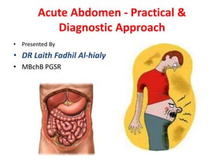
  • 1. Acute Abdomen - Practical & Diagnostic Approach • Presented By • DR Laith Fadhil Al-hialy • MBchB PGSR
  • 2. The 'acute abdomen' is a clinical condition characterized by severe abdominal pain, requiring the clinician to make an urgent therapeutic decision- surgical or none surgical Major causes                Acute appendicitis. Acute Cholecystitis Diverticulitis. or Meckel's diverticulitis Acute pancreatitis. Ectopic pregnancy. Peptic ulcer disease, perforated DU ( 80% duodenum , pyloric antrum ) Salpingitis , PID , Expelled IUCCD Intestinal obstruction, including paralytic ileus (a dynamic obstruction). Gastroenteritis. Acute intestinal ischemia/infarction or vasculitis. Renal colic or renal tract pain , acute urinary retention. Abdominal aortic aneurysm (AAA). Gastrointestinal (GI) haemorrhage Testicular torsion. Nonsurgical disease, e.g. myocardial infarction, pericarditis, pneumonia, sickle cell crisis, hepatitis, inflammatory bowel disease, opiate withdrawal, typhoid.
  • 3. acute abdomen includes a wide spectrum of disorders, ranging from life-threatening diseases to benign self-limiting conditions
  • 4. Radiological strategy  Before you perform an examination, obtain relevant information from the referring clinician. Don't let the clinician simply 'order' a plain X-ray film , sonogram or CT, but discuss the patient's age and posture, laboratory results and the number one clinical diagnosis and differential diagnosis. Based on that information and your own degree of confidence with the modalities decide for yourself whether to perform plan X-Ray film ,Sonography or CT. Sonography has the advantage of close patient contact, enabling assessment of the spot of maximum tenderness and the severity of illness without ionizing radiation. In general the diagnostic accuracy of CT is higher than Sonography. We advocate the following two-step radiological approach of an acute abdomen. 1. Confirm or exclude the most common disease 2. Screen whole abdomen for general signs of pathology
  • 5. Confirm or exclude most common disease 1st step focusing on most common causes Location of pain determined strategy • • • • RLQ : Appendicitis RUQ : Cholecystitis LLQ : Diverticulitis LUQ : rare – mostly related to gastric pathology • 2nd step is Screen the whole abdomen for the pathological signs • • • • • • Bowel wall thickening Ileus ( dilated a tonic loop ) Air ( Pneumoperitoneum ) Fluid ( ascites ) Inflamed surrounding fat Renal , uretric stone – pathology
  • 6. A plain X-Ray abdominal film A normal film does not exclude an ileus or other pathology and may falsely reassure the clinician. An ileus may not be appreciated on a plain abdominal film if bowel loops are filled with fluid only without intraluminal air (figure). Alternatively if a plain abdominal film does indicate an ileus than Sonography or CT are usually needed to identify its cause. Thus, a plain abdominal film is seldomly useful, with the exception of detection of kidney stones 70 % , GS 20 % , fecolith 10 % or a Pneumoperitoneum. For all other indications use Sonography or CT. LEFT: Plain abdominal film in a patient with an acute abdomen, showing no abnormalities. RIGHT: Subsequent CT shows distended small bowel loops (arrowheads) that are not seen on plain abdominal film because they are filled with fluid only and do not contain intraluminal air.
  • 7. RLQ : Appendicitis Pain in the RLQ, regardless of any other symptom or laboratory results, should be considered to be appendicitis until proven otherwise. If you are unable to find the appendix you cannot rule out the diagnosis of appendicitis unless a good alternative diagnosis is found. If you do not find the appendix and there is no alternative diagnosis call the results of the examination indeterminate. Do not call it:' no appendicitis'. Inflamed Appendix An inflamed appendix ( none compressible tubal lesion ) has a diameter larger than 6 mm, and is usually surrounded by inflamed fat. The presence of a fecolith or hypervascularity on power Doppler strongly supports inflammation.
  • 8. DDX of RLQ disease Mesenteric lymphadenitis. Mesenteric lymphadenitis is a common mimicker of appendicitis. It is the second most common cause of right lower quadrant pain after appendicitis. It is defined as a benign self-limiting inflammation of right-sided mesenteric lymph nodes without an identifiable underlying inflammatory process, occurring more often in children than in adults.. This diagnosis can only be made confidently when a normal appendix is found, because adenopathy also frequently occurs with appendicitis. Key finding: Lymphadenopathy with a normal appendix and normal mesenteric fat. Bacterial ileocecitis Infectious enter colitis may cause mild symptoms resembling a common viral gastroenteritis, but it may also clinically present with features indistinguishable from appendicitis especially in bacterial ileocecitis, caused by Yersinia, Campylobacter, or Salmonella. Key finding: ileocecal wall thickening without inflamed fat, adenopathy, normal appendix
  • 9. Right-sided diverticulitis Right-sided colonic diverticulitis may clinically mimic appendicitis or Cholecystitis, though the patient's history is generally more protracted. In contrast to sigmoid diverticula, right-sided colonic diverticula are usually true diverticula, that is, outpunching of the colonic wall containing all layers of the wall. This may possibly explain the essentially benign self- limiting character of right-sided diverticulitis Salphingitis Salphingitis is a common mimicker of both of appendicitis and diverticulitis. Transvaginal Sonography depicts an inhomogeneous enlarged inflamed ovary. ( adenixial mass lesion )
  • 10. Epiploic appendagitis. Epiploic appendages are small adipose protrusions from the serosal surface of the colon. An epiploic appendage may undergo torsion and secondary inflammation causing focal abdominal pain that simulates appendicitis when located in the right lower quadrant or diverticulitis when located in the left lower quadrant. The characteristic ring-sign corresponds to inflamed visceral peritoneal lining surrounding an infracted fatty epiploic appendage. Epiploic appendagitis has been reported in approximately 1% of patients clinically suspected of having appendicitis. It is very important to make a positive diagnosis of this characteristic entity since epiploic appendagitis is a self-limiting disease. Both US and CT will depict an inflamed fatty mass adjacent to the colon. Key finding: inflamed fatty mass adjacent to the colon with characteristic ring sign.
  • 11. Urolithiasis Urolithiasis often causes flank pain, but an ureteral stone (arrowhead) may occasionally present with clinical signs simulating appendicitis, Cholecystitis or diverticulitis. Appendicitis on the other hand may cause hematuria, pyuria and albuminuria in up to 25% of patients because of direct invaded ureteral inflammation Note Renal colic may associated by paralytic ileus , sever pain air swallowing commonly lead to ileus
  • 12. LLQ : Diverticulitis If the pain is located in the LLQ your main concern is sigmoid diverticulitis. In diverticulitis Sonography and CT show diverticulosis with segmental colonic wall thickening and inflammatory changes in the fat surrounding a diverticulum. LEFT: Sigmoid diverticulitis. Diverticulum (arrow) is surrounded by hyper attenuating fat. The sigmoid wall is thickened. RIGHT: Sigmoid carcinoma with limited fat stranding. Complications of diverticulitis such as abscess formation or perforation, can best be excluded with CT.
  • 13. As DDX of LLQ lesion Ruptured Aneurysm Most abdominal aortic aneurysms rupture into the left retro peritoneum (4). Clinically this may simulate sigmoid diverticulitis or renal colic due to impingement of the hematoma on adjacent structures. However most patient will present with the classic triad of hypotension, a pulsating mass and back pain. Continuous leakage will lead to rupture into the peritoneal cavity and eventually death. Sonography is a quick and convenient modality, but it is much less sensitive and specific for the diagnosis of aneurysmal rupture than CT. The absence of sonographic evidence of rupture does not rule out this entity if clinical suspicion is high
  • 14. RUQ : Cholecystitis Cholecystitis occurs when a calculus obstructs the cystic duct. The trapped bile causes inflammation of the gallbladder wall. As gallstones are often occult on CT, Sonography is the preferred imaging method for the evaluation of Cholecystitis, Some gallbladders happen to be small and others are large. The imaging appearance of Cholecystitis consists of an enlarged hydropic (meaning non-compressible) gallbladder with a thickened wall in the region of maximum tenderness (the so-called 'Murphy sign‘)
  • 15. Epigastric or upper abdominal pain Pancreatitis CT depicts fat-stranding (arrowheads) surrounding the primary focus of the inflammation: the pancreas BY US focal edema on the pancreatic fat , enlarge hypoechoic texture pancreas By X-ray , duodenal ileus , Lt plural effusion , obliteration Lt poses shadow
  • 16. Screen the whole abdomen for general signs of pathology 1. 2. 3. 4. 5. Bowel wall thickening Air ( Pneumoperitoneum ) Ileus ( dilated a tonic loop ) Fluid ( ascites ) Renal , uretric stone and pathology 6. Inflamed surrounding fat Bowel wall thickening Thickening of bowel wall indicates inflammation or tumor, and has an extensive differential diagnosis. Thickening of small bowel loops usually indicates regional inflammation, as small bowel tumors (carcinoid, lymphoma, GIST) are relatively infrequent. In patients with local colonic wall thickening a carcinoma is a prime concern.
  • 17. Free air The presence of free intraperitoneal air is proof of bowel perforation, and indicates a surgical emergency. A Pneumoperitoneum has only two frequent causes: - Perforation of a duodenal , gastric ulcer 80% - Perforation of colonic diverticulitis Free air is usually not seen in perforated appendicitis). Always examine the images in lung setting for better detection of free intraabdominal air (figure).
  • 18. Ileus Pathologic distention of bowel loops may be caused by obstruction or paralysis. Firstly determine which parts of the gut are affected: small bowel, large bowel, or both. Look for normal non distended bowel loops, which, if present, strongly suggest an obstructive cause for the ileus Causes of paralytic Ileus • post operative • Peritonitis • Inflammation ( appendicitis , cholecystis , pancreatitis ) • Trauma • CHF , Uremia • Hypokalaemia • Pneumonia •Drugs – morphine •Renal colic Causes of SBO • adhesion 70-80 % • Hernia • volvulus • intussusception • GS Ileus • tumor – lymphoma • mesenteric ischemia Causes of LBO • Ca 60% - mostly sigmoid • volvulus • diverticulitis • fecal impaction • IBD - toxic mega colon
  • 19. Distinction between small and Large bowel dilation • No. of loops • distribution of loops • diameter • Sold feces • structure • Radius of curvature • Many • Central • 3-5 cm • absent • valvule conniventes • small • Few • peripheral • 5 cm + • present • haustra • large
  • 20. Ascites Asymptomatic person do not have a detectable amount of free intraperitoneal fluid, with the exception of an incidental drop of fluid in Douglas in fertile women. The presence of ascites is a nonspecific sign of abdominal pathology, indicating that 'something is wrong'. You may want to perform a US-guided diagnostic puncture of the ascites, in order to investigate whether it is sterile reactive fluid, pus, blood, urine, or bile.