1. CHAPTER 1: INTRODUCTION
1.1 Coagulation
Coagulation is brought about primarily by the reduction of the repulsive potential of
electric double layer (1). The chemical coagulation of turbid and/or naturally colored
waters involves the interaction of particulates and/or colloids with a destabilizing agent.
The essential purpose of coagulation is to aggregate these particles into larger sizes that
will settle quickly within an hour or two, which will then be removed by sedimentation
(3). Several inorganic salts of iron and aluminum such as alum, lime, ferric chloride,
ferric sulfate, copper-as, sodium aluminate and aluminum chloride are available
commercially for coagulation. But the common coagulants used are alum and ferric
chloride (2, 3). These coagulants can effectively remove broad range of impurities from
water, such as colloidal particles and dissolved organic substances. There modes of action
can be generally explained in terms of two distinct mechanisms: charge neutralization of
negatively charged particles by cationic hydrolysis products and incorporation of
impurities in an amorphous hydroxide precipitate.
The Electrostatic stability of particles is of concern in coagulation of natural waters.
In natural waters, particles and microbes are predominantly negatively charged due to a
variety of negative functional groups on their surfaces. This negative charge was first
reported in 1929 (4). Hence, these particles may be stable as a result of electrical
repulsion. So if the surface of the particle has a net negative charge, and the bulk of
solution is electrically neutral, a potential difference exists between the surface and a
point at a distance in the bulk solution (5). In water treatment practice, such particles are
destabilized by the addition of salts causing their aggregation for rapid sedimentation and
2. 2
filtration. Destabilization of particles occur in two different mechanisms: 1) compression
of double layer (Diffuse Layer + Stern Layer) by charge neutralization and 2) adsorption
for inter particle bridging (6).
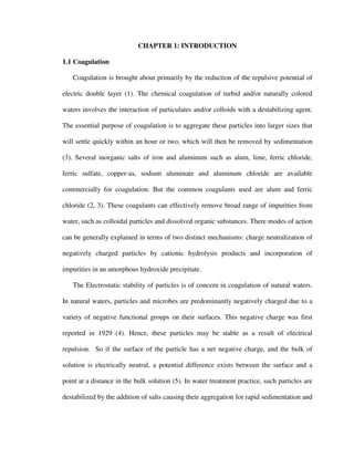
FIG. 1.1. Source of Zeta Potential and distribution of charges around a particle (7).
The electric double layer consists of two layers called the Stern Layer and the Diffuse
Layer. The stern layer (rigid layer) at the surface of the particle, consists of positively
charged counterions which may have originated from the particle or adsorption from the
solution. The total potential of this layer is called Nernst Potential. The second layer
called the Diffuse Layer is formed by the boundary between the stern layer and the bulk
solution. It is also known as the Gouy-Chapman Layer. The diffuse layer results from the
electrostatic attraction of oppositely charged ions to the particles, electrostatic repulsion
3. 3
of ions of the same charge as the particle and thermal or molecular diffusion acting
against the concentration gradients due to electrostatic effects (19). The potential gradient
over this layer is the zeta potential. The zeta potential has a maximum value at the
particle surface and decreases with distance from the surface. This decrease in the
potential is attributed to the type and concentration of ions in the bulk solution.
Addition of an electrolyte decreases the double layer thickness and hence the zeta
potential. The decrease in the repulsive electrostatic force allows short-range attractive
physiochemical interactions such as Van der Waals forces between the particles to
become significant (8). Such attractive forces cause aggregation of the particles resulting
in coagulation. This effect increases greatly with the valence of the electrolyte, which
acts as the counter ion. Aggregation increases dramatically when valence is raised from
one to two and from two to three, etc (2). Hence a trivalent cation such as Al3+ and Fe3+
will require smaller concentrations than divalent and monovalent ions to cause
aggregation (3, 9). Coagulation occurs with either a soluble hydrolytic species (i.e. Al3+)
or a solid compound (i.e. Al(OH)3) that carries positive charge in solution, under the
correct conditions of dosage and pH.
1.2 Particle-Particle Interaction
Gouy and Chapman mathematically developed the double layer theory, the results of
which has been presented by Verwey and Overbeek (1948). The double layer theory has
been developed considering the interaction between two particles in terms of potential
energy.
4. 4
FIG.1.2. Particle-Particle interaction energies in electrostatically stabilized colloidal
systems (19).
In the above figure, ΨR is the repulsive potential energy and ΨA is the attractive
potential energy between the particles. When the particles approach each other, the
diffuse layers overlap causing a repulsive potential, ΨR, which increases in magnitude as
the distance of separation between the particles decreases. The magnitude of the
attractive energy of interactions, ΨA, which are primarily due to van der Waals forces
decreases as the distance of separation increases. The sum of potentials, ΨR and ΨA,
gives the net interaction energy between the particles. When the ΨR is greater than ΨA, an
energy barrier, Ψmax, exists between the particles resulting in electrostatic stabilization of
particles. When coagulants are added, the thickness of the double layer decreases with the
decay of zeta potential, resulting in a greater attractive potential, ΨA, between the
particles. This decreases the energy barrier, Ψmax, resulting in better aggregation and
precipitation of the particles.
5. 5
1.3 Hypothesis
This research is based on the hypothesis that the addition of coagulants to a
suspension of microorganisms and inorganic colloidal particles, reduces repulsive
electrostatic effects such that attractive van der Waals and/or hydrophobic interactions
become significant and particles are drawn together. In other words, coagulants changes
the balance of physiochemical interactions between colloidal particles facilitating
adhesion and the subsequent floc formation.
1.4 Research Objectives
As stated earlier, coagulation is one of the critical steps in drinking water treatment
processes that causes colloids, fine particles and microbes present in water to aggregate
and collect into larger particles which settle to the bottom of the sedimentation tank
before filtration. The particle-particle, particle-microbe and microbe-microbe interactions
are governed by physiochemical forces such as, hydrophobic, van der Waals, and
electrostatic forces, which in turn are determined by the interfacial surface properties of
the interacting entities (10). In addition, the physiochemical properties of water such as
pH and salt concentrations also play a significant role in these physiochemical
interactions (2).
Aluminum Sulfate and Ferric Chloride are considered non-toxic and hence widely
used as coagulants in drinking water industry. Both Aluminum and iron are trivalent
cations which form cationic and anionic complexes when added to water. These trivalent
cations form strong bonds with the oxygen atoms associated with water molecules
releasing hydrogen atoms in to the solution and thus forming aluminum and ferric
6. 6
hydroxide species (20). The solubility of metal hydroxide precipitate is an important
factor to be considered for improving coagulant performance. Amorphous Al(OH)3(am)
and Fe(OH)3(s) are formed when aluminum and ferric salts are added to water. When the
salt concentrations are less than the solubility product constants of such amorphous
hydroxides, soluble hydroxometal complexes are formed which results in coagulation of
turbid waters.
Traditionally, coagulation processes in drinking water industry are optimized by
direct inference such as Jar Tests, rather than direct analysis of physiochemical forces in
the system. Jar Tests are conducted to determine optimum coagulant dosages for the
removal of particulate matter. The results are based on rate of agglomeration, settleability
of the flocs and clarity of the supernatant water. Many variables such as temperature,
intensity and duration of mixing can affect the test results. As a result, aluminum and iron
levels in finished water are highly variable, reflecting a need for better optimization of
coagulation process by direct analysis of physiochemical forces in the system.
The atomic force microscope (AFM) is the best suited tool for the measurement of
interactive forces between colloidal particles in solution (11). Rapid advances in AFM
applications have facilitated the imaging of bacterial protein crystals, biofilms, and
microbial cells in physiological conditions (26, 12). Moreover the AFM has been used to
quantify and analyze physiochemical interactions between colloidal particles, most
notably bacteria (23). The AFM based methodology consists of immobilizing bacteria
directly onto the tip of standard AFM cantilevers, which are then used to perform force
measurements in physiological solutions.
7. 7
The Atomic Force Microscope was used in this study to evaluate bacteria-bacteria
interactions in varying concentrations of NaCl (Sodium Chloride), alum (Aluminum
Sulfate) and Ferric Chloride. The goal of this study was to evaluate the effect of the
aforementioned agents on electrostatic interactions and stability of bacterial particles in
solutions. Results from this study will ultimately be used to optimize current coagulation
processes in industry.
This research is an innovative nano-scale approach to understand and optimize
coagulation. Although coagulation is used to treat large volumes of water, the removal of
pathogens depends on the interfacial interactions of individual colloidal particles. From
an engineering perspective, this work may build a foundation of knowledge that will
facilitate the design of coagulation processes effective against current and future
pathogens. This research project is the application of a nano-scale experimental technique
to characterize and ultimately manipulate a large-scale environmental process.
8. CHAPTER 2: BACTERIAL STRAINS AND GROWTH CONDITIONS
2.1 Description of Bacterium
Bacteria are the simplest microorganisms that represents the most common and
ubiquitous organisms found in the environment. For example, about 1010 number of
bacteria can be found in a gram of soil (13). Bacteria are prokaryotes and they lack a
defined nucleus (25). Bacteria are classified as gram positive and gram negative based on
cell wall structure. The gram positive cell wall consists of the cell membrane (a single
lipid bilayers) surrounded by peptidoglycan and polysaccharides including teichoic acids.
The gram negative cell wall is more complex than the gram positive cell walls and
contains both inner as well as the outer lipid bilayer membranes. Between the two lipid
bilayers (periplasmic space), gram negative cell walls contain a thin layer of
peptidoglycan compared to the thick layer found in gram positives (Fig. 2.1 and Fig. 2.2).
The Gram Staining technique, developed by Hans Christian Gram, is used to classify
bacteria as gram positive or gram negative. Since gram positive bacteria have a relatively
thick peptidoglycan layer, they retain crystal violet color. whereas gram negative bacteria
have a thinner peptidoglycan component and an additional phospholipid bilayer outside
the cell wall. So, they fail to retain the violet stain and appear red.
10. 10
This research focuses on the gram negative bacteria Eschericia coli (E. coli). E. coli
was used as a representative bacteria to perform this study because it is a very well
studied microbe and is used as an indicator organism in water industry. The Cell wall
structure of E. coli has three distinct layers, they are, Inner membrane, periplasmic space
and the outer membrane. As shown in Fig. 2.1, lipopolysaccharides (LPS) forms the
outermost part of the outer membrane. The LPS molecule is anchored to the outer
membrane by a hydrophobic fatty acid region known as the Lipid A. Attached to Lipid A
is ketodeoxyoctonate (KDO), a core polysaccharide region and a relatively larger O-
antigen region (Fig. 2.3). The core and the O-antigen region of the LPS primarily consists
of six and seven carbon sugars. The hydrophilic portion of the LPS, consisting of (KDO)
through the O-antigen, extends into the surrounding aquatic environment (15). The LPS
of the E. coli K-12 strains used in this study contains only the Lipid A, KDO and the core
polysaccharide. K-12 bacteria do not synthesize O-antigens.
11. 11
FIG. 2.3. Structure of Bacterial Lipopolysaccharide showing the Lipid A, Core
polysaccharide and O-antigens (16).
2.2 Bacterial Strains and Growth Conditions
E. coli was used in this study because it is the most extensively studied
microorganism. Moreover, previous AFM studies on bacterial adhesion of E. coli K-12
strains served as a basis for data analysis in this research. E. coli K-12 D21, which is a
wild type strain, was acquired from the E. coli Genetic Stock Center (Dept. Biology, Yale
University, New Haven CT USA). This strain synthesizes the core LPS molecules but no
O-antigen (17, 18). Previous AFM-based studies found LPS molecules to play an
important role in the bacteria-surface interactions (23).
Previous studies have characterized surface properties of E.coli D21, such as
hydrophobicity (contact angle measurements) and surface charge density (ζ potential
measurements). According to Ong Y. L. et al. (51), the contact angle measurement was
12. 12
found to be 19.4±3.0 and ζ potential was -28.8±1.7. Therefore E. coli D21 was very
strongly hydrophilic and negatively charged.
In this study, bacteria were grown in Luria Broth at 37oC and harvested in the mid-
exponential phase (Optical Density, OD600 0.4 to 0.6). The cells were washed twice with
phosphate buffer saline (PBS; 136 mM NaCl, 2.68 mM KCl, 10.1 mM Na2PO4, 1.37 mM
KH2PO4; pH 7.5). Cells were then stirred with 2.5%v/v gluteraldehyde solution for three
hours at 4oC to a final concentration of 0.6-0.8 mg dry cell weight/ml. Gluteraldehyde
working solution was prepared from a 25%v/v aqueous stock solution, diluted to 2.5%v/v
in PBS buffer and then purified by stirring with 50 mg/ml charcoal at 4oC for 24hrs. The
pH of the gluteraldehyde solution was maintained at 7.5 before the addition of charcoal.
After treating the cells with gluteraldehyde, they were rinsed at least 10-15 times in 1mM
Tris buffer [tris(hydroxymethyl)aminomethane], pH 7.5 and stored at 4oC.
2.3 Bacteria Immobilization Protocol
Gluteraldehyde treated bacteria were irreversibly immobilized onto a planar glass
substrate as well as on the tip of AFM cantilever according to the protocol developed by
Razatos et al. (23). To immobilize bacteria on the planar glass substrate, the planar glass
were first cut into 1x1 cm slides, soaked in 1M HNO3 overnight, rinsed with ddH2O
followed by MeOH for a period of 30 minutes each, and dried with sterile air. Clean glass
was then coated with a positively charged polymer, 1%v/v polyethyleneimmine, (PEI;
MW = 1,200; Polysciences Inc., Warrington, PA USA) and allowed to adsorb for 2 – 3
hrs. The glass slides coated with 1%v/v PEI were then rinsed in distilled de-ionized water
(ddH2O) and stored at 4oC. 4%v/v PEI was used to coat AFM cantilevers. Both 4%v/v
13. 13
and 1%v/v PEI were prepared by diluting distilled de-ionized water (ddH2O) and pH was
adjusted to 8 with HCl. The negatively charged bacterial cells adhere the positively
charged PEI, causing their immobilization on PEI coated cantilever tip as well as on PEI
coated glass substrate.
To immobilize bacteria onto the planar glass substrate, glutaraldehyde-treated cells in
1mM Tris were placed on PEI-coated glass substrates and incubated at room temperature
for 12 – 15 hrs.
To immobilize bacteria onto the tip of AFM cantilevers, gluteraldehyde-treated cells
in 1mM Tris were manually deposited on PEI-coated tips of AFM cantilevers. The
cantilevers were dried for 2 – 3hrs at room temperatures. Bacteria coated cantilever were
then rinsed in ddH2O and air dried.
14. CHAPTER 3: CRYPTOSPORIDIUM AND ITS IMMOBILIZATION PROTOCOL
3.1 Cryptosporidium
Cryptosporidium is a coccidian protozoan parasite known to infect humans. For
several decades, cryptosporidium was thought to be a rare, opportunistic animal
pathogen. However, this belief has changed over the past 20 years as cryptosporidium has
been designated as a large asymptomatic infectious parasite and an important cause of
enterocolitis and diarrhea in humans (45, 46). It is ubiquitous in the environment as per
the reports of surface water contamination all across the world (47, 48).
The parasite is protected by an outer shell that allows it to survive outside the body
for long periods of time. It infects humans, but has also been shown to infect animals like
dogs, cattle, pigs and sheep. Cryptosporidium is classified as a sporozoa, since its oocyst
releases four sporozoites upon ecxystation. Its life cycle includes both sexual and asexual
cycles (Fig. 3.1). They are;
Excystation of the orally ingested oocyst and release of the sporozoites.
Sporozoites attach to the walls of the intestine and initiate asexual intracellular
multiplication.
Production of merozoites which are again able to infect intestinal cells.
Initiation of sexual replication and fertilization causing development of the
oocysts, and
Development of infectious sporozoites within the oocysts.
15. 15
FIG. 3.1. Cryptosporidium life cycle (49).
Oocysts have been found in lakes and reservoirs, raw and treated surface waters. It
can be transmitted from fecally contaminated food and water, from animal-person contact
or person-person contact. Several water borne outbreaks of cryptosporidiosis have been
reported in recent years, most notably of which was the outbreak in Milwaukee, USA,
where up to 400 000 people were infected and 100 deaths of immunocompromised
individuals were attributed to the disease (50). No safe and effective therapy for
16. 16
cryptosporidiosis has been successfully developed. Oocysts are resistant to conventional
treatment processes such as chlorination for disinfection and this has focused attention on
the need for enhanced physical and chemical removal of oocysts from water supplies.
Since cryptosporidiosis is a self-limiting illness in immunocompromised individuals,
general, supportive care is the only treatment for the illness.
3.2 Oocyst Immobilization
Cryptosporidium oocysts were obtained from the sterling parasitology laboratory at
the University of Arizona, Tucson, Arizona. The oocysts were stored in antibiotic
solution (100 µg/ml penicillin and 100 µg/ml gentamicin) containing 0.01% tween 20.
The oocysts were washed atleast five times in phosphate buffer saline (PBS; 136 mM
NaCl, 2.68 mM KCl, 10.1 mM Na2PO4, 1.37 mM KH2PO4; pH 7.5). Oocysts were then
stirred with 2.5%v/v gluteraldehyde solution for three hours at 4oC. After treating the
oocysts with gluteraldehyde, they were rinsed atleast 10-15 times in 1mM Tris buffer
[tris(hydroxymethyl)aminomethane], pH 7.5 and stored at 4oC.
To immobilize Cryptosporidium oocysts on the planar glass substrate, the planar
glass were first cut into 1x1 cm slides, soaked in 1M HNO3 overnight, rinsed with ddH2O
followed by MeOH for a period of 30 minutes each, and dried with sterile air. Clean glass
was then coated with a positively charged polymer, 1%v/v polyethyleneimmine, (PEI;
MW = 1,200; Polysciences Inc., Warrington, PA USA) and allowed to adsorb for 2 – 3
hrs. The glass slides coated with 1%v/v PEI were then rinsed in distilled de-ionized water
(ddH2O) and stored at 4oC. 4%v/v PEI was used to coat AFM cantilevers. Both 4%v/v
17. 17
and 1%v/v PEI were prepared by diluting distilled de-ionized water (ddH2O) and pH was
adjusted to 8 by adding HCl.
To immobilize Cryptosporidium onto the planar glass substrate, glutaraldehyde-
treated oocysts in 1mM Tris were placed on PEI-coated glass substrates and was
incubated at room temperature for 12 – 15 hrs.
To immobilize Cryptosporidium onto the tip of AFM cantilevers, gluteraldehyde-
treated oocysts in 1mM Tris were placed over PEI-coated tip of AFM cantilevers. The
cantilevers were dried for 2 – 3hrs at room temperatures. Cryptosporidium coated
cantilever tips were then rinsed in ddH2O and air dried.
18. 18
FIG. 3.2. AFM image of Cryptosporidium oocyst immobilized on a planar glass in
solution.
19. CHAPTER 4: MATERIALS AND METHODS
4.1 Bacteria Cell Immobilization
In order to use the AFM to evaluate bacteria-bacteria interactions in solution, the
bacteria must be immobilized on both the AFM cantilever tip and the planar substrate
probed by the tip. Razatos et al recently developed an immobilization protocol that
reproducibly and reliably immobilizes bacteria on the aforementioned surfaces.
Glutaraldehyde was found to be critical to the stability of bacterial lawns on AFM
cantilever tips and on planar glass slides (22, 23, 24).
Glutaraldehyde is a small molecule with two aldehyde groups separated by a flexible
chain of 3 methyl groups (HCO-(CH2)3-CHO) (28). In aqueous solutions, glutaraldehyde
polymerize through the aldehyde groups into molecules of variable size (27, Fig. 4.1). In
biological samples, the aldehyde group reacts with free amine groups (the amino
terminus or lysine residues) of proteins via a Schiff base reaction (Fig. 4.2). Each of the
two aldehyde groups in glutaraldehyde can form a Schiff base with different amino
groups of proteins resulting in cross-linking of the proteins (51).
20. 20
FIG. 4.1. (A) Different schematics of a monomeric glutaraldehyde molecule (28). (B)
Polymerization reaction of gluteraldehyde, showing an aldehyde side-chain on each unit
of the polymer (28).
FIG. 4.2. Schiff base reaction of glutaraldehyde with amino groups of proteins (28).
21. 21
As a result of this reaction, glutaraldehyde increases cell rigidity. Moreover it is
believed that the glutaraldehyde reacts with the polyethyleneimmine during the
immobilization of bacteria resulting in stable bacterial lawns. The glutaraldehyde does
not react with the lipopolysaccharide or exopolysaccharide molecules coating the
bacterial cell surface, which are devoid of amino acids. Extensive control studies found
that glutaraldehyde does not alter the physiochemical properties of the bacterial cell
surface and does not affect AFM force measurements (52).
4.2 Bacterial Growth and Immobilization Protocol
Bacteria-bacteria interactions were evaluated for E. coli K-12 D21 acquired from the
E. Coli Genetic Stock Center (Dept. Biology, Yale University, New Haven CT USA).
This is a typical gram negative bacteria that synthesizes the full core lipopolysaccharide
molecule but is devoid of exopolysaccharide and cell surface appendages. Bacteria were
grown overnight at 37oC in Luria Broth (LB). About 0.25ml of the overnight culture were
added to 25ml of LB and grown in a shaker at 37oC. The culture was grown to an optical
density (OD600) of 0.4-0.6. The 25ml suspension was then centrifuged for 10 minutes at
5,000 RPM. The supernatant was eliminated and the cells were rinsed with phosphate
buffered saline (PBS, pH 7.4). The cells were then added to a 2.5 vol% glutaraldehyde
(pH 7.5) solution to a final concentration of 3-6 mg dry cell weight/mL. This solution
was constantly stirred for 2-3 hrs at 4oC. After fixing the cells with glutaraldehyde, they
were rinsed 10-15 times and resuspended in 1mM Tris buffer (pH 7.4).
Standard Nanoprobe cantilevers (Digital Instruments, Santa Barbara, CA) were pre-
soaked in 4 vol% polyethyleneimmine (Sigma Chemical Co., St. Louis, MO USA)
22. 22
solution (pH 8.0) for 2 to 3 hours and then rinsed with ddH2O (distilled de-ionized
water). The bacterial cells suspended in Tris buffer (pH 7.4) described above were
centrifuged for 10 minutes at 5,000 RPM into a pellet. Bacteria from the pellet were then
manually transferred onto the polyethyleneimmine coated silicon nitride tips. The
bacteria coated tips were incubated at room temperatures for 10 minutes, rinsed with
ddH2O, and stored overnight at 4oC.
Glass microscope slides were cut into 1x1 cm slips, soaked in 1M HNO3 overnight
and rinsed for 30 minutes with ddH2O followed by MeOH. Clean glass slides were then
coated with 1 vol% polyethyleneimmine, which was allowed to adsorb for 2-3 hrs and
then rinsed with ddH2O. The bacterial cells suspended in Tris buffer were transferred
onto the PEI coated glass slides. The bacteria were incubated on the glass at room
temperatures for 12-15 hrs until excess liquid had evaporated resulting in a bacterial film
immobilized on the glass slide.
23. 23
FIG. 4.3. AFM image of E. coli D21 bacteria immobilized on a planar glass in
solution.
24. CHAPTER 5: ATOMIC FORCE MICROSCOPY
5.1 Introduction
The atomic force microscope is a primary form of scanning probe microscope (SPM),
developed by Binning, Quate, and Gerber in 1986. It is an instrument that can provide
nanometer-scale analysis to sample surface. Therefore AFM is broadly used in many
fields such as chemistry, physics, microbiology and material science. Imaging under
atomic force microscope has opened a range of novel applications in the field of
microbiology. Since 1980, AFM has been useful in the imaging of biomolecules, lipid
membranes, two dimensional protein crystals and cells (29, 30, 31). Recently AFM has
been used to visualize the surface structure of two-dimensional bacterial protein crystals,
biofilms and individual cells in physiological conditions. It was also used to evaluate
bacterial adhesion to biomaterials in physiological solutions by Razatos et al. (22, 24).
Progress has been made to measure biomolecular interactions and physical properties
of microbial surfaces using force spectroscopy (26). Spatially resolved AFM force
spectroscopy, combined with topographic imaging, is emerging as a valuable technique
to map physical heterogeneities of the cell surfaces. The capacity of the AFM to measure
surface forces as well as topography is well established, with measurements of van der
Waals, electric double layer, solvation, steric and hydrophobic forces (32, 33).
Sometimes modifications have to be made for the AFM probes with defined chemical
groups for quantitative measurements of the surface forces (34). Recently, Razatos et al.
(24), developed the AFM-based methodology to evaluate bacterial adhesion on
biomaterials in physiological solutions.
25. 25
5.2 Components of AFM
FIG. 5.1. Components of an Atomic Force Microscope (AFM).
The atomic force microscope (AFM) is a surface imaging technique, which operates
by sensing the force between a sharp tip and a sample surface (12). The different
components of an AFM are shown in Fig. 5.1. It consists of a laser arrangement, a mirror,
a piezo electric scanner, photodiode detector and a standard nano-probe cantilever.
Silicon or silicon nitride tips are integrated in the cantilever. The tips have a pyramidal
profile with a curvature radius ranging between 5 and 30nm. The sample is placed over a
sample holder on top of a piezo scanner. To image the samples, probe and the sample are
brought in contact and the surface of the sample is scanned by the probe. A laser beam
focused on the free end of a cantilever is reflected into a sensitive photodiode. This
optical lever is used to monitor the position of the cantilever. The light intensities
incident on the photodiode detector are converted into voltages and amplified by the
amplifier. The feedback amplifier is connected to a computer, where the image of the
sample is displayed.
26. 26
5.3 AFM Force Measurements
The AFM can also be used to measure interaction forces between the cantilever tip
and planar substrates. In this case the surface is advanced towards and retracted away
from the stationary cantilever. Tip deflection is monitored as a function of distance of
separation (Fig. 5.2)
3 1
Cantilever
Deflection 2
(nm) 4 5
Distance of Separation (nm)
1,2 and 3 represent approach curve
4 and 5 represent retraction curve
FIG. 5.2. AFM operation in Force Mode. (1) Distance between the cantilever and the
surface is sufficiently large. (2) Cantilever deflecting towards the substrate due to force
exerted on the cantilever by the substrate (Attraction). (3) Cantilever and the substrate
moving at the same rate in the constant compliance region. (4) Surface retracted away
from the cantilever. (5) Tip detaches from the substrate as the surface moves away from
the cantilever.
When the tip-sample interaction is negligible (i.e. when the distance of separation
between the cantilever and the substrate is sufficiently large) no force is exerted on the
cantilever by the substrate. So, the cantilever is not deflected towards or away from the
substrate (Fig. 5.2, line1). As the surface approaches the cantilever, at a critical distance
of separation the cantilever tip will deflect towards or away from the surface depending
on the presence of an attractive or repulsive interaction between them. In Fig. 5.2, line 2
is pointing downwards depicting an attractive interaction between the tip and the surface.
27. 27
This completes the approach curve reflecting the long-range, reversible, physiochemical
interactions between the sample and the probe. At this point, the cantilever and the
surface move at the same rate in the constant compliance region to achieve maximum
limit of compression of the sample surface (Fig. 5.2, line3). The substrate is then
retracted away from the AFM cantilever to measure the pull-off force which reflects the
strength of adhesion between the cantilever and surface (Fig. 5.2, lines 4 & 5). This
completes the retraction curve reflecting short-range, irreversible interactions between the
sample and the AFM cantilever tip.
5.4 AFM Data Analysis
AFM force measurement data are initially acquired and displayed in terms of tip
deflection (nm) versus relative distance of separation (nm) (Fig. 5.2). The deflection
curves were normalized so that the tip deflection is zero where there is negligible
interaction between the tip and the surface. Also the slope of the portion of the curve
where the cantilever moves with the surface to achieve maximum limit of compression of
the sample surface (constant compliance region) was set equal to the rate of piezo
displacement. The distance of separation (nm) was calculated as the sum of tip deflection
and piezo position relative to zero distance of separation (11). To convert the data from
tip deflection (nm) versus relative distance of separation (nm) to force (nN) versus
distance of separation (nm), tip deflection (nm) was multiplied by the spring constant of
the cantilever according to Hooke’s law, F = k * ∆X. F is force (nN), ∆X is the tip
deflection (nm) and k is the spring constant. The spring constant used in this study was
28. 28
0.06nN/nm for long, thin standard cantilevers as reported by the manufacturer (Digital
Instruments, Santa Barbara, CA).
5.5 Atomic Force Microscope Operation
AFM force measurements were performed using a Nanoscope III Contact Mode AFM
and standard Nanoprobe cantilevers with silicon nitride tips (Digital Instruments, Santa
Barbara, CA). The experiments were conducted in a fluid cell (Digital Instruments, Santa
Barbara, CA), in which bacteria coated cantilevers were placed. The planar glass slides
coated with bacteria were mounted on a piezo. AFM force measurements were carried
out by manually approaching the surface towards the AFM cantilever without touching
the two entities prior to the force measurement. To ensure the presence of confluent
bacterial lawns on the glass surface, AFM images of the bacteria coated substrate were
taken following every force measurement.
29. CHAPTER 6: RESULTS AND DISCUSSION
6.1 Experimental Results
The model organism used for initial AFM- based coagulation studies was the E. coli
K-12 strain D21. This strain is a wild type in the synthesis of core lipopolysaccharide
molecules that coat the bacterial surface and influence bacterial adhesion (23, 24). The
bacterial strain E. coli D21 was chosen because of its extensive use in previous AFM-
based studies. Zeta potential and contact angle measurements have found D21 to be
negatively charged and hydrophilic. The measured zeta potential was 28.8 ± 1.7 mV and
the contact angle measured with water was 19.3 ± 3.0o for E. coli D21 (51). This bacterial
strain has been successfully immobilized on AFM cantilevers and on planar glass
according to protocols by Razatos et al. (24).
In addition to E. coli, the AFM was also used to evaluate adhesion forces of
Cryptosporidium Parvum, a protozoan parasite that represents a series threat to
contamination of drinking water. Cryptosporidium oocysts are known to be negatively
charged from the electrophoresis experiments (32). The zeta potential measurements for
Cryptosporidium parvum oocysts typically vary from –15mV ± 10 mV to around –35 ±
10 mV (37, 38, 39, 40, 41).
6.2 Control Experiments
AFM based coagulation experiments were conducted in PBS buffer and in PBS +
NaCl to determine the role of electrostatic interactions between bacteria. The addition of
an electrolyte such as NaCl, decreases the double layer thickness and hence the zeta
potential resulting in destabilization of negatively charged particles in solution. This
decreases the repulsive electrostatic forces allowing short-range attractive
30. 30
physiochemical interactions such as Van der Waals forces between the particles to
become significant. Fig. 6.1 is a representative plot of tip deflection versus relative
distance of separation between D21 bacteria immobilized on to the AFM cantilevers and
D21 immobilized on clean glass in PBS buffer. The black curve represents the initial
physiochemical interactions between the D21 bacteria during approach. The gray curve
represents the pull-off force between the bacteria-coated tip and the bacteria-coated
substrate during retraction. During AFM operation, as the surface advanced towards the
tip (approach), the cantilever deflected towards the surface when the relative distance of
separation between bacteria on the tip and bacteria on substrate was 30nm. This indicates
that there is an attractive force between bacteria on tip and bacteria on substrate in PBS
buffer (Fig. 6.1). During retraction, the cantilever deflected due to a strong binding force
between bacteria in PBS buffer (Fig. 6.1).
Fig. 6.2 is a representative plot of tip deflection versus relative distance of separation
between D21 bacteria immobilized on the AFM cantilevers and D21 bacteria
immobilized on clean glass in PBS buffer + 100mM NaCl. The black curve represents
initial physiochemical interactions between D21 bacteria during approach. The gray
curve represents pull-off force between bacteria-coated tip and bacteria-coated substrate
during retraction. During AFM operation, as the surface advanced towards the tip
(approach), the cantilever deflected towards the surface when the relative distance of
separation between bacteria on tip and bacteria on substrate was 70nm. In this case the
initial attraction between the D21 bacteria becomes significant over longer distances of
separation in comparison to PBS buffer alone (Fig. 6.1, black curve). The gray curve
31. 31
represents pull-off force between bacteria due to binding. This pull-off force measured in
PBS + NaCl was greater than in PBS buffer alone (Fig. 6.2, gray curve). These results
indicate that the addition of an electrolyte such as NaCl reduces the repulsive effect of
electrostatic interactions such that attractive (hydrophobic and van der Waals) forces
dominate over longer distances of separation. Therefore, NaCl has a destabilizing effect
on colloid particles in solution.
32. 32
15
10
Tip Deflection (nm)
5
Approach
0
Retraction
-5 0 20 40 60 80 100
-10
-15
-20
Relative Distance of Separation (nm)
-25
FIG. 6.1. Tip deflection versus relative distance of separation reflecting interaction
between bacteria on glass surface and bacteria on cantilever tip in PBS Buffer.
33. 33
15
10
Tip Deflection (nm)
5
Approach
0
Retraction
-5 0 20 40 60 80 100
-10
-15
-20
-25 Relative Distance of Separation (nm)
FIG. 6.2. Tip deflection versus relative distance of separation reflecting interaction
between bacteria on glass surface and bacteria on cantilever tip in PBS Buffer + NaCl.
34. 34
The deflection curves during the approach for both the experiments i.e., experiment in
PBS buffer and in PBS buffer + 100mM NaCl solution, were converted in to force
curves. These force curves are obtained by multiplying the tip deflection during approach
with the spring constant of the cantilever (5.4, Fig. 6.3). The black curve represents the
force curve for experiments in PBS only, whereas the gray curve is the force curve for
experiments in PBS + NaCl.
0.1
PBS+NaCl
0
PBS only
0 10 20 30 40 50
Force (nN)
-0.1
-0.2
-0.3
-0.4
-0.5 Relative Distance of Separation (nm)
FIG.6.3. Force curves between bacteria on glass surface and bacteria on cantilever tip
in PBS buffer and in PBS buffer + NaCl during approach portion of AFM cycle.
TABLE 6.1. Summary of AFM force measurements between E. coli bacteria.
Configurations E. coli bacteria on tip and on E. coli bacteria on tip
glass surface and on glass surface
Experiments In PBS Buffer only In PBS buffer + NaCl
Force values in nN -0.35 ± 0.06 -0.45 ± 0.02
35. 35
For bacteria-bacteria interaction in PBS buffer, the force value was 0.35nN whereas
for bacteria-bacteria interaction in PBS buffer + 100mM NaCl, the force value was
0.45nN. The negative sign for the force values indicate downward deflection of the
cantilever due to attractive interactions between bacteria (Fig. 6.3 and Table 6.1). These
force values indicate that NaCl reduces the long-range repulsive interactions such that
attractive (hydrophobic and van der Waals) forces dominate.
6.3 Experiments with Coagulants
Different experiments were conducted to determine bacteria-bacteria interaction in
different concentrations of alum and Ferric Chloride dissolved in PBS buffer. Alum and
ferric chloride are the most common chemical coagulants used for water treatment.
During the coagulation process in water treatment, destabilization/charge neutralization
of negatively charged particles is caused by the cationic hydrolysis products of aluminum
and iron, and floc formation of impurities with amorphous hydroxide precipitate of
aluminum and iron. The optimum pH value for alum is variable from 5.4 - 8.0, whereas
for ferric chloride the pH value is variable between 4.5 – 12 (2). The typical dosage of
alum and ferric chloride used in water treatment varies between 5 – 50 mg/l and 10 – 80
mg/l respectively, for the removal of impurities including inorganic particles, pathogenic
microbes and dissolved organic matter (2, 42, 43, 44). The different concentrations of
alum and ferric chloride selected for our experiments were, 12, 18, 24mg/l and 20, 40, 60
mg/l respectively. These concentrations were used to closely model the conditions used
in municipal water treatment plants.
36. 36
Fig. 6.4 is a representative plot of tip deflection versus relative distance of separation
between D21 bacteria immobilized on the standard AFM cantilevers and D21 bacteria
immobilized on a clean glass in different concentrations of alum in PBS Buffer. The
black curves, gray curves and the light gray curves represent the deflection curves for
bacteria-bacteria interaction in 12mg/l, 18mg/l and 24mg/l of alum in PBS buffer
respectively. Fig. 6.5 is a representative plot of tip deflection versus relative distance of
separation between D21 bacteria immobilized on the standard AFM cantilevers and D21
bacteria immobilized on a clean glass in different concentrations of ferric chloride in PBS
buffer. The black curves, gray curves and the light gray curves represent the deflection
curves for bacteria-bacteria interaction in 20mg/l, 40mg/l and 60mg/l of ferric chloride in
PBS buffer respectively. In both Fig. 6.4 & Fig. 6.5, thick curves represent the approach
curves and the dotted curves represent retraction curves.
37. 37
Tip Deflections with 5nm offsets
(a)
(b)
(c)
0 10 20 30 40 50 60 70 80
Relative Distance of Separation (nm)
FIG. 6.4. Plots of tip deflection versus relative distance of separation for bacteria-
bacteria interaction in different concentrations of alum in PBS buffer. Black curves, gray
curves and light gray curves are for 12mg/l, 18mg/l and 24 mg/l of alum respectively.
Tip deflections with 5nm offsets
0 10 20 30 40 50 60 70 80
Relative distance of separation (nm)
FIG. 6.5. Plots of tip deflection versus relative distance of separation for bacteria-
bacteria interaction in different concentrations of ferric chloride in PBS buffer. Black
curves, gray curves and light gray curves are for 20mg/l, 40mg/l and 60mg/l of ferric
chloride respectively.
38. 38
For 12mg/l of alum in PBS buffer, the cantilever deflected towards the surface during
the approach cycle when the relative distance of separation between bacteria coated tip
and substrate was 35 nm. (Fig. 6.4(a)). With 18mg/l of alum in PBS buffer, the cantilever
deflected when the relative distance of separation was 45nm (Fig. 6.4(b)) and for 24mg/l
of alum in PBS buffer, the cantilever deflected when the relative distance of separation
was 55 nm (Fig. 6.4 (c)). The approach curves for all the three experiments indicate that
as the concentration of alum is increased, attractive interactions between bacterial cells
are occurring at a greater distance of separation. Even during the retraction cycle, as
shown by the dotted curves in Fig. 6.4, cantilever deflections increase due to greater
binding forces between bacteria on the tip and on the surface as a function of alum
concentration.
Experiments with ferric chloride coagulant showed similar results. With 20mg/l of
ferric chloride in PBS buffer solution, the cantilever was observed to deflect towards the
surface when the relative distance of separation between the bacteria coated tip and
bacteria coated surface was 20nm (Fig. 6.5(a)). With 40mg/l of ferric chloride in PBS
buffer, the cantilever deflected towards the surface at 25nm and with a larger tip
deflection (stronger force) compared with 20mg/l of ferric chloride. (Fig. 6.5(b)). For
60mg/l of ferric chloride, the cantilever deflected at 35nm and the deflection of the
cantilever was larger in comparison to 20 and 40mg/l of ferric chloride in PBS buffer
solution (Fig. 6.5(c)). The tip deflection during retraction also increased with the increase
in ferric chloride concentration reflecting stronger binding interactions between the
bacteria.
39. 39
0.5 12 mg/l
0 18 mg/l
0 10 20 30 40 24 mg/l
Force (nN)
-0.5
-1
-1.5
-2
-2.5
Relative Distance of Separation (nm)
FIG. 6.6. Representative plots of force versus relative distance of separation between
bacteria on tip of the cantilever and bacteria on surface in different concentrations of
Alum in PBS buffer.
TABLE 6.2. Summary of AFM force measurements for experiments with different
concentrations of alum in PBS buffer.
Alum Conc. in (mg/l) 12 18 24
Force in (nN) -0.78 ± 0.06 -0.81 ± 0.02 -1.98 ± 0.2
The deflection curves during the approach for all the three experiments with alum,
were converted in to force curves by multiplying the tip deflections during approach
cycle, with the cantilever spring constant. (5.4, Fig. 6.5). The negative sign indicates
downward deflection of the cantilever due to attractive interactions. For alum
concentrations of 12 and 18 mg/l, the force values were the same. But the attractive force
of interaction between bacteria more than doubled for the alum concentration of 24 mg/l.
40. 40
The results demonstrate that alum coagulant reduces repulsive electrostatic interactions
between bacterial cells, such that attractive interactions dominate over greater distance of
separation between them. Moreover, there is a critical alum concentration that will yield
significantly stronger interaction forces between bacteria. This concentration and its
effect on interactions forces can only be elucidated using the AFM-based coagulation
methodology.
0.2 20 mg/l
0 40 mg/l
-0.2 0 10 20 30 40 60 mg/l
Force (nN)
-0.4
-0.6
-0.8
-1
-1.2
-1.4 Relative Distance of Separation (nm)
FIG. 6.7. Representative plots of force versus relative distance of separation between
bacteria on tip of the cantilever and bacteria on surface in different concentrations of
ferric chloride in PBS buffer.
TABLE 6.3. Summary of AFM force measurements for experiments with different
concentrations of ferric chloride in PBS buffer.
Ferric Chloride Conc. in mg/l 20 40 60
Force in nN -0.22 ± 0.05 -0.45 ± 0.08 -1.16 ± 0.01
41. 41
The deflection curves during the approach for all the three experiments with ferric
chloride, were converted in to force curves (Fig. 6.7). The negative sign indicates
downward deflection of the cantilever due to attractive interactions between bacteria. For
ferric chloride concentrations of 20 mg/l, 40 mg/l and 60 mg/l, the attractive force values
were 0.22nN, 0.45nN and 1.16nN respectively, showing a progressive increase in force
as a function of ferric chloride concentration. These results demonstrate that ferric
chloride coagulant reduces repulsive electrostatic interactions between bacterial cells,
such that attractive interactions dominate over greater distance of separation between
them.
The greater attractive forces between bacterial cells may be due to the decrease in
double layer thickness and hence the zeta potential due to coagulants. The decrease in
repulsive electrostatic forces allows short-range attractive physiochemical interactions
such as Van der Waals forces between the bacterial cells to become significant. Such
attractive forces are the cause for particle aggregation and coagulation.
42. CHAPTER 7: SUMMARY
The AFM was used to directly measure bacteria-bacteria interactions in different
concentrations of Alum and Ferric chloride coagulants. The different concentrations of
both alum and ferric chloride used in our experiments reflect the conditions used in
municipal water treatment plants.
The bacteria used for this study was the E. coli K-12 strain, D21, which is a wild type
strain that synthesizes core Lipopolysaccharide molecules but no O-antigen. For the
purpose of force measurements, bacteria were immobilized on planar glass and on the
tips of standard AFM cantilevers. In all the experiments, the addition of coagulants
reduced the long-range stabilizing electrostatic interactions between bacteria resulting in
strong attractive interactions over longer distances of separation.
The results of this study can be summarized by the following conclusions:
Control studies (i.e., experiments in PBS buffer and in PBS buffer + NaCl,)
demonstrate that the electrostatic interactions play a dominant role in bacterial
adhesion.
Coagulants such as Aluminum Sulfate (Alum) and Ferric Chloride reduces
repulsive electrostatic interactions such that attractive force (i.e., van der
Waals or hydrophobic) become stronger over greater distances of separation
between bacterial cells.
The AFM-based coagulation Methodology makes it possible to optimize
coagulation conditions by providing quantitative data in the form of force
versus distance curves.
43. 43
Future studies should focus on investigating specific interactions between i) microbes
that are commonly found in surface waters, other than E. coli, ii) microbes-particle
interactions and iii) particle-particle interactions in different concentrations of commonly
used coagulants in drinking water treatment processes.
44. REFERENCES
1. Baker, M. N. 1981. The Quest for Pure Water. American Water Works
Association. Vol. I, 2nd Ed., Denver, Colorado.
2. Faust, S. D., and O. M. Aly. Chemistry of Water Treatment. Second Edition.
3. Craun, G.F. 1979. Waterborne giardiasis in the United States: a review. Am. J.
Public Health. 69: 817.
4. Mahdy, M. S. 1979. J. Am. Water Works Assoc. 71: 445.
5. Craun, G. F. 1976. J. Am. Water Works Assoc. 68: 420.
6. Geldreich, E. E. 1970. J. Am. Water Works Assoc. 62: 113.
7. Biomedx, [Online.] http://biomedx.com/zeta/page2.html
8. Gristina, A. G., C. D. Hobgood, L. X. Webb, Q. N. Myrvik. 1987. Biomaterials 8:
423-426.
9. Fenters, J.D. and J.M. Reed. 1977. J. Am. Water Works Assoc. 69:328.
10. Weber, Jr., and J. Walter. 1972. Physicochemical Processes for Water Quality
Control. Wiley-Interscience, New York.
11. Ducker, W. A., Z. Xu, J. N. Israelachvili. 1994. Langmuir. 10:3279-3289.
12. Binning, G., C. F. Quate, C. Gerber. 1986. Atomic Force Microscope. Phys Rev
Lett. 56:930-933
13. Paul, E. A., and F. E. Clark. 1989. Soil Microbiology and Biochemistry. Academic
Press, San Diego, p. 76.
14. Madigan, T. Michael et al. 2002. Brock Biology of Microorganisms, 10th Edition.
Prentice Hall. Upper Saddle River, NJ.
45. 45
15. Burks, G. A., B. S. Velegol, E. Paramonova, B. E. Lindenmuth, J. D. Feick, and
B. E. Logan. 2003. Macroscopic and nano-scale measurements of the adhesion of
bacteria with varying outer layer surface composition. Langmuir. 19:2366-2371.
16. Dr. Alvin Fox., Medical Microbiology. MBIM. 650-720, www.med.sc.edu:85/fox/
cell_envelope.htm
17. Boman, H. G., and D. A. Monner. 1975. J. Bacteriol. 121:455-464.
18. Havekes, L., J. Tommassen, W. Hoekstra, and B. Lungtenberg. 1977. J. Bacteriol
129:1-8.
19. Letterman, R. D. c1990. Water Quality and treatment. American Water Works
Association, New York, 4th Edition, Mc Graw-hill.
20. Letterman, R. D., and S. G. Vanderbrook. 1983. Effect of Solution chemistry on
coagulation with hydrolyzed Al III. Water Research. 17:195-204.
21. Letterman, R. D. 1991. Filtration Strategies to Meet the Surface Water Treatment
Rule. American Water Works Association. Denver, CO.
22. Razatos, A., Y. L. Ong, F. Boulay, D. L. Elbert, J. A. Hubbell, M. M. Sharma,
and G. Georgiou. 2000. Force Measurements between Bacteria and PEG-coated
surfaces. Langmuir. 16:9155-9158.
23. Razatos, A., Y. L. Ong, M. M. Sharma, and G. Georgiou. 1998. Molecular
Determinants of Bacterial Adhesion Monitored by Atomic Force Microscopy.
Proceedings of the National Academy of Sciences U.S.A. 95(19):11059-11064.
24. Razatos, A., Y. L. Ong, M. M. Sharma, and G. Georgiou. 1998. Evaluating the
Interaction of Bacteria with Biomaterials Using Atomic Force Microscopy. Journal of
Biomaterials Science, Polymer Edition.9(12):1361-1373.
46. 46
25. Environmental Microbiology, Raina M. Maier, Ian L. Pepper, Charles P. Gerba.
26. Dufrene, Y. F. 2003. Recent Progress in the application of Atomic Force Microscopy
imaging and force spectroscopy to microbiology. Current opinion in Microbiology.
6:317-323.
27. Monsan, P., G. Puzo, and H. Marzarguil. 1975. Etude du mecanisme
d'etablissement des liaisons glutaraldehyde-proteines. Biochimie. 57:1281-1292.
28. Kiernan, J. A. 2000. Formaldehyde, Formalin, paraformaldehyde and
Gluteraldehyde: What they are and what they do. Article published in Microscopy
Today. 1:8-12.
29. Morris, V. J., A. R. Kirby, and A. P. Gunning. 1999. Atomic Force Microscopy for
biologists. London: Imperial College Press.
30. Ikai., A. 1996. STM and AFM of bio/organic molecules and structures. Surf Sci Rep.
26:261-332.
31. Shao, Z., J. Mou, D. M. Czajkowsky, J. Yang. 1996. Biological Atomic Force
Microscopy: What is Achieved and What is needed. Adv Physics. 45:1-86.
32. Considine, R. F., D. R. Dixon, C. J. Drummond. 2002. Oocysts of Cryptosporidium
parvum and model sand surfaces in aqueous solutions: an atomic force microscope
(AFM) study. Water Research. 36:3421-3428.
33. Cappella, B., G. Dietler. 1999. Force-Distance curves by atomic force microscopy.
Surf Sci Rep. 34:1-104.
34. Ahimou, F., F. A. Denis, A. Touhami, Y. F. Dufrene. 2002. Probing microbial cell
surface charges by atomic force microscopy. Langmuir. 18:9937-9941.
47. 47
35. Ducker, W. A., T. J. Senden, and R. M. Pashley. 1991. Direct measurements of
colloidal forces using an atomic force microscope. Nature. 353:239-241.
36. Considine, R. F., D. R. Dixon, and C. J. Drummond. 2000. Laterally-Resolved
Force Microscopy of Biological Microspheres–Oocysts of Cryptosporidium Parvum.
Langmuir. 16:1323-1330.
37. Lucas, J., J. Moran. 2000. Optimizing old technology to solve a new problem,
Cryptosporidium in enviro 2000. Water Technol Proc. 9-13.
38. Brush, C. F., M. F. Walter, L. J. Anguish, and W. C. Ghiorse. 1998. Influence of
pretreatment and experimental conditions on electrophoretic mobility and
hydrophobicity of Cryptosporidium parvum oocysts. Appl Environ Microbiol.64:
4439-4445.
39. Drozd, C., and J. Schwartzbrod, 1996. Hydrophobic and electrostatic cell surface
properties of Cryptosporidium parvum. Appl Environ Microbiol. 62:1227-32.
40. Karaman, M. E., R. M. Pashley, H. Bustamante, and S. R. Shankar. 1999.
Microelectrophoresis of Cryptosporidium parvum oocysts in aqueous solutions of
inorganic and surfactant cations. Colloids Surf A. 146:217-225.
41. Ongerth, J. E., and J. P. Pecoraro. 1996. Electrophoretic mobility of
Cryptosporidium oocysts and Giardia cysts. J Environ Eng. 122:228-31.
42. Volk, C., K. Bell, E. Ibrahim, D. Verges, G. Amy, and M. Lechevallier. 2000.
Impact of Enhanced and Optimized Coagulation on Removal of Organic Matter and
its Biodegradable Fraction in Drinking Water. Wat. Res. 34(12):3247-3257.
48. 48
43. Mark, C. W., J. D. Thompson, G. W. Harrington, and P. C. Singer. 1997.
Evaluating Criteria for enhanced coagulation compliance. AWWA. 89(5).
44. Duan, J., and J. Gregory. 2003. Coagulation by hydrolyzing metal salts. Advances
in colloid and Interface Science. 100-102:475-502.
45. Okhuysen, P. C., C. L. Chappell, C. R. Sterling, W. Jakubowski, and H. L.
Dupont. 1998. Infect. Immun. 66:441-443.
46. Frost, F. J., and G. F. Craun. 1998. Infect. Immun. 66:4008-4009.
47. Ionas, G., J. J. Learmonth, E. A. Keys, and T. J. Brown. 1998. Distribution of
Giardia and Cryptosporidium in natural water systems in New Zealand – a nation
wide survey. Water Sci Technol. 38:57-60.
48. Lisle, J. T., and J. B. Rose. 1995. Cryptosporidium Contamination of water in the
USA and UK: a mini-review. Water Supply Res Technol – Aqua. 44:103 –117.
49. Cryptosporidium & Cryptosporidiosis; Centers for diseases control and prevention.
[Online.] www.germology.com/ cryptosporidium.htm
50. Eisenber, J. N. S., E. y. W. Seto, J. M. Colford, A. Olivieri, and R. Spear. 1998.
Epidemiology. 9:255-263.
51. Ong, Y. L., A. Razatos, G. Georgious, and M. M. Sharma. 1999. Evaluation of the
Significance of Bacterial Cell and Biomaterial Surface Properties to Bacterial
Adhesion as Monitored by Atomic Force Microscopy. Langmuir. 15:2719-2725.
49. 49
52. Razatos, A., and G. Georgiou. 2000. Evaluating Bacterial Adhesion Using Atomic
Force Microscopy". in An, Y.H., Friedman, R.J. (eds.): Handbook of Bacterial
Adhesion: Principles, Methods, and Applications. Humana Press, Totowa, NJ, 285-
296.