SlideShare une entreprise Scribd logo
1  sur  177
Télécharger pour lire hors ligne
PHYLOGENETIC RELATIONSHIPS AND
CLASSIFICATION OF DIDELPHID MARSUPIALS,
AN EXTANT RADIATION OF NEW WORLD
METATHERIAN MAMMALS

ROBERT S. VOSS
Department of Mammalogy
American Museum of Natural History
SHARON A. JANSA
Department of Ecology, Evolution, and Behavior; and
J.F. Bell Museum of Natural History
University of Minnesota

BULLETIN OF THE AMERICAN MUSEUM OF NATURAL HISTORY

Number 322, 177 pp., 54 figures, 18 tables
Issued June 30, 2009

Copyright

E American Museum of Natural History 2009

ISSN 0003-0090
Representative opossums (top to bottom, not to the same scale): Caluromys derbianus, Monodelphis
brevicaudata, Marmosa robinsoni, Chironectes minimus, Metachirus nudicaudatus.
CONTENTS
Abstract . . . . . . . . . . . . . . . . . . . . . . . . . . . . . . . . . . .
Introduction . . . . . . . . . . . . . . . . . . . . . . . . . . . . . . . .
Materials and Methods . . . . . . . . . . . . . . . . . . . . . . .
Comparative Morphology . . . . . . . . . . . . . . . . . . . . . . .
Size and External Features . . . . . . . . . . . . . . . . . . . .
Cranium and Mandible . . . . . . . . . . . . . . . . . . . . . . .
Dentition . . . . . . . . . . . . . . . . . . . . . . . . . . . . . . . . .
Summary . . . . . . . . . . . . . . . . . . . . . . . . . . . . . . . . .
Gene Sequences . . . . . . . . . . . . . . . . . . . . . . . . . . . . . .
Breast Cancer Activating 1 Gene . . . . . . . . . . . . . . . .
Dentin Matrix Protein 1 . . . . . . . . . . . . . . . . . . . . . .
Interphotoreceptor Retinoid Binding Protein . . . . . . . .
Recombination Activating 1 Gene . . . . . . . . . . . . . . .
von Willebrand Factor . . . . . . . . . . . . . . . . . . . . . . .
Summary, Sequence Characteristics, and Model Fitting
Phylogenetic Relationships . . . . . . . . . . . . . . . . . . . . . .
Analyses of Nonmolecular Data. . . . . . . . . . . . . . . . .
Single-gene Analyses . . . . . . . . . . . . . . . . . . . . . . . . .
Analyses of Concatenated Genes . . . . . . . . . . . . . . . .
Analyses of Combined Datasets . . . . . . . . . . . . . . . . .
Discussion . . . . . . . . . . . . . . . . . . . . . . . . . . . . . . . .
Classification . . . . . . . . . . . . . . . . . . . . . . . . . . . . . . . .
A Revised Phylogenetic System . . . . . . . . . . . . . . . . .
Family Didelphidae Gray, 1821 . . . . . . . . . . . . . . . . .
Subfamily Glironiinae, new . . . . . . . . . . . . . . . . . . . .
Glironia Thomas, 1912 . . . . . . . . . . . . . . . . . . . . . .
Subfamily Caluromyinae Reig et al., 1987 . . . . . . . . . .
Caluromys J.A. Allen, 1900 . . . . . . . . . . . . . . . . . .
Caluromysiops Sanborn, 1951 . . . . . . . . . . . . . . . . .
Subfamily Hyladelphinae, new . . . . . . . . . . . . . . . . . .
Hyladelphys Voss et al., 2001 . . . . . . . . . . . . . . . . .
Subfamily Didelphinae Gray, 1821 . . . . . . . . . . . . . . .
Tribe Marmosini Hershkovitz, 1992 . . . . . . . . . . . .
Marmosa Gray, 1821 . . . . . . . . . . . . . . . . . . . . .
Monodelphis Burnett, 1830 . . . . . . . . . . . . . . . . .
Tlacuatzin Voss and Jansa, 2003 . . . . . . . . . . . . .
Tribe Metachirini Hershkovitz, 1992 . . . . . . . . . . . .
Metachirus Burmeister, 1854 . . . . . . . . . . . . . . . .
Tribe Didelphini Gray, 1821. . . . . . . . . . . . . . . . . .
Chironectes Illiger, 1811 . . . . . . . . . . . . . . . . . . .
Didelphis Linnaeus, 1758 . . . . . . . . . . . . . . . . . .
Lutreolina Thomas, 1910 . . . . . . . . . . . . . . . . . .
Philander Brisson, 1762 . . . . . . . . . . . . . . . . . . .
Tribe Thylamyini Hershkovitz, 1992 . . . . . . . . . . . .
Chacodelphys Voss et al., 2004 . . . . . . . . . . . . . .
Cryptonanus Voss et al., 2005 . . . . . . . . . . . . . . .
Gracilinanus Gardner and Creighton, 1989 . . . . . .
Lestodelphys Tate, 1934 . . . . . . . . . . . . . . . . . . .
Marmosops Matschie, 1916 . . . . . . . . . . . . . . . . .
Thylamys Gray, 1843 . . . . . . . . . . . . . . . . . . . . .
Acknowledgments . . . . . . . . . . . . . . . . . . . . . . . . . . . .
3

.
.
.
.
.
.
.
.
.
.
.
.
.
.
.
.
.
.
.
.
.
.
.
.
.
.
.
.
.
.
.
.
.
.
.
.
.
.
.
.
.
.
.
.
.
.
.
.
.
.
.

.
.
.
.
.
.
.
.
.
.
.
.
.
.
.
.
.
.
.
.
.
.
.
.
.
.
.
.
.
.
.
.
.
.
.
.
.
.
.
.
.
.
.
.
.
.
.
.
.
.
.

.
.
.
.
.
.
.
.
.
.
.
.
.
.
.
.
.
.
.
.
.
.
.
.
.
.
.
.
.
.
.
.
.
.
.
.
.
.
.
.
.
.
.
.
.
.
.
.
.
.
.

.
.
.
.
.
.
.
.
.
.
.
.
.
.
.
.
.
.
.
.
.
.
.
.
.
.
.
.
.
.
.
.
.
.
.
.
.
.
.
.
.
.
.
.
.
.
.
.
.
.
.

.
.
.
.
.
.
.
.
.
.
.
.
.
.
.
.
.
.
.
.
.
.
.
.
.
.
.
.
.
.
.
.
.
.
.
.
.
.
.
.
.
.
.
.
.
.
.
.
.
.
.

.
.
.
.
.
.
.
.
.
.
.
.
.
.
.
.
.
.
.
.
.
.
.
.
.
.
.
.
.
.
.
.
.
.
.
.
.
.
.
.
.
.
.
.
.
.
.
.
.
.
.

.
.
.
.
.
.
.
.
.
.
.
.
.
.
.
.
.
.
.
.
.
.
.
.
.
.
.
.
.
.
.
.
.
.
.
.
.
.
.
.
.
.
.
.
.
.
.
.
.
.
.

.
.
.
.
.
.
.
.
.
.
.
.
.
.
.
.
.
.
.
.
.
.
.
.
.
.
.
.
.
.
.
.
.
.
.
.
.
.
.
.
.
.
.
.
.
.
.
.
.
.
.

.
.
.
.
.
.
.
.
.
.
.
.
.
.
.
.
.
.
.
.
.
.
.
.
.
.
.
.
.
.
.
.
.
.
.
.
.
.
.
.
.
.
.
.
.
.
.
.
.
.
.

.
.
.
.
.
.
.
.
.
.
.
.
.
.
.
.
.
.
.
.
.
.
.
.
.
.
.
.
.
.
.
.
.
.
.
.
.
.
.
.
.
.
.
.
.
.
.
.
.
.
.

.
.
.
.
.
.
.
.
.
.
.
.
.
.
.
.
.
.
.
.
.
.
.
.
.
.
.
.
.
.
.
.
.
.
.
.
.
.
.
.
.
.
.
.
.
.
.
.
.
.
.

.
.
.
.
.
.
.
.
.
.
.
.
.
.
.
.
.
.
.
.
.
.
.
.
.
.
.
.
.
.
.
.
.
.
.
.
.
.
.
.
.
.
.
.
.
.
.
.
.
.
.

.
.
.
.
.
.
.
.
.
.
.
.
.
.
.
.
.
.
.
.
.
.
.
.
.
.
.
.
.
.
.
.
.
.
.
.
.
.
.
.
.
.
.
.
.
.
.
.
.
.
.

.
.
.
.
.
.
.
.
.
.
.
.
.
.
.
.
.
.
.
.
.
.
.
.
.
.
.
.
.
.
.
.
.
.
.
.
.
.
.
.
.
.
.
.
.
.
.
.
.
.
.

.
.
.
.
.
.
.
.
.
.
.
.
.
.
.
.
.
.
.
.
.
.
.
.
.
.
.
.
.
.
.
.
.
.
.
.
.
.
.
.
.
.
.
.
.
.
.
.
.
.
.

.
.
.
.
.
.
.
.
.
.
.
.
.
.
.
.
.
.
.
.
.
.
.
.
.
.
.
.
.
.
.
.
.
.
.
.
.
.
.
.
.
.
.
.
.
.
.
.
.
.
.

.
.
.
.
.
.
.
.
.
.
.
.
.
.
.
.
.
.
.
.
.
.
.
.
.
.
.
.
.
.
.
.
.
.
.
.
.
.
.
.
.
.
.
.
.
.
.
.
.
.
.

.
.
.
.
.
.
.
.
.
.
.
.
.
.
.
.
.
.
.
.
.
.
.
.
.
.
.
.
.
.
.
.
.
.
.
.
.
.
.
.
.
.
.
.
.
.
.
.
.
.
.

.
.
.
.
.
.
.
.
.
.
.
.
.
.
.
.
.
.
.
.
.
.
.
.
.
.
.
.
.
.
.
.
.
.
.
.
.
.
.
.
.
.
.
.
.
.
.
.
.
.
.

.
.
.
.
.
.
.
.
.
.
.
.
.
.
.
.
.
.
.
.
.
.
.
.
.
.
.
.
.
.
.
.
.
.
.
.
.
.
.
.
.
.
.
.
.
.
.
.
.
.
.

.
.
.
.
.
.
.
.
.
.
.
.
.
.
.
.
.
.
.
.
.
.
.
.
.
.
.
.
.
.
.
.
.
.
.
.
.
.
.
.
.
.
.
.
.
.
.
.
.
.
.

.. 5
.. 5
.. 7
. 11
. 12
. 26
. 46
. 60
. 61
. 61
. 61
. 63
. 63
. 64
. 64
. 65
. 66
. 69
. 76
. 76
. 78
. 81
. 84
. 86
. 88
. 89
. 91
. 92
. 95
. 97
. 97
100
100
101
105
108
110
110
113
113
116
118
121
124
124
126
128
131
134
138
141
4

BULLETIN AMERICAN MUSEUM OF NATURAL HISTORY

References . . . . . . . . . . . . . . . . . . . . . . . . . . . . . . .
Appendix 1: Morphological Specimens Examined . . .
Appendix 2: Specimens Sequenced for Nuclear Genes
Appendix 3: Nonmolecular Character Descriptions . .
Appendix 4: Nonmolecular Data Matrix . . . . . . . . .
Appendix 5: Nonmolecular Apomorphy List . . . . . .
Appendix 6: Key to the Didelphid Genera . . . . . . . .

.
.
.
.
.
.
.

.
.
.
.
.
.
.

.
.
.
.
.
.
.

.
.
.
.
.
.
.

.
.
.
.
.
.
.

.
.
.
.
.
.
.

.
.
.
.
.
.
.

.
.
.
.
.
.
.

.
.
.
.
.
.
.

.
.
.
.
.
.
.

.
.
.
.
.
.
.

.
.
.
.
.
.
.

.
.
.
.
.
.
.

.
.
.
.
.
.
.

.
.
.
.
.
.
.

.
.
.
.
.
.
.

.
.
.
.
.
.
.

NO. 322

.
.
.
.
.
.
.

.
.
.
.
.
.
.

.
.
.
.
.
.
.

.
.
.
.
.
.
.

.
.
.
.
.
.
.

.
.
.
.
.
.
.

.
.
.
.
.
.
.

142
157
160
161
170
171
174
ABSTRACT
This report summarizes a decade of morphological and molecular research on the
phylogenetic relationships of didelphid marsupials (opossums), a substantially intact radiation
of New World metatherian mammals. We review the comparative morphology of Recent
opossums, emphasizing those anatomical systems from which taxonomically useful information
is available for the majority of living genera and species, namely the integument, cranium, and
dentition. Morphological similarities and differences among didelphids and other plesiomorphic
marsupials (caenolestids, microbiotheriids, dasyurids, and peramelids) are also described. These
observations, representing evolved differences in diverse functional-morphological systems,
together with karyotypic information gleaned from the literature, provide the basis for coding
129 phylogenetic characters that we scored for 44 ingroup and seven outgroup taxa.
Published information about the size, internal organization, chromosomal location, and
physiological properties of five nuclear genes (BRCA1, DMP1, IRBP, RAG1, vWF) sequenced
for this study suggest that these loci are unlinked, exist as single copies, are active in different
tissues, and encode protein products with widely divergent functions. All of the sequenced
fragments are long (.900 bp), free of ingroup alignment ambiguities, and translate to open
reading frame. Nucleotide data from a total of 7320 aligned sites were obtained from 43 ingroup
and seven outgroup taxa.
Separate parsimony, likelihood, and Bayesian analyses of these six data partitions
(morphology + karyotypes, five genes) resulted in highly congruent estimates of didelphid
phylogeny with few examples of conflict among strongly supported nodes. Analyses of
concatenated sequences and combined (nonmolecular + sequence) datasets effectively summarize
all of the common signal recovered from separate analyses: a completely resolved ingroup
phylogeny with high support statistics at most nodes. Remaining problems (not conclusively
resolved in this study) include the position of the ingroup root and the relationships of three
genera (Chacodelphys, Cryptonanus, Tlacuatzin) within their respective suprageneric clades.
The history of didelphid classification is reviewed, and all previous systems are found to
contain nonmonophyletic groups. A revised phylogenetic classification consistent with our
analytic results includes the following higher taxa: Glironiinae (for Glironia), Caluromyinae
(Caluromys and Caluromysiops), Hyladelphinae (Hyladelphys), Didelphinae (Marmosini,
Metachirini, Didelphini, and Thylamyini), Marmosini (Marmosa, Monodelphis, and Tlacuatzin),
Metachirini (Metachirus), Didelphini (Chironectes, Didelphis, Lutreolina, and Philander), and
Thylamyini (Chacodelphys, Cryptonanus, Gracilinanus, Lestodelphys, Marmosops, and Thylamys). The probable relationships of several Neogene fossil genera are also discussed. To
facilitate identifications, all Recent genera are redescribed, representative crania are illustrated,
and a key is provided.

INTRODUCTION

Asia and western North America (Rougier et
al., 1998; Luo et al., 2003; Kielan-Jaworowska et al., 2004), and other early metatherians are present in the Tertiary record of
Europe and Africa (Crochet, 1980; Kurz,
2007; Hooker et al., 2008). The metatherian
crown group Marsupialia, however, probably
evolved in South America and subsequently
dispersed across Antarctica to Australia and
New Guinea (Muizon et al., 1997; Springer et
al., 1997a; Rougier et al., 1998). Living
marsupials comprise seven major groups,
currently ranked as orders in the Linnaean
hierarchy, each of which is endemic to either
the New World or the Old World (table 1).
Although the extant New World marsupial
fauna consists of three orders, two of these

They affirme that there are trees of suche
byggenes, that xvi men ioyninge handes togyther
and standinge in coompasse, can scarsely embrase sum of them. Emonge these trees is fownde
that monstrous beaste with a snowte lyke a foxe,
a tayle lyke a marmasette, eares lyke a batte,
handes lyke a man, and feete lyke an ape, bearing
her whelpes abowte with her in an owtwarde
bellye much lyke vnto a greate bagge or purse. —
Richard Eden (1555), describing the first
European encounter with a marsupial, by Vicente Yanez Pinzon and his men on the coast of
´˜
´
Brazil in the year 1500

The oldest known metatherian mammals
occur in Cretaceous sediments of eastern
5
6

BULLETIN AMERICAN MUSEUM OF NATURAL HISTORY

TABLE 1
Higher Classification and Geographic Distribution of
Recent Marsupialsa
Genera Species Distributionb
DASYUROMORPHIA
Dasyuridae
Myrmecobiidae
Thylacinidae

20
1
1

69
1
1

OW
OW
OW

18c

91c

NW

2
2
1
11
3
6
1
4
6
1
2

2
5
1
65
11
27
1
10
17
1
3

OW
OW
OW
OW
OW
OW
OW
OW
OW
OW
OW

1

1

NW

1

2

OW

1
6
1

1
18
2

OW
OW
OW

3

6

NW

DIDELPHIMORPHIA
Didelphidae
DIPROTODONTIA
Acrobatidae
Burramyidae
Hypsiprymnodontidae
Macropodidae
Petauridea
Phalangeridae
Phascolarctidae
Potoroidae
Pseudocheiridae
Tarsipedidae
Vombatidae
MICROBIOTHERIA
Microbiotheriidae
NOTORYCTEMORPHIA
Notoryctidae
PERAMELEMORPHIA
Chaeropodidae
Peramelidae
Thylacomyidae
PAUCITUBERCULATA
Caenolestidae
a

Numbers of genera and species after Wilson and
Reeder (2005) except as noted.
b
NW 5 New World; OW 5 Old World (Sahul).
c
After Gardner (2008).

are represented by a mere handful of living
forms: Microbiotheria by a single genus and
species (Dromiciops gliroides), and Paucituberculata by six species in three genera
(Caenolestes, Lestoros, Rhyncholestes). Both
groups inhabit wet-temperate Patagonian
forests, but paucituberculates also occur in
climatically similar montane habitats of the
tropical Andes (Bublitz, 1987; Patterson and
Gallardo, 1987; Albuja and Patterson, 1996;
Hershkovitz, 1999; Lunde and Pacheco,
2003). Because microbiotherians and paucituberculates were more diverse and widely

NO. 322

distributed in the Tertiary than they are
today (Marshall, 1980, 1982b; Bown and
Fleagle, 1993; Goin, 1997; Goin and Candela, 2004), these orders are appropriately
regarded as relictual elements in modern
faunas.
Instead, the only substantially intact radiation of New World marsupials is represented by the family Didelphidae, commonly
known as opossums, of which 91 Recent
species in 18 genera are currently recognized
(Gardner, 2008). Although didelphids were
the first metatherians to be encountered by
European explorers (Eden, 1555), the first to
be described scientifically (Tyson, 1698), and
the first to be classified by taxonomists
(Linnaeus, 1758), zoological interest in opossums was soon eclipsed by the discovery of
Australasian marsupials. Nevertheless, steady
advances in didelphid taxonomy (mostly
involving the description of new species)
were made throughout the 19th and 20th
centuries. Although this progress was periodically summarized by authors (e.g., Thomas,
1888; Winge, 1893; Cabrera, 1919, 1958),
there is no modern monographic treatment of
the family, and the current suprageneric
classification (McKenna and Bell, 1997;
Gardner, 2005, 2008) does not reflect current
knowledge about opossum evolutionary relationships.
The history of didelphid phylogenetic
research was reviewed by Jansa and Voss
(2000), who analyzed DNA sequence data
from the nuclear Interphotoreceptor Retinoid Binding Protein (IRBP) gene for 21
species. Subsequent reports were focused on
analyses of morphological and karyotypic
character data in combination with IRBP
sequences for larger sets of species (Voss and
Jansa, 2003; Voss et al., 2005), on phylogenetic problems associated with individual
taxa (Voss et al., 2004a; Jansa and Voss,
2005), or on the evolutionary dynamics of
newly sequenced nuclear loci (Jansa et al.,
2006; Gruber et al., 2007). However, no
comprehensive phylogenetic synthesis has yet
been attempted, and much new morphological and molecular character data remain
unanalyzed.
This monograph summarizes our collaborative research to date on didelphid phylogenetic relationships and concludes the first
2009

VOSS AND JANSA: DIDELPHID MARSUPIALS

phase of our ongoing evolutionary study of
the family. Although we take this opportunity to review information that has previously been published elsewhere, many new
results are reported herein. Among these,
we describe morphological comparisons of
didelphids with other plesiomorphic (polyprotodont) marsupial clades; we describe and
illustrate integumental and craniodental
traits not previously discussed in the literature; we report new sequence data from the
Breast Cancer Activating 1 gene (BRCA1)
and the von Willebrand Factor gene (vWF);
we analyze didelphid phylogenetic relationships based on a new set of morphological
and karyotypic characters together with
sequence data from five protein-coding nuclear loci; we propose a new suprageneric
classification consistent with our analytic
results; and we provide morphological descriptions and cranial illustrations of all
currently recognized genera to facilitate
taxonomic identifications.
Materials and Methods

PHYLOGENETIC ASSUMPTIONS AND TAXON
SAMPLING: Our phylogenetic analyses are
based on the assumption of didelphid monophyly, which is strongly supported by a
variety of nuclear-gene sequence datasets
(e.g., Jansa and Voss, 2000; Amrine-Madsen
et al., 2003; Meredith et al., 2008). Our
ingroup terminals (table 2) consist of 44
species representing every currently recognized didelphid genus and subgenus, including all of the species previously analyzed by
Jansa and Voss (2000, 2005), Voss and Jansa
(2003), Voss et al. (2004a, 2005), Jansa et al.
(2006), and Gruber et al. (2007). Altogether,
this is by far the most taxon-dense and
character-rich didelphid dataset ever analyzed.
The outgroups for this study include
representatives of every extant ordinal-level
clade that has ever been recovered within one
or two nodes of Didelphidae in previous
analyses of marsupial relationships, including
caenolestids (Paucituberculata), dasyurids
(Dasyuromorphia), Dromiciops (Microbiotheria), and peramelids (Peramelemorphia).
We did not include Notoryctes (Notoryctemorphia) or diprotodontians because these

7

taxa have never been considered close to
didelphids, and because their highly derived
morphologies raise many unresolved issues of
homology that are beyond the scope of this
study.
COMPARATIVE MORPHOLOGY: Our morphological comparisons are based on skins,
skulls, and fluid-preserved specimens (appendix 1), which we examined for discretely
varying traits that could be scored for most
of the terminal taxa in our analysis. Additionally, we summarize information about
other phenotypic characters that, although
not suitable for phylogenetic analysis due to
continuous variation (e.g., body size), provide taxonomically useful descriptors. Information about morphological character variation among nondidelphid marsupials is
largely based on our scoring of outgroup
terminal taxa, but we also include remarks on
the morphology of other Old World marsupials where these seem relevant or interesting.
To provide a maximally user-friendly
reference, our morphological observations
are summarized in two different formats.
First, we describe taxonomic comparisons
using normal (nontelegraphic) prose in the
body of the text. These accounts, organized
organ-system-by-organ system, are accompanied as necessary by illustrations of representative specimens. Second, we summarize
morphological descriptors taxon-by-taxon
using telegraphic prose in the generic accounts of our classification. Although largely
redundant with our data matrix (appendix 4),
these generic descriptions are easier to use for
taxonomic identifications, and they provide
an opportunity to summarize relevant observations that were not encoded as characters.
Formal character descriptions (in appendix 3) are accompanied by technical details
about coding procedures and ordering criteria. Our criteria for character choice were
described at length by Voss and Jansa (2003),
so they are not repeated here. The only
noteworthy methodological change in this
study concerns our coding of polymorphisms. As in our previous study, we ignored
rare variants (on the assumption that all
characters are polymorphic given sufficiently
large samples) and coded only those polymorphisms represented by nearly equal
frequencies of alternative states (e.g., when
8

BULLETIN AMERICAN MUSEUM OF NATURAL HISTORY

NO. 322

TABLE 2
Marsupial Terminal Taxa Scored for Phylogenetic Analysisa
Percent complete character datab
Nonmolecular

Molecular

Combined

99.2
99.2
93.7
86.0
99.2
98.4
93.0
100
100
100
93.0
93.0
95.3
95.3
95.3
95.3
97.6
99.2
96.1
99.2
99.2
99.2
92.2
99.2
96.1
99.2
98.4
96.8
98.4
95.3
95.3
98.4
97.7
96.1
92.3
89.9
97.7
100
99.2
96.1
96.1
98.4
96.1
99.2

100
100
16.5
0
99.8
95.2
99.8
99.1
99.1
100
100
99.8
99.7
99.8
95.1
100
30.4
100
100
98.8
98.6
98.8
99.7
98.5
95.7
100
98.0
99.0
100
98.8
99.7
98.9
100
100
100
100
100
99.2
99.1
99.8
98.3
95.7
99.5
97.5

100
100
17.8
1.5
99.8
95.3
99.7
99.1
99.1
100
99.9
99.7
99.6
99.7
95.1
99.9
31.6
100
99.9
98.8
98.6
98.8
99.6
98.5
95.7
100
98.0
99.0
100
98.7
99.6
98.9
100
99.9
99.9
99.8
100
99.2
99.1
99.7
98.3
95.7
99.4
97.5

86.9
85.3

63.3
62.5

63.7
62.9

INGROUP:
Didelphidae
Caluromys lanatus
Caluromys philander
Caluromysiops irrupta
Chacodelphys formosa
Chironectes minimus
Cryptonanus chacoensis
Cryptonanus unduaviensis
Didelphis albiventris
Didelphis marsupialis
Didelphis virginiana
Glironia venusta
Gracilinanus aceramarcae
Gracilinanus agilis
Gracilinanus emiliae
Gracilinanus microtarsus
Hyladelphys kalinowskii
Lestodelphys halli
Lutreolina crassicaudata
Marmosa (Marmosa) lepida
Marmosa (Marmosa) mexicana
Marmosa (Marmosa) murina
Marmosa (Marmosa) robinsoni
Marmosa (Marmosa) rubra
Marmosa (Micoureus) demeraraec
Marmosa (Micoureus) paraguayanac
Marmosa (Micoureus) reginac
Marmosops impavidus
Marmosops incanus
Marmosops noctivagus
Marmosops parvidens
Marmosops pinheiroi
Metachirus nudicaudatus
Monodelphis brevicaudata
Monodelphis emiliae
Monodelphis peruvianad
Monodelphis theresa
Philander frenatus
Philander mcilhennyi
Philander opossum
Thylamys macrurus
Thylamys pallidior
Thylamys pusillus
Thylamys venustus
Tlacuatzin canescens
OUTGROUPS:
Caenolestidae
Caenolestes fuliginosus
Rhyncholestes raphanurus
2009

VOSS AND JANSA: DIDELPHID MARSUPIALS

9

TABLE 2
(Continued )
Percent complete character datab
Nonmolecular
Dasyuridae
Murexia longicaudata
Sminthopsis crassicaudata
Microbiotheriidae
Dromiciops gliroides
Peramelidae
Echymipera kalubu
Perameles gunnii

Molecular

Combined

95.3
96.1

57.9
42.1

58.5
43.0

99.2

63.3

63.9

95.3
93.0

63.2
63.3

63.8
63.8

a

Nomenclature follows Wilson and Reeder (2005) and Gardner (2008) except as noted.
Number of filled (total minus empty) cells in the corresponding row of each data matrix, divided by the total number
of cells (N5 129, 7320, and 7449 for nonmolecular, molecular, and combined data, respectively) 3 100. For the
nonmolecular data, empty cells include those scored as missing (‘‘?’’) and those scored as inapplicable (‘‘-’’). For the
molecular data, only unsequenced base pairs were counted as missing (‘‘?’’); gaps (‘‘-’’) were counted as filled data cells.
c
Micoureus was formerly treated as a full genus.
d
Formerly identified as Monodelphis adusta (e.g., by Jansa and Voss, 2000; Voss and Jansa, 2003), our material is
referable to M. peruviana, a distinct species recently resurrected from synonymy by Solari (2007).
b

presence and absence were both commonly
observed for a given taxon). However,
whereas we formerly (Voss and Jansa, 2003)
treated such polymorphism as a separate state
intermediate to the two fixed conditions, with
transformations to and from the polymorphic
state weighted as half-steps (the ‘‘scaled’’
option discussed by Wiens, 2000), we here
conform with the prevailing custom of coding
polymorphisms as taxonomic ambiguities
(e.g., as ‘‘0/1’’ for a binary character). This
coding change has minimal impact on our
results (only weakly supported relationships
are affected, even in separate analyses of
morphology) but it facilitates likelihood
modeling of morphological character evolution (Lewis, 2001) in Bayesian analyses of our
combined datasets (see below).

MOLECULAR

SEQUENCING AND HOMOLO-

GY COMPARISONS:

The laboratory procedures
we used for DNA amplification and sequencing (including the names and locations of
primers used in PCR reactions) have already
been described for three of the protein-coding
nuclear loci analyzed in this report: IRBP
(Jansa and Voss, 2000), Dentin Matrix
Protein 1 (DMP1; Jansa et al., 2006), and
Recombination Activating 1 Gene (RAG1;
Gruber et al., 2007). In addition, we
sequenced two other protein-coding nuclear
genes as described below.

We amplified 2.1 kb of BRCA1 exon 11
from genomic DNA in two fragments using
the primers listed in table 3. The first
fragment, comprising the upstream 1.2 kb
of the exon, was amplified using primers F1
paired with R1218 or F47 paired with R1343.
The second, downstream fragment (ca. 1 kb)
was amplified with primers F1163 or F1163a
paired with R2078 or R2151. These amplification products were then used in a second
round of PCR to generate smaller pieces of
suitable size for sequencing. For the upstream fragment, either F1 or F47 was paired
with R743, and F593 was paired with either
R1218 or R1343. For the downstream
fragment, F1163 or F1163a was paired with
R1780, and F1697 was paired with either
R2078 or R2151. A single fragment of ca.
1 kb was amplified from vWF exon 28 using
either F104 or F120 paired with R1141. This
product was then used in a second round of
PCR in which F1 or F47 was paired with
either R665 or R742, and F557 was paired
with R1141.
Initial amplifications using genomic DNA
as template were performed as 20 ml reactions using Ampli-Taq Gold polymerase
(Perkin-Elmer Corp.) and recommended
concentrations of primers, nucleotides, buffer, and MgCl2. These genomic amplifications
were performed using a four-stage touch-
10

BULLETIN AMERICAN MUSEUM OF NATURAL HISTORY

NO. 322

TABLE 3
Primers Used to Amplify and Sequence BRCA1 and vWF
Primer name
BRCA1BRCA1BRCA1BRCA1BRCA1BRCA1BRCA1BRCA1BRCA1BRCA1BRCA1BRCA1VWFVWFVWFVWFVWFVWF-

F1
F47
F593
F1163
F1163a
F1697
R743
R1218
R1343
R1780
R2078
R2151

F104
F120
F557
R655
R743
R1141

Sequence
59
59
59
59
59
59
59
59
59
59
59
59

TCATTACTGCCTGAGATCACCAG
TATTGCCTAACACAGACAGCAT
CAACAATATTGAAGACAAAATATTAGGAAA
ATGARACWGAACTACWGATCGATAG
AATGAGACTGAACTACAGATCGAT
TTWGATGRTTGTTCATCYRAAAACAC
TTGATGAAATCCTCAGGCTGYAGGT
GAAGYCTTCTGCTGCGTCTGA
CTAACATTTGATCACTATCAGTAG
TAAATAYTGGGTRTCRAGTTCACT
GAAATTTCCTGGTTGTTTCCAGCAA
TCCTTTTGATYAGGAACTTGTGAAATT

59
59
59
59
59
59

GGTGTGATGGAGCGTTTACACATCTC
GACTTGGCYTTYCTSYTGGATGGCTC
CCTGGGCTACCTCTGTGACCTGGT
CTTCTAGCACAAACACCACATCCAGAACCA
CTCACATCCATYCGTTGCATCA
ATCTCATCSGTRGCRGGATTGC

down protocol as described in Jansa and
Voss (2000). Reamplification reactions were
performed using Taq DNA polymerase (Promega Corp.) in 25 ml reactions for 30 PCR
cycles. The resulting PCR products were
sequenced in both directions using amplification primers and dye-terminator chemistry on
an AB 3700 automated sequencer.
We searched the draft Monodelphis domestica genome using the BLAT (modified
BLAST; Kent, 2002) algorithm to determine
the copy number and chromosomal location
of query sequences from M. brevicaudata. As
reported on the Ensemble database (www.
ensemble.org/index.html), the available reference genome (assembly MonDom5) was
released on October 2006 and has a base
coverage of approximately 7.33.

ALIGNMENT AND PHYLOGENETIC ANALYWe aligned DNA sequences with reference to translated amino-acid sequences
using MacClade 4.08 (http://macclade.org).
Aligned sequences were then analyzed phylogenetically using maximum parsimony
(MP) as implemented by PAUP* ver.
4.0b10 (Swofford, 1998), maximum likelihood (ML) as implemented by GARLI ver.
0.95 (Zwickl, 2006), and Bayesian inference
as implemented by MrBayes ver. 3.1.1
(Ronquist and Huelsenbeck, 2003). For MP
SIS:

analyses, all molecular characters were treated as unordered and equally weighted, and
all tree searches were heuristic with at least 10
replicates of random stepwise taxon addition
followed by tree bisection-reconnection
(TBR) branch swapping. To choose the best
models of nucleotide substitution for ML and
Bayesian analyses, we examined the fit of
various models separately for each of our five
gene partition (IRBP, vWF, BRCA1, DMP1,
and first and second codon positions of
RAG1) based on neighbor-joining trees of
Jukes-Cantor-corrected distances using both
hierarchical likelihood-ratio tests (hLRTs)
and the Akaike Information Criterion (AIC)
as implemented in ModelTest 3.7 (Posada
and Crandall, 1998). Where the two approaches disagreed on model choice, we used
the model selected by the AIC for reasons
outlined by Posada and Buckley (2004). For
both ML and Bayesian searches, the best-fit
model was specified, but model parameter
values were not fixed. For ML analyses, we
conducted three independent runs of geneticalgorithm searches in GARLI, with random
starting topologies and automatic termination after 10,000 generations with no improvement in log-likelihood scores. For
Bayesian analysis of each gene partition, we
conducted two independent runs of Metrop-
2009

VOSS AND JANSA: DIDELPHID MARSUPIALS

olis-coupled Markov-chain Monte Carlo
(MCMCMC), each with one cold and three
incrementally heated chains. For each run, we
assumed uniform-interval priors for all parameters, except base composition, which
assumed a Dirichlet prior. Runs were allowed
to proceed for 5 3 106 generations, and trees
were sampled every 100 generations. We
evaluated the burn-in for each run, and
pooled the post-burn-in trees to calculate
estimated parameter distributions and posterior probabilities for each node. We assessed
nodal support from MP and ML analyses
using nonparametric bootstrapping (Felsenstein, 1985). Bootstrap values for the parsimony analyses (MPBS) were calculated in
PAUP* from 1000 pseudoreplicated datasets,
each of which was analyzed heuristically with
10 random-addition replicates with TBR
branch swapping. Bootstrap values for the
likelihood analysis (MLBS) were calculated in
GARLI using genetic-algorithm searches of
1000 pseudoreplicated datasets, allowing
model parameters to be estimated for each
pseudoreplicate.
We analyzed the combined-gene dataset
using ML as implemented in RAxML-VIHPC (ver. 2.2.3; Stamatakis, 2006). We
specified the GTRMIX model, which performs initial tree inference using a GTRCAT
approximation, with final topology evaluation performed under a GTRGAMMA
model. We allowed parameters to be estimated independently across the five gene partitions. To evaluate nodal support for this
combined-gene, mixed-model analysis, we
performed 1000 bootstrap replicates, again
allowing model parameters to be estimated
independently across the five genes. We also
performed a Bayesian analysis of the combined-gene dataset, using the same MCMCMC
settings given above. For this analysis, we
specified the best-fit model for each gene,
decoupled estimation of substitution parameters across the partitions, and allowed each
gene to assume a separate rate.
We analyzed the nonmolecular (morphological + karyotypic) data alone and in
combination with the molecular data using
MP and Bayesian approaches. Due to the
large number of suboptimal trees recovered
from parsimony analysis of the nonmolecular
dataset, we first performed 1000 random-

11

taxon-addition replicates with TBR branch
swapping, but saved only 10 trees per
replicate. We then used this pool of 10,000
trees as the starting point for an unbounded
heuristic search. Parsimony analysis of the
combined (molecular + nonmolecular) dataset did not exhibit this problem; therefore, we
analyzed the combined dataset using unbounded heuristic searches with 1000 replicates of random-taxon addition and TBR
branch swapping. For Bayesian analysis of
the morphological dataset alone, we specified
the Mkv model (Lewis, 2001) with a Cdistributed rate parameter and ascertainment
bias corrected for omission of constant characters (lset coding 5 variable). For Bayesian
analysis of the combined (molecular + nonmolecular) dataset, we specified this same
model for the morphological data and applied
the best-fit model to each of the five gene
partitions. As above, we allowed parameters to
be estimated independently across all partitions and used the same MCMCMC settings.
To assess the impact of the large amount of
missing data from Chacodelphys, we performed all analyses that included nonmolecular characters with and without this taxon.
ONLINE DATA ARCHIVES: All of the new
molecular sequences produced for this study
have been deposited in GenBank with
accession numbers FJ159278–FJ159314 and
FJ 159316–FJ159370 (for a complete list of
GenBank accession numbers of all analyzed
sequences, old and new, see table 9). All of
our datasets, selected ML and Bayesian
analyses, and associated trees have been
deposited on TreeBase (http://www.treebase.
org) with accession numbers S2164, M4107,
and M4108. Our nonmolecular data matrix
has also been deposited on MorphoBank
(http://morphobank.geongrid.org) with accession number X600.
COMPARATIVE MORPHOLOGY
The literature on didelphid comparative
morphology is widely scattered, and no
adequate review of this topic has yet been
published. Although the following accounts
are far from comprehensive, they include
most of the anatomical features that have
been surveyed widely among extant genera
and that provide relevant taxonomic infor-
12

BULLETIN AMERICAN MUSEUM OF NATURAL HISTORY

mation.1 In effect, such information comes
from commonly available materials that can
be examined without dissection or other
special preparations. Therefore, microscopic
features (e.g., those of the spermatozoa;
Temple-Smith, 1987) and visceral characters
(e.g., of the digestive tract; Santori et al.,
2004) are not reviewed below, nor are
osteological traits that require X-ray computed tomography, serial sectioning, or
destructive methods for their study.
Size and External Features
Most opossums are externally unremarkable mammals with pointed muzzles, large
rhinaria, well-developed vibrissae, prominent
eyes, membranous ears, nonspinous pelage,
subequal limbs, pentadactyl feet, and naked
tails. In many of these respects they resemble
other plesiomorphic marsupials (e.g., Dromiciops and dasyurids) as well as certain
unspecialized placentals (e.g., solenodontids,
rice tenrecs, gymnures, and tree shrews).
Closer inspection, however, reveals numerous
distinctive and phylogenetically informative
details of didelphid external morphology.
SIZE: Didelphids are small to mediumsized mammals. The smallest Recent species
is probably Chacodelphys formosa, the young
adult holotype of which had a head-andbody length of 68 mm and probably weighed
about 10 g (Voss et al., 2004a). By contrast,
the largest living opossum, Didelphis virginiana, can measure almost 500 mm in headand-body length and weigh more than 3000 g
(Hamilton, 1958). Most didelphids, however,
range in head-and-body length from about
100 to 300 mm and weigh between about 20
and 500 g (table 4). Although some authors
have recognized ‘‘large’’, ‘‘medium’’, and
‘‘small’’ opossums, taxonomic assignments
to discrete size categories are often arbitrary
and sometimes misleading. For example,
Reig et al. (1987: character 25) scored
Metachirus as ‘‘large’’ and Caluromys as
‘‘medium’’ in their phylogenetic analysis,
but linear measurements and weights that
we compiled suggest that these taxa are
indistinguishable in size. More taxonomically
1
An important exception is postcranial skeletal morphology,
the topic of an independent study (Flores, 2009).

NO. 322

comprehensive compilations of morphometric data will probably document a continuum
of didelphid size distributions.
By comparison, Old World marsupials
include some species that are smaller and
others that are much larger than any
didelphid. The smallest living Australian
species, for example, is said to be Planigale
ingrami, with an adult weight of only about
4 g, and the largest is Macropus rufus, males
of which are said to weigh as much as 85 kg
(Dawson et al., 1989). The extinct Pleistocene
species Diprotodon optatum, however, may
have weighed almost 2800 kg (Wroe et al.,
2004).
RHINARIUM AND MOUTH: The rhinarium,
a prominent pad of naked glandular skin
surrounding the nostrils, is divided by a
median crease or sulcus (sulcus medianus;
Ade, 1999) that extends from between the
nares to the upper lip in all examined
didelphids. The part of the rhinarium that
borders the upper lip (pars supralabialis) is
broad, and its ventral margin is notched by
one or two distinct grooves on each side of
the median sulcus (fig. 1). Two ventrolateral
grooves are present on each side in most
examined taxa, but only a single groove is
present in Chironectes, Didelphis, Lestodelphys, Lutreolina, Metachirus, Monodelphis,
Philander, and Thylamys pallidior. Dasyurids
and Dromiciops have rhinaria that are
essentially similar in gross morphology to
those of didelphids, with a pars supralabialis
that makes broad contact with the upper lip;
however, only a single ventrolateral groove is
present in these taxa (Pocock, 1926: figs. 26–
28). By contrast, the supralabial part of the
rhinarium is reduced to a narrow philtrum in
caenolestids and peramelids, such that the
groove-bearing ventral rhinarial margin of
other marsupials is effectively absent.
The mouth is large in all didelphids, with a
posterior angle (angulis oris; Brown, 1971)
that extends posteriorly to a point below the
eye; the upper and lower oral margins are
smooth or irregularly wrinkled and anatomically featureless. Most other marsupials have
anatomically featureless oral margins like
those of didelphids, but caenolestids are
uniquely provided with reciprocating fleshy
lappets of unknown function on the upper
and lower lips (Osgood, 1921: pl. 2; Bublitz,
2009

VOSS AND JANSA: DIDELPHID MARSUPIALS

13

TABLE 4
External Measurements (mm) and Weights (g) of Exemplar Didelphid Speciesa
Nb
e

Caluromys (Caluromys) philander
Caluromys (Mallodelphys) lanatusf
Caluromysiops irruptag
Chironectes minimush
Cryptonanus unduaviensisi
Didelphis marsupialise
Glironia venustaj
Gracilinanus agilisk
Hyladelphys kalinowskiie
Lestodelphys hallil
Lutreolina crassicaudatam
Marmosa (Marmosa) murinan
Marmosa (Micoureus) reginaf
Marmosops noctivagusf
Marmosops pinheiroie
Metachirus nudicaudatuse
Monodelphis emiliaef
Philander opossume
Thylamys karimiio
Thylamys macruruso
Tlacuatzin canescensp

7
8
1
6
8
9
2
11
3
1
11
13
41
16
11
9
6
11
33
6
7

Head and Bodyc
261
278
260
289
105
419
194
98
77
132
295
133
180
138
103
262
107
301
104
112
137

(224–279)
(270–296)
(276–307)
(97–121)
(405–446)
(188–201)
(86–109)
(76–78)
(241–342)
(118–152)
(142–209)
(118–155)
(94–121)
(249–287)
(97–113)
(264–346)
(78–129)
(101–126)
(126–149)

Taild
390
422
310
348
122
434
208
137
111
88
283
173
262
183
149
345
50
306
80
144
137

(373–410)
(400–446)
(316–362)
(112–135)
(366–497)
(201–215)
(121–162)
(107–113)
(242–336)
(156–195)
(238–294)
(154–202)
(137–156)
(326–370)
(45–53)
(280–333)
(69–106)
(136–153)
(131–145)

Weight
330
412
496
605
25
1351
130
25
16
76
530
51
118
51
27
380
30
549
28
39
—

(220–390)
(349–500)
(520–700)
(15–40)
(1025–1700)
(129–130)
(18–34)
(13–18)
(300–800)
(35–80)
(70–164)
(30–70)
(21–33)
(260–480)
(20–38)
(380–695)
(16–43)
(30–55)
(38–60)

a
Tabulated statistics are the sample mean (rounded to the nearest whole unit) and the observed range (in parentheses)
of measurements and weights recorded from dentally mature specimens; male and female data were combined to increase
sample size despite apparent sexual dimorphism in some species. Most genera are represented by a single exemplar
species, but several genera with recognized subgenera or that include taxa differing conspicuously in size or body:tail
ratios are represented by additional species.
b
Sample size.
c
Obtained by subtracting length of tail from total length following the standard American protocol.
d
Basal flexure to fleshy tip.
e
French Guianan specimens measured by Voss et al. (2001).
f
Measurements and weights from western Brazilian specimens (Patton et al., 2000).
g
Measurements and weight of AMNH 208101; because this was a zoo specimen that may have been obese, the weight
datum is suspect.
h
From Paraguayan specimens (UMMZ 126289, 134022, 134023, 134025, 134559, 134560).
i
From Bolivian specimens measured by Voss et al. (2005).
j
Measurements and weight of MMD 607 (collected near Iquitos, Peru; to be deposited in the Museo de Historia
´
Natural de la Universidad Nacional Mayor de San Marcos, Lima; M.M. Dıaz, personal commun.) and INPA 5237
(collected near Mirassol d’Oeste, Mato Grosso, Brazil; Santos Filho et al., 2007).
k
From Paraguayan specimens (UMMZ 124675, 126104, 133998–134006).
l
Measurements and weight from MVZ 173727.
m
From Paraguayan specimens (UMMZ 126109–126111, 126113, 134010, 134011, 134017–134021).
n
From Surinamese specimens measured by Voss et al. (2001).
o
From Carmignotto and Monfort (2006).
p
External measurements from Oaxacan specimens unaccompanied by weight data (AMNH 3111/2433, 3111/2434,
3114/2437, 148969, 149104, 165651, 165653); range of weights from Zarza et al. (2003).

1987: fig. 4; Patterson and Gallardo, 1987:
fig. 3).
FACIAL VIBRISSAE AND MARKINGS: Didelphid facial vibrissae are grouped into discrete
tracts that are easily homologized with those
described and illustrated by Pocock (1914),

Lyne (1959), and Brown (1971), whose
terminology is followed here. All of the taxa
we examined (including representative species
from every genus) exhibit well-developed
mystacial, submental, interramal, superciliary (supraorbital), and genal vibrissae. La-
14

BULLETIN AMERICAN MUSEUM OF NATURAL HISTORY

Fig. 1. Ventral view of rhinarium in Thylamys
pallidior (A, UMMZ 156349) and Marmosa
robinsoni (B, UMMZ 117236). Both species have
a broad pars supralabialis (psl) that contacts the
upper lip. Only a single groove (g) is present along
the ventral margin of the pars supralabialis on
each side of the median sulcus (ms) in T. pallidior,
whereas two ventrolateral grooves are present in
M. robinsoni. Scale bars 5 2 mm.

beled illustrations of didelphid facial vibrissae are provided by Pocock (1914: fig. 1A)
and Lyne (1959: fig. 2). Most other plesiomorphic marsupials closely resemble didelphids in cranial vibrissal traits, but caenolestids lack interramal vibrissae (Lyne, 1959).
Didelphids exhibit conspicuous taxonomic
variation in facial markings (frontispiece;
Voss and Jansa, 2003: fig. 3). In Caluromys,
a median streak of dark fur, unconnected
with any other dark marking, extends from

NO. 322

the rostrum to the frontal region. In other
didelphids, a chevron of dark coronal fur
sometimes extends anteriorly between the
ears and down the rostral midline, but the
condition seen in Caluromys is distinctive and
apparently nonhomologous. No nondidelphid marsupial that we examined has a dark
midrostral stripe.
The fur surrounding the eye is not
distinctively colored in Caluromysiops, Lutreolina, or Monodelphis, but most other
didelphids have masklike markings. A circumocular mask or ring of dark (usually
blackish) fur that contrasts sharply with the
paler (usually brownish, whitish, or grayish)
color of the crown and cheeks is present in
Glironia, Lestodelphys, and ‘‘marmosines’’
(taxa formerly included in ‘‘Marmosa’’ sensu
Tate, 1933). Species of Caluromys have
essentially similar reddish-brown eye rings
that contrast with grayish cheeks and crowns.
A blackish mask is likewise present in all
examined species of Didelphis, but this
marking is inconspicuous in D. marsupialis
and D. virginiana. A blackish mask is also
present in Metachirus, Chironectes, and
Philander, but the dark circumocular fur in
these taxa is usually continuous with dark fur
on the crown of the head. Among nondidelphid marsupials, dark circumocular masks
are present in Dromiciops, some dasyurids
(e.g., Sminthopsis crassicaudata), a few peramelemorphians (e.g., Echymipera kalubu),
and some small arboreal diprotodontians
(e.g., Petaurus breviceps); only one examined
outgroup taxon, Echymipera kalubu, has a
mask that is more or less continuous with
dark coronal fur.
A distinct whitish supraocular spot is
consistently present in species of Metachirus
and Philander, resulting in the ‘‘four-eyed’’
marking by which these animals are commonly known. Most other didelphids lack
pale supraocular markings. (An indistinct
pale bar above each eye in Chironectes
appears to be part of the unique transverse
banding pattern in that taxon rather than a
homologue of the condition seen in Metachirus and Philander.) We have not examined
any nondidelphid marsupial with pale supraocular spots.
GULAR GLAND: As described by Tate
(1933: 30), many didelphids have a cutaneous
2009

VOSS AND JANSA: DIDELPHID MARSUPIALS

gular (throat) gland, the presence of which is
indicated on dried skins and fluid-preserved
specimens by a bare median patch of skin;
often, but not invariably, the surrounding fur
is discolored. According to Barnes (1977:
390), this secretory region contains ‘‘hypertrophied apocrine sudoriferous glands and
sebaceous glands, both confined to the
thickened dermis.’’ External signs of glandular activity tend to be maximally developed in
fully mature males (loc. cit.).
No unambiguously glandular throat patch
was observed in any examined specimens of
Caluromys, Caluromysiops, Chironectes, Hyladelphys, Lutreolina, Philander, or Tlacuatzin. By contrast, fully adult male specimens
of Cryptonanus, Gracilinanus, Lestodelphys,
and Thylamys usually exhibit well-developed
gular glands. A gular gland is also present on
the young adult male holotype (and only
known skin) of Chacodelphys formosa. Other
didelphid genera (Marmosa, Marmosops, and
Monodelphis) include some species that consistently develop adult male gular glands and
others that just as consistently show no trace
of such organs (Voss and Jansa, 2003).
Although adult male Didelphis often have
discolored gular fur, no glandular skin is
macroscopically distinguishable. Because we
were not able to examine any fully adult male
specimens of Glironia, the occurrence of gular
glands in this taxon is unknown.
Gular (or sternal) glands that are macroscopically and histologically similar to those
of didelphids are present in most dasyurids
(Cooper et al., 2005), but other plesiomorphic outgroup taxa (e.g., caenolestids,
peramelids, Dromiciops) seem to lack all
external traces of glandular activity on the
throat or chest.
BODY PELAGE: All didelphids have one or
more tracts of postcranial vibrissae (Brown
and Yalden, 1973), including ulnar-carpal
vibrissae (at the wrist), medial antebrachial
vibrissae (at or near the middle of the
forearm), anconeal vibrissae (at the elbow),
and/or calcaneal vibrissae (on the ankle).
Lyne (1959) reported the occurrence of
postcranial vibrissae in several didelphid
species based on his examination of pouch
young, whose sensory hair follicles are easily
seen because they are not obscured by coat
hairs. Unfortunately, postcranial vibrissae

15

are much harder to observe on fully furred
adult specimens, the only material commonly
available for most species. We found ulnarcarpal and medial antebrachial vibrissae on
most examined didelphids, whereas anconeal
and calcaneal vibrissae were often inapparent.
All didelphids have soft (nonspinous) fur
consisting of two or more distinct types of
hairs whose density and morphology determine the appearance and texture of the coat.
Some taxa (e.g., Caluromys) have somewhat
woolly fur that does not lie flat or exhibit the
glossy highlights typically seen in the pelts of
many other taxa, but textural differences are
hard to define by objective criteria that can
be used for character-state definitions or
taxonomic diagnoses. The only structural
(nonpigmental) feature of didelphid body
pelage that seems useful in this context is
the presence of uniquely long, coarse guard
hairs that project conspicuously from the
underfur in species of Didelphis.
The dorsal body pelage of most didelphids
is uniformly colored and unpatterned, usually some shade of brownish or grayish, but
some taxa are distinctively marked (see
illustrations in Eisenberg, 1989; Redford
and Eisenberg, 1992; Perez-Hernandez et
´
´
al., 1994; Reid, 1997; Eisenberg and Redford,
1999). Blackish transverse bars connected
middorsally on a pale-grayish background,
for example, characterize Chironectes; dark
scapular stripes are unique to Caluromysiops;
three longitudinal stripes are present in
several species of Monodelphis (e.g., M.
theresa); a grayish middorsum contrasting
with reddish flanks is exhibited by other
species in that genus (e.g., M. brevicaudata);
and a grayish midbody contrasting with
reddish head and rump is seen in others
(e.g., M. emiliae). The subtle but consistently
diagnostic ‘‘tricolor’’ shading of Thylamys
and Lestodelphys was described by Tate
(1933: 209):
Instead of the usual bicolor system composed of
a dorsal color, paling a little on the sides, which
is replaced at a generally well-marked transition
line by a distinct ventral color, the elegans group
[5 Thylamys] displays three distinct shades,
separated from each other along each side by
two lines of transition. The additional lines are
subdorsal, running from a point at the center of
16

BULLETIN AMERICAN MUSEUM OF NATURAL HISTORY

the frons [forehead] past the inner edge of each
ear (not including it), and straight backward
through scapulae and hips, where they again
approach the median line of the body and
merge with the tail. This pair of lines encloses
the major part of the dorsal area of head and
body, the color of the area being very dark
brownish-gray or grayish fuscous. The fuscous
area is pointed at front, projecting forward
between the ears, and narrows again to a point
as it merges with the dark color of the upper
surface of the tail. The second [lateral] area,
light gray in color, frequently tinged with buffy
or yellowish, extends [on each side] between the
dark dorsal region and the edge of the belly
color at the normal transition line. Ventral color
either buffy, grayish, or snowy white.

The hair bases of the dorsal fur are heavily
pigmented, usually dark gray (or grayish), in
most didelphids, but species of Didelphis
uniquely exhibit white dorsal underfur.
The ventral pelage also exhibits noteworthy taxonomic variation among didelphids.
In some species the ventral fur is ‘‘graybased,’’ a descriptor that applies when the
individual hairs are grayish basally and
abruptly paler (usually whitish, yellowish,
or brownish) distally; the overall color of the
ventral fur then depends on the degree to
which the dark basal pigmentation shows
through the paler superficial hue. In other
species, the ventral fur is partially or entirely
‘‘self-colored,’’ a descriptor that applies when
the individual hairs have the same pigmentation (usually whitish) from root to tip.
Because marked differences in the patterning
of gray-based versus self-colored ventral pelage can occur among closely related (congeneric) species, such variation is often described
and illustrated in the revisionary taxonomic
literature (e.g., Patton et al., 2000: fig. 41).
However, the existence of numerous intermediate conditions spanning the entire range of
taxonomic variation in ventral color patterns
(e.g., from entirely gray-based to completely
self-white ventral fur) precludes meaningful
phylogenetic scoring of this character.
Whereas most of the pelage colors (and
color patterns) described above are preserved
on museum skins that have been protected in
dark cabinets from the bleaching effects of
light, other colors that can be striking in life
fade quickly after death. The ventral pelage

NO. 322

of live Monodelphis emiliae, for example, has
been described as ‘‘bright glowing violaceous
pink’’ (Emmons, 1997: 34), a lurid hue that is
not retained in any examined museum
specimens. What little is known about such
fugitive pigments (some of which fluoresce
under ultraviolet light) was summarized by
Pine et al. (1985).
Most other marsupials have unremarkable
body pelage that essentially resembles the
common didelphid condition as described
above, but postcranial vibrissae are reduced
or absent in a few clades (Lyne, 1959) and
peramelemorphians have stiff, dorsally grooved
guard hairs (illustrated in cross section by
Lyne and McMahon, 1951: figs. 51, 60) that
impart a distinctively harsh, spinous texture
to their fur. Most other marsupials also
resemble didelphids in having unpatterned
dorsal fur, although there are noteworthy
examples of apparently convergent markings
(e.g., those of Dromiciops somewhat resemble
Chironectes’; Marshall, 1978b) and some
strikingly divergent ones (e.g., the transverse
sacral barring seen in Perameles gunnii; Lyne,
1951: pl. 1B). All examined outgroup taxa
have dark dorsal underfur.
WRIST: The wrists of males and females
are morphologically similar and externally
featureless in most didelphids, but striking
sexual dimorphism is present in certain small
arboreal and scansorial forms (Lunde and
Schutt, 1999). Grossly enlarged glabrous
tubercles supported internally by carpal
ossifications are exhibited by large adult
males of Cryptonanus, Gracilinanus, Marmosops, Tlacuatzin, and some species of Marmosa. Two distinct kinds of tubercles can be
distinguished, consisting of lateral (‘‘ulnar’’)
tubercles supported internally by the pisiform, and medial (‘‘radial’’) tubercles supported by the prepollex (op. cit.). Although
some intraspecific variation in the development of carpal tubercles has been documented, most of it can be attributed to ontogeny:
tubercles are consistently present in the
largest adult male specimens of species in
which such structures occur, whereas they
may be lacking in some smaller (presumably
younger) conspecific males. Lunde and
Schutt (1999) plausibly suggest that these
structures function as clasping devices during
copulation.
2009

VOSS AND JANSA: DIDELPHID MARSUPIALS

17

Fig. 2. Sexually dimorphic wrist morphology of Marmosops pinheiroi. Adult males (left, AMNH
267346) possess an externally obvious lateral carpal tubercle that is supported internally by an enlarged
pisiform bone (arrows); females (right, AMNH 267342) do not exhibit this trait.
18

BULLETIN AMERICAN MUSEUM OF NATURAL HISTORY

Lateral carpal tubercles (fig. 2) are more
widespread than medial carpal tubercles,
occurring in all taxa that exhibit any externally obvious sexual dimorphism in the wrist.
We found these structures to be consistently
present in large adult male specimens of
Cryptonanus, Gracilinanus, Marmosops, Tlacuatzin, and most species of Marmosa. In
addition, we observed medial carpal tubercles
(Lunde and Schutt, 1999: fig. 3) in Marmosa
demerarae, M. mexicana, M. paraguayana,
M. regina, M. robinsoni, and M. rubra. Other
didelphids appear to lack sexually dimorphic
carpal tubercles,2 but we were not able to
determine whether or not such structures are
present in Chacodelphys and Glironia because
no fully adult male specimens of either genus
are currently available for study.
The water opossum Chironectes has carpal
tubercles that, uniquely, are neither sexually
dimorphic nor ontogenetically variable. In
this taxon, juveniles and adults of both sexes
possess a large, fleshy process on the outside
of the wrist, resembling a sixth finger, that is
supported internally by the pisiform (Augustiny, 1942: fig. 16; Mondolfi and Medina,
1957: fig. 14; Oliver, 1976: fig. 1b).
No examined nondidelphid marsupial has
carpal tubercles of any kind.
MANUS: The didelphid hand is provided
with five well-developed clawed digits (fig. 3).
When the digits are flexed, their tips converge
toward the center of the palm, and it seems
likely that at least the arboreal and scansorial
forms—which tend to have relatively longer
fingers than terrestrial taxa (Lemelin, 1999;
Kirk et al., 2008)—are capable of manual
prehension. In most didelphids, the manual
digits are more or less evenly spaced, but in
some arboreal taxa (e.g., Caluromys, Caluromysiops, Marmosa) the gap between dII and
dIII is somewhat larger than the gaps
separating other pairs of adjacent fingers,
and it is possible that these taxa are
incipiently schizodactylous (sensu Haines,
2
´
Prochel and Sanchez-Villagra (2003) reported that adult
males of Monodelphis domestica, a species that lacks external
evidence of sexual dimorphism in the wrist, nevertheless have
significantly larger and more robust pisiforms than females. This
condition could obviously be interpreted as a state intermediate
to complete absence of sexual dimorphism in the wrist on the
one hand and the very marked dimorphism exhibited by species
with male carpal tubercles on the other.

NO. 322

1958). The pollex (dI) of Chironectes is set
off from the other manual digits by a wide
gap and appears to be pseudo-opposable
(sensu Napier, 1961); in fluid-preserved
specimens it is often folded across the palm
(Augustiny, 1942: fig. 16).
According to Tate (1947), the third and
fourth digits of the didelphid manus are
subequal and longer than the other fingers
(fig. 3C), proportions that correspond to the
paraxonic morphotype defined by Brown
and Yalden (1973). Not all didelphids have
paraxonic forefeet, however. Instead, many
have a mesaxonic manus in which dIII is
distinctly longer than the other fingers; taxa
that exhibit this condition include Chacodelphys, Chironectes, Didelphis, Lestodelphys,
Lutreolina, Marmosops (fig. 3B), Metachirus,
Monodelphis (fig. 3A), Philander, and Thylamys. Yet another alternative condition is seen
in Caluromys and Caluromysiops, in which
dIV is slightly but distinctly longer than dIII
(fig. 3D).
Many didelphids have small, weakly recurved manual claws that do not extend
much (if at all) beyond the fleshy apical pad
of each digit (fig. 3B, C), but some arboreal
taxa such as Caluromys (fig. 3D) and others
that are strictly terrestrial such as Monodelphis (fig. 3A) have large manual claws that
extend well beyond the apical pads. The large
manual claws of Glironia (not illustrated) are
unusually deep, laterally compressed, and
strongly recurved. Although illustrated taxonomic differences in claw length are striking,
intermediate morphologies observed among
other forms (e.g., Hyladelphys) make it
difficult to recognize discrete character states
for taxonomic analysis.
Like most other plantigrade mammals
(Whipple, 1904; Brown and Yalden, 1973),
didelphids usually have two well-developed
metacarpal pads (thenar, hypothenar) and
four interdigital pads on the hairless ventral
(plantar or volar) surface of the manus. These
six pads encircle a central palmar region that
is either smooth or sparsely tubercular (as in
most didelphids; see Hershkovitz, 1997: fig.
6A) or densely covered with small convex
tubercles (as in Chacodelphys, Lestodelphys,
and most species of Thylamys; see Carmignotto and Monfort, 2006: fig. 3C). In
almost all species, the epidermis that covers
2009

VOSS AND JANSA: DIDELPHID MARSUPIALS

19

Fig. 3. Dorsal views of right forefeet of Monodelphis brevicaudata (A, AMNH 140466), Marmosops
incanus (B, MVZ 197629), Marmosa robinsoni (C, AMNH 259983), and Caluromys philander (D, AMNH
7433) illustrating generic differences in claw size and digital proportions. Monodelphis and Marmosops
both have mesaxonic forefeet in which the third digit (dIII) is longest, whereas Marmosa has a paraxonic
forefoot in which dIII and dIV are subequal. In Caluromys, dIV is the longest manual digit.

the plantar pads is sharply differentiated from
that of the surrounding plantar surface because
it is provided with dermatoglyphs (friction or
papillary ridges; Hamrick, 2001) resembling
those on human fingertips. Plantar pads tend

to be larger and to have more pronounced
dermatoglyphs in arboreal opossums (e.g.,
Caluromys) than in terrestrial forms (e.g.,
Monodelphis), but a few didelphids exhibit
qualitatively different morphologies.
20

BULLETIN AMERICAN MUSEUM OF NATURAL HISTORY

The plantar pads of two Brazilian species
of Thylamys (illustrated by Carmignotto and
Monfort, 2006: figs. 3a, 3b) are fused
together and fill the entire palmar region
rather than encircling a central palmar
surface as in other didelphids. Additionally,
only the apices of the plantar pads are
provided with dermatoglyphs in T. velutinus,
whereas the pads of T. karimii lack dermatoglyphs completely. Instead, the plantar
pads are mostly (T. velutinus) or entirely (T.
karimii) covered with small convex tubercles
like those that occur on the center of the
palm in other species of Thylamys.
The ventral surface of the manus in the
water opossum uniquely lacks any trace of
plantar pads. Instead, the palm of Chironectes is essentially flat and densely covered
with microscopically dentate tubercles whose
fine structure was described and illustrated
by Brinkmann (1911) and Hamrick (2001).
Numerous smooth, hemisperical papillae
(not mentioned by Hamrick, 2001), however,
are scattered at regular intervals among the
dentate tubercles and presumably have a
different function (Brinkmann, 1911).
Among other plesiomorphic marsupials,
dasyurids and Dromiciops most closely resemble didelphids in manual morphology,
both taxa having five well-developed clawed
digits. However, whereas dIII and dIV are
subequal in Dromiciops, dIII is distinctly
longer than dIV in dasyurids. By contrast,
dI and dV are conspicuously reduced and
nail bearing in caenolestids (Osgood, 1921:
pl. 2, fig. 4), and the same digits are
vestigial—lacking claws or nails—in peramelemorphians (Lyne, 1951: fig. 11); of the
remaining manual digits, dIII is distinctly the
longest in both of these groups. Manual
plantar pads are indistinct or absent in all
examined peramelemorphians but they are
distinct in dasyurids, caenolestids, and Dromiciops. Whereas the plantar pads of Dromiciops and at least some dasyurids (e.g.,
Murexia) have dermatoglyphs, the plantar
pads of other dasyurids (e.g., Sminthopsis
crassicaudata) are tubercular, and those of
caenolestids are smooth. The central palmar
surface is essentially smooth (or irregularly
creased) in caenolestids and Dromiciops, but
it is densely tubercular in examined dasyurids
and peramelids.

NO. 322

Fig. 4. Plantar view of right hind foot of
Gracilinanus marica (redrawn from Boas, 1918: pl.
1, fig. 2). Pedal characters that distinguish
didelphids from most other marsupials include
eleuthrodactyly, a large opposable hallux (digit I),
a grooming claw (Putzkralle) on digit II, and
prominent dermatoglyph-bearing plantar pads.

PES: Most didelphids have an eleuthrodactylous hind foot with five well-developed
digits that are all separate and freely movable
(fig. 4). The only exception is Chironectes,
whose pedal digits are bound together by
webs of skin to form a paddlelike swimming
organ (illustrated by Augustiny, 1942: Mondolfi and Medina, 1957; Oliver, 1976; Hershkovitz, 1997). Although Bensley (1903), Tate
(1933), and Kirsch (1977b) suggested that
some didelphids exhibit incipient fusion of
dII and dIII, we have not observed any
specimen with a hind foot that even remotely
resembles the syndactylous condition seen in
peramelemorphians and diprotodontians.3
3
Weisbecker and Nilsson (2008) likewise concluded that
didelphids do not exhibit incipient syndactyly. The illustration
in Hall (1987: fig. 1n) that appears to show an incipiently
syndactylous foot in ‘‘Didelphis microtarsus’’ (5 Gracilinanus
microtarsus) is a grotesque cartoon that does not accurately
depict the pedal morphology of that species or any other
opossum (for an anatomically accurate illustration of the hind
foot of Gracilinanus, see fig. 4).
2009

VOSS AND JANSA: DIDELPHID MARSUPIALS

The didelphid hallux (dI) is well developed,
set off at a wide angle from the other pedal
digits, and fully opposable. As in other
marsupials, the hallux is claw- and nailless
in opossums. Although this digit tends to be
maximally developed in arboreal forms, it is
always long enough to contact the tips of the
other digits when they are flexed, even in
such strictly terrestrial taxa as Lestodelphys,
Lutreolina, and Monodelphis.
The remaining pedal digits bear welldeveloped claws that are laterally compressed
and dorsoventrally recurved like those of
most other plantigrade mammals (Brown
and Yalden, 1973). The claws on dIV and
dV are more or less symmetrically crescentic,
with an unguis that is equally extensive on
the axial and abaxial surfaces, and a subunguis that is only exposed ventrally. Digit
II, however, bears a strikingly asymmetrical
grooming claw (Putzkralle; Boas, 1918) with
an abaxially exposed subunguis and a rounded, spoon-shaped apex. The subunguis is
sometimes also exposed abaxially on the claw
of dIII, but the asymmetry is consistently less
than that of the second digital claw, and the
third claw never has a rounded tip.
In the strictly terrestrial opossums Lestodelphys, Lutreolina, and Monodelphis, the
third pedal digit is longer than the adjacent
second and fourth digits, and the hind foot is
therefore mesaxonic (sensu Brown and Yalden, 1973). By contrast, dII, dIII, and dIV
are subequal (none distinctly longer than the
others) in Didelphis albiventris and D. virginiana. Among all other examined didelphid
taxa (including Didelphis marsupialis), the
second, third, and fourth pedal digits progressively increase in length, such that dIV is
the longest toe opposing the hallux. It is
noteworthy that the relative lengths of pedal
digits do not strictly covary with those of the
manual digits, as they might be expected to
do if transformations of the pes and manus
were functionally or developmentally determined by the same factors. For example,
whereas dIV is the longest pedal digit in
Marmosops, dIII is the longest manual digit
in that genus.
In most didelphids (e.g., Philander; Hershkovitz, 1997: fig. 6B) the entire ventral
surface of the hind foot is macroscopically
naked from the apex of the heel to the tips of

21

the toes. Under high magnification, a fine
plantar pelage of very short hairs partially or
completely covers the heel in some taxa (e.g.,
Marmosops), but intermediate conditions in
other forms and variation among conspecific
individuals suggest that such microscopic
details are not a reliable basis for either
phylogenetic analysis or taxonomic diagnosis. By contrast, a morphologically distinctive
condition occurs in Lestodelphys and Thylamys (see Carmignotto and Monfort, 2006:
fig. 3), whose heels are entirely covered by
coarse (macroscopically visible) fur.
Most didelphids have six separate plantar
pads like many other plantigrade mammals
(Brown and Yalden, 1973), but several
patterns of taxonomic variation are noteworthy: (1) The position normally occupied by
the hypothenar (lateral tarsal) and fourth
interdigital pads in most didelphids is occupied by a single elongate pad in Caluromys,
Caluromysiops, and Glironia; although this
large structure is plausibly interpreted as the
result of fusion (hypothenar + interdigital 4),
this scenario is complicated by the occasional
presence (e.g., in AMNH 273038) of a small
proximal metatarsal pad that might be a
vestigial hypothenar. (2) The hypothenar is
absent or variably present but clearly vestigial in Monodelphis, Lutreolina, Philander,
and Didelphis. (3) The thenar (medial tarsal)
and first interdigital pads are often in contact
or indistinguishably fused among arboreal
taxa (e.g., Caluromys and Marmosa), but a
continuum of intermediate conditions precludes any unambiguous distinction between
these morphologies and the separate conditions seen in terrestrial forms (e.g., Monodelphis and Thylamys). (4) Interdigital 2 is
much larger than interdigital 3 in most
arboreal/scansorial forms (e.g., Marmosa,
Marmosops), but they are subequal in some
terrestrial taxa (e.g., Monodelphis and Lutreolina).
The pedal plantar pads of most didelphids
are provided with dermatoglyphs like those
of the manual plantar pads. However, the
dermatoglyph-bearing epithelium is much
reduced in Thylamys velutinus, and in T.
karimii the pedal plantar pads are entirely
tubercular (lacking dermatoglyphs completely; Carmignotto and Monfort, 2006: fig. 3).
As in so many other aspects of its appendic-
22

BULLETIN AMERICAN MUSEUM OF NATURAL HISTORY

ular morphology, Chironectes is unique
among didelphids in lacking any trace of
pedal plantar pads.
The eleuthrodactylous hind foot of Dromiciops resembles the common didelphid
morphotype in having a large opposable
hallux, a grooming claw on dII, dIV . dIII,
and dermatoglyph-bearing plantar pads
(Hershkovitz, 1999: fig. 21). By contrast, the
hind foot of peramelids is syndactylous (with
fused dII and dIII; Hall, 1987; Weisbecker
and Nilsson, 2008); the hallux is small and
effectively nonopposable in caenolestids (Osgood, 1921: pl. 2, fig. 3), dasyurids (Thomas,
1888: pl. 23, fig. 8), and peramelids (Lyne,
1951: fig. 12); dIII and dIV are subequal in
examined caenolestids and dasyurids, both of
which groups also lack a grooming claw on
dII; peramelids do not have distinct plantar
pads; the plantar pads of caenolestids are
smooth; and the plantar pads of some
dasyurids are tubercular. The plantar epithelium of the heel is macroscopically naked in
most of these outgroup taxa, but the underside of the heel is coarsely furred in some
dasyurids (e.g., Sminthopsis crassicaudata).
POUCH AND MAMMAE: Based on our firsthand examination of parous adult female
specimens, pouchlike enclosures for nursing
young are unequivocally present or absent
among didelphids. Although our sample sizes
were always small, we observed no intraspecific variation in this aspect of female
reproductive morphology, nor did we observe any intermediate condition between
absence and presence of a pouch. Despite
the fact that several distinctly different pouch
configurations can be recognized among
opossums, we provisionally recognize all
pouches as homologous in the absence of a
priori evidence to the contrary.
We found no trace of a pouch in suitable
material (fluid-preserved specimens and carefully prepared skins) of parous adult female
Cryptonanus, Glironia, Gracilinanus, Hyladelphys, Lestodelphys, Marmosa, Marmosops,
Metachirus, Monodelphis, Thylamys, and
Tlacuatzin. By contrast, well-developed
pouches were consistently found to be
present in suitably prepared parous adult
females of Caluromys, Chironectes, Didelphis,
and Philander. Although Lutreolina was
described as pouchless by Thomas (1888),

NO. 322

Cabrera (1919), and Marshall (1978a), two
fluid-preserved parous females that we examined (UMMZ 166634, USNM 536827)
had pouches exactly resembling the morphology illustrated and described by Krieg (1924:
fig. 11). Caluromysiops is said to have a
pouch (Izor and Pine, 1987; Reig et al., 1987),
but no explicit description or illustration of
the female reproductive anatomy of this
genus is available, nor were we able to
examine suitably preserved parous female
specimens. The presence or absence of a
pouch remains undocumented for many
opossums, notably Chacodelphys. Contradictory literature accounts of a pouch as present
or absent in Metachirus were discussed by
Voss and Jansa (2003).
The marsupium of Caluromys philander
uniquely consists of deep lateral skin folds
that enclose the nursing young and open in
the midline (resembling the morphology that
Tyndale-Biscoe and Renfree [1987: fig. 2.8]
incorrectly attributed to didelphids in general). In Caluromys lanatus, Didelphis, and
Philander, however, the lateral pockets are
joined posteriorly, forming a more extensive
enclosure that opens anteriorly (Enders,
1937: fig. 19). Yet another condition is
exhibited by Chironectes and Lutreolina, in
which the lateral pockets are connected
anteriorly, forming a marsupium that opens
posteriorly (Krieg, 1924: fig. 11A; Oliver,
1976: fig. 1B).
In all marsupials that possess a pouch the
mammae are contained within it, but the
mammae of pouchless taxa are variously
distributed (Tate, 1933: fig. 3). In most
pouchless didelphids (e.g., Glironia, Marmosa, Metachirus) the mammae are confined
to a more or less circular inguinal/abdominal
array that occupies the same anatomical
position as the pouch in taxa that possess a
marsupium. However, other pouchless opossums (e.g., Cryptonanus guahybae, Marmosops incanus) have bilaterally paired mammae
that extend anteriorly well beyond the pouch
region. Although most of these anterior teats
are not actually located on the upper chest,
they are usually referred to as ‘‘pectoral’’ or
‘‘thoracic’’ mammae (e.g., by Tate, 1933;
Reig et al., 1987).
Most didelphids have, in addition to
bilaterally paired mammae, an unpaired
2009

VOSS AND JANSA: DIDELPHID MARSUPIALS

median teat that occupies the ventral midline
approximately in the center of the circular
abdominal-inguinal array (fig. 5B). Mammary counts for didelphids are therefore usually
odd-numbered, an allegedly diagnostic attribute previously noted by Bresslau (1920),
Osgood (1921), and Tate (1933, 1947, 1948a).
The only exceptions that we have encountered are Caluromys lanatus, Glironia venusta,4 and Hyladelphys kalinowskii (fig. 5A),
examined females of which have four mammae in two abdominal-inguinal pairs with no
trace of a median teat. By convention,
didelphid mammary complements are summarized by formulae representing the rightside (R), median (M), left-side (L), and total
(T) teat counts in the format R–M–L 5 T.
Thus, the mammary complement of the
illustrated specimen of Marmosops parvidens
could be written as 4–1–4 5 9, whereas that
of Hyladelphys kalinowskii would be 2–0–2 5
4. Occasionally, lateral teats are unpaired,
resulting in even-numbered totals for taxa
that possess an unpaired median teat, but
such anomalies are easily distinguished by
formulae (e.g., 3–1–4 5 8 if the anteriormost
right teat of AMNH 267344 were really
missing in fig. 5B).
Among other plesiomorphic marsupials
that we examined, a marsupium is absent
only in caenolestids and some dasyurids (e.g.,
Murexia). The pouch of Dromiciops consists
of lateral skin folds opening medially (Hershkovitz, 1999: fig. 14), whereas the pouches of
other dasyurids are variously configured
(Woolley, 1974), and those of peramelids
open posteriorly. Most nondidelphid marsupials lack an unpaired median teat, and
therefore normally have even-numbered
mammary counts, but an unpaired median
teat is said to be present in Rhyncholestes
(e.g., by Osgood, 1924; Patterson and Gallardo, 1987).
CLOACA AND MALE GENITALIA: In most
didelphids the openings of the urogenital and
rectal ducts are inguinal, closely juxtaposed,
4
A parous adult female specimen with 2–0–2 5 4 mammae
´
was recently collected near Iquitos, Peru, by M. Monica Dıaz,
whose observations confirm Marshall’s (1978c) report of four teats
based on an old museum skin (FMNH 41440). The Iquitos
specimen (with field number MMD 607) will be deposited in the
Museo de Historia Natural de la Universidad Nacional Mayor de
´
San Marcos (M.M. Dıaz, personal commun.).

23

Fig. 5. Mammary morphology of adult female
specimens of Hyladelphys kalinowskii (A, AMNH
267339) and Marmosops parvidens (B, AMNH
267344). Only two pairs of inguinal-abdominal
teats are present in H. kalinowskii, which is
unusual among didelphids in lacking an unpaired
median teat; its mammary formula is 2–0–2 5 4.
By contrast, the illustrated specimen of M.
parvidens has four pairs of inguinal-abdominal
teats plus an unpaired median teat (4–1–4 5 9).
24

BULLETIN AMERICAN MUSEUM OF NATURAL HISTORY

and share a continuous mucosa. In life, both
openings are normally recessed in a common
sinus (cloaca), but this feature is obscured in
male specimens with everted genitalia. Chironectes uniquely lacks a cloaca because the
urogenital and rectal openings are widely
separated by furred inguinal skin (Nogueira
et al., 2004: fig. 1). Conflicting descriptions of
didelphid cloacal morphology by Hershkovitz (1992a, 1992b) were discussed and
refuted by Voss and Jansa (2003). All
examined nondidelphid marsupials have an
inguinal cloaca resembling the common
didelphid condition, with the conspicuous
exception of Dromiciops, in which the cloaca
is basicaudal (Hershkovitz, 1999: fig. 12).
Descriptions of didelphid male genitalia
(e.g., by Ribeiro and Nogueira, 1990; Martinelli and Nogueira, 1997; Nogueira et al.,
1999a, 1999b; Nogueira and Castro, 2003)
were recently reviewed and summarized by
Nogueira et al. (2004), who tabulated morphological comparisons among 18 species in
12 genera. All examined opossums have a
bifid penis (contra Hershkovitz, 1992b), but
didelphid male genitalia exhibit conspicuous
taxonomic variation in length, shape, disposition of the urethral grooves, presence/
absence of diverticula, and other details.
Unfortunately, the genitalic characters of
many species remain unstudied, and we were
unable to extend Nogueira et al.’s (2004)
observations for this report.
TAIL: Most didelphids have a tail that is
substantially longer than the combined
length of the head and body, but some taxa
are much shorter tailed (table 5). The known
range of relative tail length in the family is
bracketed on the one hand by Gracilinanus
emiliae, an arboreal species with a tail that
may be almost twice as long as the combined
length of its head and body (Voss et al., 2005:
table 2), and on the other by some terrestrial
forms (e.g., Monodelphis spp.) with a tail that
may be less than half as long as the head and
body. However, arboreal taxa are not always
longer tailed than terrestrial forms. Treedwelling Glironia venusta, for example, has a
tail that is subequal in length to its head and
body, whereas ground-dwelling Metachirus
nudicaudatus is much longer tailed.
Body pelage (soft fur composed of ordinary coat hairs that are not associated with

NO. 322

TABLE 5
Relative Tail Length of Exemplar Species from 18
Didelphid Genera
LT/HBLa
Caluromys (Caluromys) philander
Caluromys (Mallodelphys) lanatus
Caluromysiops irrupta
Chacodelphys formosa
Chironectes minimus
Cryptonanus unduaviensis
Didelphis marsupialis
Glironia venusta
Gracilinanus agilis
Hyladelphys kalinowskii
Lestodelphys halli
Lutreolina crassicaudata
Marmosa (Marmosa) murina
Marmosa (Micoureus) regina
Marmosops noctivagus
Marmosops pinheiroi
Metachirus nudicaudatus
Monodelphis emiliae
Philander opossum
Thylamys karimii
Thylamys macrurus
Tlacuatzin canescens

1.49
1.52
1.19
0.81b
1.20
1.16
1.04
1.07
1.40
1.44
0.67
0.96
1.30
1.46
1.33
1.45
1.32
0.47
1.01
0.77
1.29
1.00

a
Length of tail (LT) divided by length of head and
body (HBL), computed from mean values in table 4
except as noted.
b
From external measurements of the holotype (Voss et
al., 2004a).

epidermal scales) extends to a variable extent
onto didelphid tails. Body fur extends onto
the tail much farther dorsally than ventrally
in Glironia (see da Silva and Langguth, 1989:
fig. 1), Caluromysiops, and some species of
Caluromys and Monodelphis (see Voss et al.,
2001: fig. 29A). By contrast, body fur extends
onto the tail dorsally and ventrally to about
the same extent (from about one-sixth to
about one-third the length of that organ) in
Caluromys philander, Chironectes, Didelphis,
Lutreolina, Philander, and some species of
Marmosa (e.g., M. paraguayana). In most
other didelphids, however, body fur does not
extend more than a short distance, if at all,
onto the tail base.
The exposed skin of didelphid tails is
variously pigmented. Some species have
uniformly pigmented (concolored) tails that
are usually some shade of grayish or brownish, but pale markings are not uncommon.
2009

VOSS AND JANSA: DIDELPHID MARSUPIALS

Many species, for example, have parti-colored tails that are paler distally than basally.
In most taxa with pale tail tips, the basal color
is grayish or brownish and the transition is
either gradual (the basal color fading to
whitish distally; e.g., in Marmosops ocellatus)
or irregularly mottled (e.g., as in Caluromys
derbianus; frontispiece). However, in most
specimens of Chironectes (frontispiece), Didelphis, Lutreolina, and Philander, the tail is
blackish basally with an abruptly white tip.
Dorsoventrally bicolored tails (which are
grayish or brownish above and pale on the
underside) represent another widespread pattern of didelphid caudal marking that is
indistinct in some species but sharply defined
in others. Different patterns of caudal markings often distinguish closely related species
and can be used in combination with other
external traits to identify congeneric taxa in
the field (Voss et al., 2004b: table 4).
The epidermal scales that cover the unfurred nonprehensile surfaces of didelphid
tails differ taxonomically in shape and
arrangement (Tate, 1933: fig. 2). Square or
rectangular caudal scales in unambiguously
annular series occur in Chacodelphys, Cryptonanus, Thylamys, Tlacuatzin, and most
species of Gracilinanus. Although individual
cutaneous scales are indistinct in Lestodelphys and Monodelphis, the annular arrangement of caudal bristles in both taxa suggest
that they also conform to the Thylamys
pattern. By contrast, rhomboidal (diamondshaped) or hexagonal scales in spiral series
occur in Caluromys, Caluromysiops, Chironectes, Didelphis, Lutreolina, Marmosops,
Philander, and most species of Marmosa. A
few taxa, however, have caudal scales that
appear to be morphologically intermediate
between the annular and spiral morphotypes.
In these taxa (e.g., Hyladelphys, Metachirus,
Marmosa mexicana) the caudal scales are
arrayed in annular series over the vertebral
articulations and in more or less spiral series
elsewhere. Glironia cannot be assigned to any
of these caudal-scale patterns because the
dorsal and lateral surfaces of its tail are
covered by body fur and the entire ventral
surface is modified for prehension (da Silva
and Langguth, 1989: fig. 1).
Three stiff, bristlelike hairs emerge from
the posterior margin of each caudal scale in

25

most didelphids, but four or more hairs
usually emerge from each caudal scale in
Lutreolina and Philander. Although individual cutaneous scales are hard to distinguish in
Monodelphis, the caudal hairs of species that
we examined usually emerge from the skin in
triplets, so this genus appears to conform to
the widespread didelphid condition. The
number of hairs normally associated with
each caudal scale is impossible to assess in
Glironia (whose dorsal caudal surface is furry
and whose ventral caudal surface is modified
for prehension; see above and below) and
Lestodelphys (on which no discrete clustering
within rows of caudal hairs could be distinguished with confidence). The hairs that
emerge from the posterior margin of each
caudal scale are not grossly differentiated,
varying in length but subequal in thickness,
in most didelphids. However, in most species
of Marmosops the middle hair of each
caudal-scale triplet is conspicuously thicker
than the lateral hairs, and it is often more
darkly pigmented (Gardner and Creighton,
1989).
Although all didelphids are perhaps capable of caudal prehension to some extent,
external morphological features associated
with this behavior are variably developed in
the family. The unfurred caudal surfaces of
Chacodelphys, Chironectes, Lestodelphys, Lutreolina, Metachirus, and Monodelphis are
covered with unmodified scales from base to
tip (for illustrations of Chironectes and
Lutreolina, see Hershkovitz, 1997: fig. 7). In
taxa conforming to this morphology, the tail
tip is sometimes provided with a smooth
terminal button but never with a ventrally
expanded apical pad bearing dermatoglyphs.
The tails of all other didelphids are provided
with a distal prehensile surface that may be
smooth or covered by modified scales (conspicuously unlike those of the caudal dorsum) but is always transversely creased and
glabrous; in taxa conforming to this morphology, the tail tip is invariably provided
with a ventrally expanded pad bearing
dermatoglyphs (for illustrations of Didelphis
and Philander, see Hershkovitz, 1997: fig. 7).
Some taxa additionally exhibit caudal modifications for basal prehension. Whereas the
unfurred ventral surface of the tail base in
most didelphids is covered by smooth flat
26

BULLETIN AMERICAN MUSEUM OF NATURAL HISTORY

scales, each of which is soft and flexible
(yielding easily to the tip of a probe on fluidpreserved material), the scales on the underside
of the base of the tail are heavily cornified,
forming hard raised tubercles in Glironia (see
da Silva and Langguth, 1989: fig 1a), Caluromysiops, and some species of Caluromys.
The tail is a slender, muscular organ in
most didelphids, but Thylamys and Lestodelphys have incrassate tails (Morton, 1980) in
which fat is seasonally deposited. Incrassate
tails can be recognized superficially by their
characteristically swollen outline and soft
texture in fresh and fluid-preserved material,
and by their flattened, grease-stained appearance in most skins. When the external aspect
of the tail leaves some room for doubt, the
presence or absence of subcutaneous adipose
tissue is easily determined by dissection. For
example, a midventral incision that we made
near the base of the tail of MZUSP 32097
provided unambiguous confirmation of Carmignotto and Monfort’s (2006) statement
that the tail of Thylamys macrurus is incrassate (contra Palma, 1997; Creighton and
Gardner, 2008).
Among other marsupials, representative
caenolestids, dasyurids, and peramelemorphians that we examined have nonprehensile
tails that are uniformly pigmented, annularly
scaled, and lack basal extensions of body
pelage; most are slender, muscular organs,
but those of Rhyncholestes and some dasyurids (e.g., Sminthopsis crassicaudata) are incrassate. The tail of Dromiciops is incrassate,
annularly scaled, and distally prehensile, with
a conspicuous extension of body pelage onto
the basal 1/4 to 1/3 of the caudal dorsum.
Cranium and Mandible
The best general description of didelphid
cranial morphology is Wible’s (2003) account
of the skull of Monodelphis brevicaudata,
which includes a review of the literature on
didelphid cranial ontogeny, myology, and
other relevant topics. Whereas Wible’s description is mostly limited to external cranial
features, Rowe et al. (2005) provided a
detailed description of internal structures of
the nasal skeleton of a closely related species,
M. domestica. Together, these publications
provide detailed and abundantly illustrated

NO. 322

accounts of the didelphid head skeleton as
exemplified by two species in a single genus
that is increasingly popular among biomedical researchers.
Other didelphids, however, differ from
Monodelphis in numerous aspects of cranial
morphology (Voss and Jansa, 2003), including some characters that remain undescribed
in the literature. Additionally, the literature
contains no comprehensive account of the
cranial features by which didelphids as a
group differ from other marsupials, although
important comparative observations were
made by Osgood (1921), Tate (1947,
1948b), Archer (1976a), Reig et al. (1987),
Marshall et al. (1995), Wroe (1997), Muizon
(1998), and Sanchez-Villagra and Wible
´
(2002), among others. Following a brief
consideration of overall cranial shape, the
following accounts summarize relevant cranial
comparisons in a roughly anterior to posterior
sequence along the dorsolateral contours of
the skull, starting with the rostrum and nasal
cavity and proceeding to the orbital region
and braincase. A second sequence describes
ventral cranial features beginning with the
palate and proceeding to the basicranium and
occiput. The principal osteological features of
the didelphid skull and mandible are illustrated in figures 6 and 7.
SHAPE VARIATION: Didelphids exhibit conspicuous variation in overall cranial shape
that is illustrated in the taxonomic accounts
below (figs. 37–54). Although some shape
variation is obviously allometric (larger
opossums tending to have relatively larger
rostra and temporal fossae but relatively
smaller orbits and braincases than smaller
opossums), size-independent shape contrasts
are also obvious. Thus, the rostrum is
relatively short and broad in both Hyladelphys and Lutreolina, whereas the rostrum is
relatively long and narrow in Marmosops and
Metachirus.5 A continuous taxonomic series
of intermediate proportions makes any qual5
We quantified rostral proportions in exemplar skulls from
taxa that were visually judged to represent morphological
extremes in this respect. We measured rostral length (RL) from
the anterior margin of one orbit to the tip of the ipsilateral nasal
bone, and we measured skull size as condylobasal length (CBL).
The values we obtained for the index (RL 4 CBL) 3 100
ranged from about 30% (in Lutreolina) to about 45% (in
Metachirus).
2009

VOSS AND JANSA: DIDELPHID MARSUPIALS

27

Fig. 6. Dorsal and ventral cranial views of Marmosa murina showing principal osteological features
mentioned in the text. Abbreviations: als, alisphenoid; atp, alisphenoid tympanic process; bo, basioccipital;
bs, basisphenoid; cc, carotid canal; ect, ectotympanic; fm, foramen magnum; fpj, frontal process of jugal;
fro, frontal; gf, glenoid fossa; gpa, glenoid process of alisphenoid; ip, interparietal; jug, jugal; lac, lacrimal;
max, maxillary; mp, mastoid process (of petrosal); nas, nasal; occ, occipital condyle (of exoccipital); of,
orbital fossa; pal, palatine; par, parietal; pcf, paracanine fossa; pogp, postglenoid process (of squamosal);
pop, postorbital process; pre, premaxillary; prgp, preglenoid process (of jugal); pro, promontorium (of
petrosal); ps, presphenoid; pt, pterygoid; rtp, rostral tympanic process (of petrosal); sq, squamosal; tcf,
transverse canal foramen; tf, temporal fossa; za, zygomatic arch.

itative coding of such variation an arbitrary
exercise, but shape differences are visually
conspicuous and are sometimes useful for
diagnosing clades discovered by analyzing
other data. The nascent literature on didelphid cranial morphometrics is not large, but
it includes studies of intraspecific ontogenetic
variation (Abdala et al., 2001; Flores et al.,
2003), multivariate comparisons of conspecific species (e.g., Ventura et al., 1998, 2002;
Cerqueira and Lemos, 2000), analyses of
evolutionary rates (Lemos et al., 2001),
geometric modeling of taxonomic variation
(Astua de Moraes et al., 2000), and prelim´

inary attempts to explain shape differences in
´
functional terms (e.g., Medellın, 1991).
ROSTRUM: The premaxillae of many didelphids (Caluromysiops, Chacodelphys, Chironectes, Cryptonanus, Didelphis, Glironia,
Hyladelphys, Lestodelphys, Lutreolina, Metachirus, Monodelphis, Philander, Thylamys,
Tlacuatzin) are short, terminating abruptly
in front of the incisors, and often lack a
definitive suture between the left and right
elements (fig. 8B). By contrast, in other
didelphids the premaxillae are produced
anteriorly as a more or less acutely pointed
shelflike process that extends the bony
28

BULLETIN AMERICAN MUSEUM OF NATURAL HISTORY

NO. 322

Fig. 7. Left lateral cranial and mandibular views of Marmosa murina showing principal osteological
features mentioned in the text. Abbreviations: als, alisphenoid; ap, angular process; atp, alisphenoid
tympanic process; conp, condylar process; corp, coronoid process; ect, ectotympanic; exo, exoccipital; fpj,
frontal process of jugal; fr, foramen rotundum; fro, frontal; hpp, hamular process of pterygoid; iof,
infraorbital foramen; ip, interparietal; jug, jugal; lac, lacrimal; lc, lambdoid crest; lf, lacrimal foramina;
maf, masseteric fossa; max, maxillary; mef, mental foramina; nas, nasal; pal, palatine; par, parietal; pc, pars
cochlearis (of petrosal); pcf, paracanine fossa; pm, pars mastoideus (of petrosal); pop, postorbital process;
pre, premaxillary; rmf, retromolar fossa; sq, squamosal; ssf, subsquamosal foramen; sup, supraoccipital;
zps, zygomatic process of squamosal.

rostrum beyond I1 and contains a distinct
suture between the right and left bones
(fig. 8A). Taxa that exhibit a well-developed
premaxillary rostral process include Caluromys, Gracilinanus, Marmosa, and most
examined species of Marmosops. Aspects of
rostral process development that impact
phylogenetic character scoring, including
some discrepancies and ambiguities in the
published literature on didelphid premaxillary morphology, were previously discussed
by Voss and Jansa (2003: 22–23).
Didelphid nasal bones always extend
anteriorly beyond the vertically oriented
facial processes of the premaxillae to form a
well-defined median apex, and they always
extend posteriorly between the lacrimals.
Despite such consistency, taxonomic differences in nasal length can be defined with
respect to other anatomical landmarks. The
tips of the nasal bones are produced anteriorly beyond I1 in most opossums, and the

nasal orifice is consequently not visible from
a dorsal perspective (figs. 37–44, 49–54). In
Chironectes, Didelphis, Lutreolina, and Philander, however, the tips of the nasals do not
extend so far anteriorly, and the nasal orifice
is dorsally exposed (figs. 45–48).
The nasals are wider posteriorly than
anteriorly in most didelphids, but they are
not so wide posteriorly as to contact the
lacrimals (except as rare, often unilateral,
variants). The transition from the narrow
anterior part of each nasal to the broader
posterior part may be gradual or abrupt and
usually occurs at or near the maxillaryfrontal suture. By contrast with this widespread condition, a few taxa (Chacodelphys,
Thylamys, Marmosops incanus) have uniformly narrow nasals with subparallel lateral
margins (e.g., fig. 54). Due to the usual
absence of nasolacrimal contact, the maxillae
and frontals are normally in contact on both
sides of the rostrum.
2009

VOSS AND JANSA: DIDELPHID MARSUPIALS

Fig. 8. Ventral view of rostrum in Marmosa
rubra (A, MVZ 153280) and Monodelphis brevicaudata (B, AMNH 257203) illustrating taxonomic
differences in premaxillary morphology. A broad,
shelflike rostral process (rp) extends the suture
between right and left bones well anterior to the
incisors in M. rubra, but the premaxillae form only
narrow alveolar rims anterior to I1 in M. brevicaudata, where the left and right bones are separated
by a small tissue-filled gap (not a distinct suture).

Most of the lateral surface of the didelphid
rostrum is formed by the maxilla, which is
prominently perforated above P2 or P3 by the
infraorbital foramen. The foramen is notably
larger in Chironectes than in other didelphids,
presumably to accommodate the hypertrophied infraorbital nerve that conducts trigeminal sensory fibers from the well-developed
mystacial vibrissae of this semiaquatic taxon
(Sanchez-Villagra and Asher, 2002). Aside
´
from small nutrient foramina that may be
variably present or absent in some taxa, there
are no other lateral rostral openings in

29

didelphids; in particular, no fenestra is ever
present at or near the conjunction of the
maxillary, nasal, and frontal bones.
Other marsupials exhibit a wide range of
rostral morphologies. In all examined dasyuromorphians, for example, the nasals are
very short and do not extend anteriorly
beyond the facial processes of the premaxillae; rather than forming an acute apex, they
are notched medially, and the incisive foramina (in the floor of the nasal orifice) are
exposed to dorsal view (e.g., in Dasyurus;
Flores et al., 2006: fig. 4B). In Recent
peramelemorphians, the nasals do not extend
posteriorly between the lacrimals (Freedman,
1967: fig. 2), whereas Recent caenolestids
have a prominent fenestra on each side of the
rostrum at or near the conjunction of the
nasal, frontal, and maxillary bones (Thomas,
1895; Osgood, 1921: pl. 20, fig. 2). In a few
Old World marsupials (e.g., Lasiorhinus,
Tarsipes) the nasals contact the lacrimals
(separating the maxillae from the frontals),
whereas the premaxillae contact the frontals
(separating the nasals from both the lacrimals
and the maxillae) in dactylopsiline petaurids
(Tate, 1948a). By contrast, many other
marsupials (e.g., Dromiciops) essentially resemble didelphids in rostral morphology.
NASAL CAVITY: The complex morphology
of the bony turbinals inside the didelphid
nasal cavity is difficult to visualize without Xray computed tomography, which has only
been used to describe the endonasal skeleton
of Monodelphis domestica (see Rowe et al.,
2005). However, some turbinal structures are
accessible to direct inspection. The maxilloturbinals, for example, are thin, dorsally
scrolling sheets of bone that occupy the
lower part of the didelphid nasal fossa, where
they arise from the inner surface of the
maxilla on each side. In most didelphids, the
greater curvature of each maxilloturbinal
scroll throws off secondary lamellae, which
branch to form tertiary lamellae, some of
which branch again to form an elaborate
dendritic mass that fills much of the ventrolateral part of the nasal lumen. By contrast,
the maxilloturbinals are simple, slender,
unbranched (or sparsely ornamented) scrolls
in all examined species of Monodelphis.
The morphology of the nasal cavity
remains to be surveyed among other marsu-
Voss & jansa 2009
Voss & jansa 2009
Voss & jansa 2009
Voss & jansa 2009
Voss & jansa 2009
Voss & jansa 2009
Voss & jansa 2009
Voss & jansa 2009
Voss & jansa 2009
Voss & jansa 2009
Voss & jansa 2009
Voss & jansa 2009
Voss & jansa 2009
Voss & jansa 2009
Voss & jansa 2009
Voss & jansa 2009
Voss & jansa 2009
Voss & jansa 2009
Voss & jansa 2009
Voss & jansa 2009
Voss & jansa 2009
Voss & jansa 2009
Voss & jansa 2009
Voss & jansa 2009
Voss & jansa 2009
Voss & jansa 2009
Voss & jansa 2009
Voss & jansa 2009
Voss & jansa 2009
Voss & jansa 2009
Voss & jansa 2009
Voss & jansa 2009
Voss & jansa 2009
Voss & jansa 2009
Voss & jansa 2009
Voss & jansa 2009
Voss & jansa 2009
Voss & jansa 2009
Voss & jansa 2009
Voss & jansa 2009
Voss & jansa 2009
Voss & jansa 2009
Voss & jansa 2009
Voss & jansa 2009
Voss & jansa 2009
Voss & jansa 2009
Voss & jansa 2009
Voss & jansa 2009
Voss & jansa 2009
Voss & jansa 2009
Voss & jansa 2009
Voss & jansa 2009
Voss & jansa 2009
Voss & jansa 2009
Voss & jansa 2009
Voss & jansa 2009
Voss & jansa 2009
Voss & jansa 2009
Voss & jansa 2009
Voss & jansa 2009
Voss & jansa 2009
Voss & jansa 2009
Voss & jansa 2009
Voss & jansa 2009
Voss & jansa 2009
Voss & jansa 2009
Voss & jansa 2009
Voss & jansa 2009
Voss & jansa 2009
Voss & jansa 2009
Voss & jansa 2009
Voss & jansa 2009
Voss & jansa 2009
Voss & jansa 2009
Voss & jansa 2009
Voss & jansa 2009
Voss & jansa 2009
Voss & jansa 2009
Voss & jansa 2009
Voss & jansa 2009
Voss & jansa 2009
Voss & jansa 2009
Voss & jansa 2009
Voss & jansa 2009
Voss & jansa 2009
Voss & jansa 2009
Voss & jansa 2009
Voss & jansa 2009
Voss & jansa 2009
Voss & jansa 2009
Voss & jansa 2009
Voss & jansa 2009
Voss & jansa 2009
Voss & jansa 2009
Voss & jansa 2009
Voss & jansa 2009
Voss & jansa 2009
Voss & jansa 2009
Voss & jansa 2009
Voss & jansa 2009
Voss & jansa 2009
Voss & jansa 2009
Voss & jansa 2009
Voss & jansa 2009
Voss & jansa 2009
Voss & jansa 2009
Voss & jansa 2009
Voss & jansa 2009
Voss & jansa 2009
Voss & jansa 2009
Voss & jansa 2009
Voss & jansa 2009
Voss & jansa 2009
Voss & jansa 2009
Voss & jansa 2009
Voss & jansa 2009
Voss & jansa 2009
Voss & jansa 2009
Voss & jansa 2009
Voss & jansa 2009
Voss & jansa 2009
Voss & jansa 2009
Voss & jansa 2009
Voss & jansa 2009
Voss & jansa 2009
Voss & jansa 2009
Voss & jansa 2009
Voss & jansa 2009
Voss & jansa 2009
Voss & jansa 2009
Voss & jansa 2009
Voss & jansa 2009
Voss & jansa 2009
Voss & jansa 2009
Voss & jansa 2009
Voss & jansa 2009
Voss & jansa 2009
Voss & jansa 2009
Voss & jansa 2009
Voss & jansa 2009
Voss & jansa 2009
Voss & jansa 2009
Voss & jansa 2009
Voss & jansa 2009
Voss & jansa 2009
Voss & jansa 2009
Voss & jansa 2009
Voss & jansa 2009

Contenu connexe

Similaire à Voss & jansa 2009

Atlas of oral disease a guide for daily practice 2016
Atlas of oral disease a guide for daily practice    2016Atlas of oral disease a guide for daily practice    2016
Atlas of oral disease a guide for daily practice 2016Soe Kyaw
 
Textbook of Traumatic Brain Injury.pdf
Textbook of Traumatic Brain Injury.pdfTextbook of Traumatic Brain Injury.pdf
Textbook of Traumatic Brain Injury.pdfAUDUABDULMALIK
 
Integrin Signalling And Cancer Front Pgs
Integrin Signalling And Cancer   Front PgsIntegrin Signalling And Cancer   Front Pgs
Integrin Signalling And Cancer Front Pgsefreiter
 
Sayings of Jesus on the Cross Musical Settings of Jesus Seven Last Words on t...
Sayings of Jesus on the Cross Musical Settings of Jesus Seven Last Words on t...Sayings of Jesus on the Cross Musical Settings of Jesus Seven Last Words on t...
Sayings of Jesus on the Cross Musical Settings of Jesus Seven Last Words on t...Sister Lara
 
Contents and front matter
Contents and front matterContents and front matter
Contents and front mattert7260678
 
1 The Biography of Langston Hughes .docx
1  The Biography of Langston Hughes .docx1  The Biography of Langston Hughes .docx
1 The Biography of Langston Hughes .docxrobert345678
 
The English language
The English languageThe English language
The English languagetranslatorale
 
Ph.D Thesis - Improvement of Conventional Leather Making Processes
Ph.D Thesis - Improvement of Conventional Leather Making ProcessesPh.D Thesis - Improvement of Conventional Leather Making Processes
Ph.D Thesis - Improvement of Conventional Leather Making ProcessesEduard Hernàndez I PMP®
 
Practical handbook of microbiology
Practical handbook of microbiologyPractical handbook of microbiology
Practical handbook of microbiologyPatricia Costa
 
The Socioeconomic and Cultural Significance of Food Gardening In the Vladimir...
The Socioeconomic and Cultural Significance of Food Gardening In the Vladimir...The Socioeconomic and Cultural Significance of Food Gardening In the Vladimir...
The Socioeconomic and Cultural Significance of Food Gardening In the Vladimir...BenBeckers
 
1 a new environmental intelligence
1 a new environmental intelligence1 a new environmental intelligence
1 a new environmental intelligenceCesar Tijerina
 
Dermatology
DermatologyDermatology
DermatologyDerma202
 
Pharmacol Rev-2014 Ratner
Pharmacol Rev-2014 RatnerPharmacol Rev-2014 Ratner
Pharmacol Rev-2014 RatnerMarcia Ratner
 
ebooksclub.org_Herbaceous_Plant_Ecology_Recent_Advances_in_Plant_Ecology.pdf
ebooksclub.org_Herbaceous_Plant_Ecology_Recent_Advances_in_Plant_Ecology.pdfebooksclub.org_Herbaceous_Plant_Ecology_Recent_Advances_in_Plant_Ecology.pdf
ebooksclub.org_Herbaceous_Plant_Ecology_Recent_Advances_in_Plant_Ecology.pdfAgathaHaselvin
 
Guide to oral lesions
Guide to oral lesionsGuide to oral lesions
Guide to oral lesionsDrAhmed Hamza
 
Biology_paper_2__HL.pdf
Biology_paper_2__HL.pdfBiology_paper_2__HL.pdf
Biology_paper_2__HL.pdfNancyKoskei
 

Similaire à Voss & jansa 2009 (20)

Atlas of oral disease a guide for daily practice 2016
Atlas of oral disease a guide for daily practice    2016Atlas of oral disease a guide for daily practice    2016
Atlas of oral disease a guide for daily practice 2016
 
Exposome
ExposomeExposome
Exposome
 
Textbook of Traumatic Brain Injury.pdf
Textbook of Traumatic Brain Injury.pdfTextbook of Traumatic Brain Injury.pdf
Textbook of Traumatic Brain Injury.pdf
 
Soil Taxonomy
Soil TaxonomySoil Taxonomy
Soil Taxonomy
 
Integrin Signalling And Cancer Front Pgs
Integrin Signalling And Cancer   Front PgsIntegrin Signalling And Cancer   Front Pgs
Integrin Signalling And Cancer Front Pgs
 
Sayings of Jesus on the Cross Musical Settings of Jesus Seven Last Words on t...
Sayings of Jesus on the Cross Musical Settings of Jesus Seven Last Words on t...Sayings of Jesus on the Cross Musical Settings of Jesus Seven Last Words on t...
Sayings of Jesus on the Cross Musical Settings of Jesus Seven Last Words on t...
 
Face handbook
Face handbookFace handbook
Face handbook
 
Botani Semester II
Botani Semester IIBotani Semester II
Botani Semester II
 
Contents and front matter
Contents and front matterContents and front matter
Contents and front matter
 
1 The Biography of Langston Hughes .docx
1  The Biography of Langston Hughes .docx1  The Biography of Langston Hughes .docx
1 The Biography of Langston Hughes .docx
 
The English language
The English languageThe English language
The English language
 
Ph.D Thesis - Improvement of Conventional Leather Making Processes
Ph.D Thesis - Improvement of Conventional Leather Making ProcessesPh.D Thesis - Improvement of Conventional Leather Making Processes
Ph.D Thesis - Improvement of Conventional Leather Making Processes
 
Practical handbook of microbiology
Practical handbook of microbiologyPractical handbook of microbiology
Practical handbook of microbiology
 
The Socioeconomic and Cultural Significance of Food Gardening In the Vladimir...
The Socioeconomic and Cultural Significance of Food Gardening In the Vladimir...The Socioeconomic and Cultural Significance of Food Gardening In the Vladimir...
The Socioeconomic and Cultural Significance of Food Gardening In the Vladimir...
 
1 a new environmental intelligence
1 a new environmental intelligence1 a new environmental intelligence
1 a new environmental intelligence
 
Dermatology
DermatologyDermatology
Dermatology
 
Pharmacol Rev-2014 Ratner
Pharmacol Rev-2014 RatnerPharmacol Rev-2014 Ratner
Pharmacol Rev-2014 Ratner
 
ebooksclub.org_Herbaceous_Plant_Ecology_Recent_Advances_in_Plant_Ecology.pdf
ebooksclub.org_Herbaceous_Plant_Ecology_Recent_Advances_in_Plant_Ecology.pdfebooksclub.org_Herbaceous_Plant_Ecology_Recent_Advances_in_Plant_Ecology.pdf
ebooksclub.org_Herbaceous_Plant_Ecology_Recent_Advances_in_Plant_Ecology.pdf
 
Guide to oral lesions
Guide to oral lesionsGuide to oral lesions
Guide to oral lesions
 
Biology_paper_2__HL.pdf
Biology_paper_2__HL.pdfBiology_paper_2__HL.pdf
Biology_paper_2__HL.pdf
 

Voss & jansa 2009

  • 1. PHYLOGENETIC RELATIONSHIPS AND CLASSIFICATION OF DIDELPHID MARSUPIALS, AN EXTANT RADIATION OF NEW WORLD METATHERIAN MAMMALS ROBERT S. VOSS Department of Mammalogy American Museum of Natural History SHARON A. JANSA Department of Ecology, Evolution, and Behavior; and J.F. Bell Museum of Natural History University of Minnesota BULLETIN OF THE AMERICAN MUSEUM OF NATURAL HISTORY Number 322, 177 pp., 54 figures, 18 tables Issued June 30, 2009 Copyright E American Museum of Natural History 2009 ISSN 0003-0090
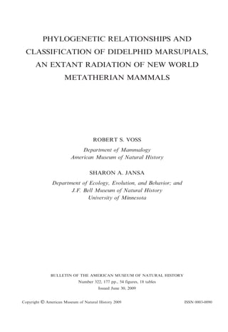
  • 2. Representative opossums (top to bottom, not to the same scale): Caluromys derbianus, Monodelphis brevicaudata, Marmosa robinsoni, Chironectes minimus, Metachirus nudicaudatus.
  • 3. CONTENTS Abstract . . . . . . . . . . . . . . . . . . . . . . . . . . . . . . . . . . . Introduction . . . . . . . . . . . . . . . . . . . . . . . . . . . . . . . . Materials and Methods . . . . . . . . . . . . . . . . . . . . . . . Comparative Morphology . . . . . . . . . . . . . . . . . . . . . . . Size and External Features . . . . . . . . . . . . . . . . . . . . Cranium and Mandible . . . . . . . . . . . . . . . . . . . . . . . Dentition . . . . . . . . . . . . . . . . . . . . . . . . . . . . . . . . . Summary . . . . . . . . . . . . . . . . . . . . . . . . . . . . . . . . . Gene Sequences . . . . . . . . . . . . . . . . . . . . . . . . . . . . . . Breast Cancer Activating 1 Gene . . . . . . . . . . . . . . . . Dentin Matrix Protein 1 . . . . . . . . . . . . . . . . . . . . . . Interphotoreceptor Retinoid Binding Protein . . . . . . . . Recombination Activating 1 Gene . . . . . . . . . . . . . . . von Willebrand Factor . . . . . . . . . . . . . . . . . . . . . . . Summary, Sequence Characteristics, and Model Fitting Phylogenetic Relationships . . . . . . . . . . . . . . . . . . . . . . Analyses of Nonmolecular Data. . . . . . . . . . . . . . . . . Single-gene Analyses . . . . . . . . . . . . . . . . . . . . . . . . . Analyses of Concatenated Genes . . . . . . . . . . . . . . . . Analyses of Combined Datasets . . . . . . . . . . . . . . . . . Discussion . . . . . . . . . . . . . . . . . . . . . . . . . . . . . . . . Classification . . . . . . . . . . . . . . . . . . . . . . . . . . . . . . . . A Revised Phylogenetic System . . . . . . . . . . . . . . . . . Family Didelphidae Gray, 1821 . . . . . . . . . . . . . . . . . Subfamily Glironiinae, new . . . . . . . . . . . . . . . . . . . . Glironia Thomas, 1912 . . . . . . . . . . . . . . . . . . . . . . Subfamily Caluromyinae Reig et al., 1987 . . . . . . . . . . Caluromys J.A. Allen, 1900 . . . . . . . . . . . . . . . . . . Caluromysiops Sanborn, 1951 . . . . . . . . . . . . . . . . . Subfamily Hyladelphinae, new . . . . . . . . . . . . . . . . . . Hyladelphys Voss et al., 2001 . . . . . . . . . . . . . . . . . Subfamily Didelphinae Gray, 1821 . . . . . . . . . . . . . . . Tribe Marmosini Hershkovitz, 1992 . . . . . . . . . . . . Marmosa Gray, 1821 . . . . . . . . . . . . . . . . . . . . . Monodelphis Burnett, 1830 . . . . . . . . . . . . . . . . . Tlacuatzin Voss and Jansa, 2003 . . . . . . . . . . . . . Tribe Metachirini Hershkovitz, 1992 . . . . . . . . . . . . Metachirus Burmeister, 1854 . . . . . . . . . . . . . . . . Tribe Didelphini Gray, 1821. . . . . . . . . . . . . . . . . . Chironectes Illiger, 1811 . . . . . . . . . . . . . . . . . . . Didelphis Linnaeus, 1758 . . . . . . . . . . . . . . . . . . Lutreolina Thomas, 1910 . . . . . . . . . . . . . . . . . . Philander Brisson, 1762 . . . . . . . . . . . . . . . . . . . Tribe Thylamyini Hershkovitz, 1992 . . . . . . . . . . . . Chacodelphys Voss et al., 2004 . . . . . . . . . . . . . . Cryptonanus Voss et al., 2005 . . . . . . . . . . . . . . . Gracilinanus Gardner and Creighton, 1989 . . . . . . Lestodelphys Tate, 1934 . . . . . . . . . . . . . . . . . . . Marmosops Matschie, 1916 . . . . . . . . . . . . . . . . . Thylamys Gray, 1843 . . . . . . . . . . . . . . . . . . . . . Acknowledgments . . . . . . . . . . . . . . . . . . . . . . . . . . . . 3 . . . . . . . . . . . . . . . . . . . . . . . . . . . . . . . . . . . . . . . . . . . . . . . . . . . . . . . . . . . . . . . . . . . . . . . . . . . . . . . . . . . . . . . . . . . . . . . . . . . . . . . . . . . . . . . . . . . . . . . . . . . . . . . . . . . . . . . . . . . . . . . . . . . . . . . . . . . . . . . . . . . . . . . . . . . . . . . . . . . . . . . . . . . . . . . . . . . . . . . . . . . . . . . . . . . . . . . . . . . . . . . . . . . . . . . . . . . . . . . . . . . . . . . . . . . . . . . . . . . . . . . . . . . . . . . . . . . . . . . . . . . . . . . . . . . . . . . . . . . . . . . . . . . . . . . . . . . . . . . . . . . . . . . . . . . . . . . . . . . . . . . . . . . . . . . . . . . . . . . . . . . . . . . . . . . . . . . . . . . . . . . . . . . . . . . . . . . . . . . . . . . . . . . . . . . . . . . . . . . . . . . . . . . . . . . . . . . . . . . . . . . . . . . . . . . . . . . . . . . . . . . . . . . . . . . . . . . . . . . . . . . . . . . . . . . . . . . . . . . . . . . . . . . . . . . . . . . . . . . . . . . . . . . . . . . . . . . . . . . . . . . . . . . . . . . . . . . . . . . . . . . . . . . . . . . . . . . . . . . . . . . . . . . . . . . . . . . . . . . . . . . . . . . . . . . . . . . . . . . . . . . . . . . . . . . . . . . . . . . . . . . . . . . . . . . . . . . . . . . . . . . . . . . . . . . . . . . . . . . . . . . . . . . . . . . . . . . . . . . . . . . . . . . . . . . . . . . . . . . . . . . . . . . . . . . . . . . . . . . . . . . . . . . . . . . . . . . . . . . . . . . . . . . . . . . . . . . . . . . . . . . . . . . . . . . . . . . . . . . . . . . . . . . . . . . . . . . . . . . . . . . . . . . . . . . . . . . . . . . . . . . . . . . . . . . . . . . . . . . . . . . . . . . . . . . . . . . . . . . . . . . . . . . . . . . . . . . . . . . . . . . . . . . . . . . . . . . . . . . . . . . . . . . . . . . . . . . . . . . . . . . . . . . . . . . . . . . . . . . . . . . . . . . . . . . . . . . . . . . . . . . . . . . . . . . . . . . . . . . . . . . . . . . . . . . . . . . . . . . . . . . . . . . . . . . . . . . . . . . . . . . . . . . . . . . . . . . . . . . . . . . . . . . . . . . .. 5 .. 5 .. 7 . 11 . 12 . 26 . 46 . 60 . 61 . 61 . 61 . 63 . 63 . 64 . 64 . 65 . 66 . 69 . 76 . 76 . 78 . 81 . 84 . 86 . 88 . 89 . 91 . 92 . 95 . 97 . 97 100 100 101 105 108 110 110 113 113 116 118 121 124 124 126 128 131 134 138 141
  • 4. 4 BULLETIN AMERICAN MUSEUM OF NATURAL HISTORY References . . . . . . . . . . . . . . . . . . . . . . . . . . . . . . . Appendix 1: Morphological Specimens Examined . . . Appendix 2: Specimens Sequenced for Nuclear Genes Appendix 3: Nonmolecular Character Descriptions . . Appendix 4: Nonmolecular Data Matrix . . . . . . . . . Appendix 5: Nonmolecular Apomorphy List . . . . . . Appendix 6: Key to the Didelphid Genera . . . . . . . . . . . . . . . . . . . . . . . . . . . . . . . . . . . . . . . . . . . . . . . . . . . . . . . . . . . . . . . . . . . . . . . . . . . . . . . . . . . . . . . . . . . . . . . . . . . . . . . . . . . . . . . . . . . . . . . . . . . . . . . NO. 322 . . . . . . . . . . . . . . . . . . . . . . . . . . . . . . . . . . . . . . . . . . . . . . . . . 142 157 160 161 170 171 174
  • 5. ABSTRACT This report summarizes a decade of morphological and molecular research on the phylogenetic relationships of didelphid marsupials (opossums), a substantially intact radiation of New World metatherian mammals. We review the comparative morphology of Recent opossums, emphasizing those anatomical systems from which taxonomically useful information is available for the majority of living genera and species, namely the integument, cranium, and dentition. Morphological similarities and differences among didelphids and other plesiomorphic marsupials (caenolestids, microbiotheriids, dasyurids, and peramelids) are also described. These observations, representing evolved differences in diverse functional-morphological systems, together with karyotypic information gleaned from the literature, provide the basis for coding 129 phylogenetic characters that we scored for 44 ingroup and seven outgroup taxa. Published information about the size, internal organization, chromosomal location, and physiological properties of five nuclear genes (BRCA1, DMP1, IRBP, RAG1, vWF) sequenced for this study suggest that these loci are unlinked, exist as single copies, are active in different tissues, and encode protein products with widely divergent functions. All of the sequenced fragments are long (.900 bp), free of ingroup alignment ambiguities, and translate to open reading frame. Nucleotide data from a total of 7320 aligned sites were obtained from 43 ingroup and seven outgroup taxa. Separate parsimony, likelihood, and Bayesian analyses of these six data partitions (morphology + karyotypes, five genes) resulted in highly congruent estimates of didelphid phylogeny with few examples of conflict among strongly supported nodes. Analyses of concatenated sequences and combined (nonmolecular + sequence) datasets effectively summarize all of the common signal recovered from separate analyses: a completely resolved ingroup phylogeny with high support statistics at most nodes. Remaining problems (not conclusively resolved in this study) include the position of the ingroup root and the relationships of three genera (Chacodelphys, Cryptonanus, Tlacuatzin) within their respective suprageneric clades. The history of didelphid classification is reviewed, and all previous systems are found to contain nonmonophyletic groups. A revised phylogenetic classification consistent with our analytic results includes the following higher taxa: Glironiinae (for Glironia), Caluromyinae (Caluromys and Caluromysiops), Hyladelphinae (Hyladelphys), Didelphinae (Marmosini, Metachirini, Didelphini, and Thylamyini), Marmosini (Marmosa, Monodelphis, and Tlacuatzin), Metachirini (Metachirus), Didelphini (Chironectes, Didelphis, Lutreolina, and Philander), and Thylamyini (Chacodelphys, Cryptonanus, Gracilinanus, Lestodelphys, Marmosops, and Thylamys). The probable relationships of several Neogene fossil genera are also discussed. To facilitate identifications, all Recent genera are redescribed, representative crania are illustrated, and a key is provided. INTRODUCTION Asia and western North America (Rougier et al., 1998; Luo et al., 2003; Kielan-Jaworowska et al., 2004), and other early metatherians are present in the Tertiary record of Europe and Africa (Crochet, 1980; Kurz, 2007; Hooker et al., 2008). The metatherian crown group Marsupialia, however, probably evolved in South America and subsequently dispersed across Antarctica to Australia and New Guinea (Muizon et al., 1997; Springer et al., 1997a; Rougier et al., 1998). Living marsupials comprise seven major groups, currently ranked as orders in the Linnaean hierarchy, each of which is endemic to either the New World or the Old World (table 1). Although the extant New World marsupial fauna consists of three orders, two of these They affirme that there are trees of suche byggenes, that xvi men ioyninge handes togyther and standinge in coompasse, can scarsely embrase sum of them. Emonge these trees is fownde that monstrous beaste with a snowte lyke a foxe, a tayle lyke a marmasette, eares lyke a batte, handes lyke a man, and feete lyke an ape, bearing her whelpes abowte with her in an owtwarde bellye much lyke vnto a greate bagge or purse. — Richard Eden (1555), describing the first European encounter with a marsupial, by Vicente Yanez Pinzon and his men on the coast of ´˜ ´ Brazil in the year 1500 The oldest known metatherian mammals occur in Cretaceous sediments of eastern 5
  • 6. 6 BULLETIN AMERICAN MUSEUM OF NATURAL HISTORY TABLE 1 Higher Classification and Geographic Distribution of Recent Marsupialsa Genera Species Distributionb DASYUROMORPHIA Dasyuridae Myrmecobiidae Thylacinidae 20 1 1 69 1 1 OW OW OW 18c 91c NW 2 2 1 11 3 6 1 4 6 1 2 2 5 1 65 11 27 1 10 17 1 3 OW OW OW OW OW OW OW OW OW OW OW 1 1 NW 1 2 OW 1 6 1 1 18 2 OW OW OW 3 6 NW DIDELPHIMORPHIA Didelphidae DIPROTODONTIA Acrobatidae Burramyidae Hypsiprymnodontidae Macropodidae Petauridea Phalangeridae Phascolarctidae Potoroidae Pseudocheiridae Tarsipedidae Vombatidae MICROBIOTHERIA Microbiotheriidae NOTORYCTEMORPHIA Notoryctidae PERAMELEMORPHIA Chaeropodidae Peramelidae Thylacomyidae PAUCITUBERCULATA Caenolestidae a Numbers of genera and species after Wilson and Reeder (2005) except as noted. b NW 5 New World; OW 5 Old World (Sahul). c After Gardner (2008). are represented by a mere handful of living forms: Microbiotheria by a single genus and species (Dromiciops gliroides), and Paucituberculata by six species in three genera (Caenolestes, Lestoros, Rhyncholestes). Both groups inhabit wet-temperate Patagonian forests, but paucituberculates also occur in climatically similar montane habitats of the tropical Andes (Bublitz, 1987; Patterson and Gallardo, 1987; Albuja and Patterson, 1996; Hershkovitz, 1999; Lunde and Pacheco, 2003). Because microbiotherians and paucituberculates were more diverse and widely NO. 322 distributed in the Tertiary than they are today (Marshall, 1980, 1982b; Bown and Fleagle, 1993; Goin, 1997; Goin and Candela, 2004), these orders are appropriately regarded as relictual elements in modern faunas. Instead, the only substantially intact radiation of New World marsupials is represented by the family Didelphidae, commonly known as opossums, of which 91 Recent species in 18 genera are currently recognized (Gardner, 2008). Although didelphids were the first metatherians to be encountered by European explorers (Eden, 1555), the first to be described scientifically (Tyson, 1698), and the first to be classified by taxonomists (Linnaeus, 1758), zoological interest in opossums was soon eclipsed by the discovery of Australasian marsupials. Nevertheless, steady advances in didelphid taxonomy (mostly involving the description of new species) were made throughout the 19th and 20th centuries. Although this progress was periodically summarized by authors (e.g., Thomas, 1888; Winge, 1893; Cabrera, 1919, 1958), there is no modern monographic treatment of the family, and the current suprageneric classification (McKenna and Bell, 1997; Gardner, 2005, 2008) does not reflect current knowledge about opossum evolutionary relationships. The history of didelphid phylogenetic research was reviewed by Jansa and Voss (2000), who analyzed DNA sequence data from the nuclear Interphotoreceptor Retinoid Binding Protein (IRBP) gene for 21 species. Subsequent reports were focused on analyses of morphological and karyotypic character data in combination with IRBP sequences for larger sets of species (Voss and Jansa, 2003; Voss et al., 2005), on phylogenetic problems associated with individual taxa (Voss et al., 2004a; Jansa and Voss, 2005), or on the evolutionary dynamics of newly sequenced nuclear loci (Jansa et al., 2006; Gruber et al., 2007). However, no comprehensive phylogenetic synthesis has yet been attempted, and much new morphological and molecular character data remain unanalyzed. This monograph summarizes our collaborative research to date on didelphid phylogenetic relationships and concludes the first
  • 7. 2009 VOSS AND JANSA: DIDELPHID MARSUPIALS phase of our ongoing evolutionary study of the family. Although we take this opportunity to review information that has previously been published elsewhere, many new results are reported herein. Among these, we describe morphological comparisons of didelphids with other plesiomorphic (polyprotodont) marsupial clades; we describe and illustrate integumental and craniodental traits not previously discussed in the literature; we report new sequence data from the Breast Cancer Activating 1 gene (BRCA1) and the von Willebrand Factor gene (vWF); we analyze didelphid phylogenetic relationships based on a new set of morphological and karyotypic characters together with sequence data from five protein-coding nuclear loci; we propose a new suprageneric classification consistent with our analytic results; and we provide morphological descriptions and cranial illustrations of all currently recognized genera to facilitate taxonomic identifications. Materials and Methods PHYLOGENETIC ASSUMPTIONS AND TAXON SAMPLING: Our phylogenetic analyses are based on the assumption of didelphid monophyly, which is strongly supported by a variety of nuclear-gene sequence datasets (e.g., Jansa and Voss, 2000; Amrine-Madsen et al., 2003; Meredith et al., 2008). Our ingroup terminals (table 2) consist of 44 species representing every currently recognized didelphid genus and subgenus, including all of the species previously analyzed by Jansa and Voss (2000, 2005), Voss and Jansa (2003), Voss et al. (2004a, 2005), Jansa et al. (2006), and Gruber et al. (2007). Altogether, this is by far the most taxon-dense and character-rich didelphid dataset ever analyzed. The outgroups for this study include representatives of every extant ordinal-level clade that has ever been recovered within one or two nodes of Didelphidae in previous analyses of marsupial relationships, including caenolestids (Paucituberculata), dasyurids (Dasyuromorphia), Dromiciops (Microbiotheria), and peramelids (Peramelemorphia). We did not include Notoryctes (Notoryctemorphia) or diprotodontians because these 7 taxa have never been considered close to didelphids, and because their highly derived morphologies raise many unresolved issues of homology that are beyond the scope of this study. COMPARATIVE MORPHOLOGY: Our morphological comparisons are based on skins, skulls, and fluid-preserved specimens (appendix 1), which we examined for discretely varying traits that could be scored for most of the terminal taxa in our analysis. Additionally, we summarize information about other phenotypic characters that, although not suitable for phylogenetic analysis due to continuous variation (e.g., body size), provide taxonomically useful descriptors. Information about morphological character variation among nondidelphid marsupials is largely based on our scoring of outgroup terminal taxa, but we also include remarks on the morphology of other Old World marsupials where these seem relevant or interesting. To provide a maximally user-friendly reference, our morphological observations are summarized in two different formats. First, we describe taxonomic comparisons using normal (nontelegraphic) prose in the body of the text. These accounts, organized organ-system-by-organ system, are accompanied as necessary by illustrations of representative specimens. Second, we summarize morphological descriptors taxon-by-taxon using telegraphic prose in the generic accounts of our classification. Although largely redundant with our data matrix (appendix 4), these generic descriptions are easier to use for taxonomic identifications, and they provide an opportunity to summarize relevant observations that were not encoded as characters. Formal character descriptions (in appendix 3) are accompanied by technical details about coding procedures and ordering criteria. Our criteria for character choice were described at length by Voss and Jansa (2003), so they are not repeated here. The only noteworthy methodological change in this study concerns our coding of polymorphisms. As in our previous study, we ignored rare variants (on the assumption that all characters are polymorphic given sufficiently large samples) and coded only those polymorphisms represented by nearly equal frequencies of alternative states (e.g., when
  • 8. 8 BULLETIN AMERICAN MUSEUM OF NATURAL HISTORY NO. 322 TABLE 2 Marsupial Terminal Taxa Scored for Phylogenetic Analysisa Percent complete character datab Nonmolecular Molecular Combined 99.2 99.2 93.7 86.0 99.2 98.4 93.0 100 100 100 93.0 93.0 95.3 95.3 95.3 95.3 97.6 99.2 96.1 99.2 99.2 99.2 92.2 99.2 96.1 99.2 98.4 96.8 98.4 95.3 95.3 98.4 97.7 96.1 92.3 89.9 97.7 100 99.2 96.1 96.1 98.4 96.1 99.2 100 100 16.5 0 99.8 95.2 99.8 99.1 99.1 100 100 99.8 99.7 99.8 95.1 100 30.4 100 100 98.8 98.6 98.8 99.7 98.5 95.7 100 98.0 99.0 100 98.8 99.7 98.9 100 100 100 100 100 99.2 99.1 99.8 98.3 95.7 99.5 97.5 100 100 17.8 1.5 99.8 95.3 99.7 99.1 99.1 100 99.9 99.7 99.6 99.7 95.1 99.9 31.6 100 99.9 98.8 98.6 98.8 99.6 98.5 95.7 100 98.0 99.0 100 98.7 99.6 98.9 100 99.9 99.9 99.8 100 99.2 99.1 99.7 98.3 95.7 99.4 97.5 86.9 85.3 63.3 62.5 63.7 62.9 INGROUP: Didelphidae Caluromys lanatus Caluromys philander Caluromysiops irrupta Chacodelphys formosa Chironectes minimus Cryptonanus chacoensis Cryptonanus unduaviensis Didelphis albiventris Didelphis marsupialis Didelphis virginiana Glironia venusta Gracilinanus aceramarcae Gracilinanus agilis Gracilinanus emiliae Gracilinanus microtarsus Hyladelphys kalinowskii Lestodelphys halli Lutreolina crassicaudata Marmosa (Marmosa) lepida Marmosa (Marmosa) mexicana Marmosa (Marmosa) murina Marmosa (Marmosa) robinsoni Marmosa (Marmosa) rubra Marmosa (Micoureus) demeraraec Marmosa (Micoureus) paraguayanac Marmosa (Micoureus) reginac Marmosops impavidus Marmosops incanus Marmosops noctivagus Marmosops parvidens Marmosops pinheiroi Metachirus nudicaudatus Monodelphis brevicaudata Monodelphis emiliae Monodelphis peruvianad Monodelphis theresa Philander frenatus Philander mcilhennyi Philander opossum Thylamys macrurus Thylamys pallidior Thylamys pusillus Thylamys venustus Tlacuatzin canescens OUTGROUPS: Caenolestidae Caenolestes fuliginosus Rhyncholestes raphanurus
  • 9. 2009 VOSS AND JANSA: DIDELPHID MARSUPIALS 9 TABLE 2 (Continued ) Percent complete character datab Nonmolecular Dasyuridae Murexia longicaudata Sminthopsis crassicaudata Microbiotheriidae Dromiciops gliroides Peramelidae Echymipera kalubu Perameles gunnii Molecular Combined 95.3 96.1 57.9 42.1 58.5 43.0 99.2 63.3 63.9 95.3 93.0 63.2 63.3 63.8 63.8 a Nomenclature follows Wilson and Reeder (2005) and Gardner (2008) except as noted. Number of filled (total minus empty) cells in the corresponding row of each data matrix, divided by the total number of cells (N5 129, 7320, and 7449 for nonmolecular, molecular, and combined data, respectively) 3 100. For the nonmolecular data, empty cells include those scored as missing (‘‘?’’) and those scored as inapplicable (‘‘-’’). For the molecular data, only unsequenced base pairs were counted as missing (‘‘?’’); gaps (‘‘-’’) were counted as filled data cells. c Micoureus was formerly treated as a full genus. d Formerly identified as Monodelphis adusta (e.g., by Jansa and Voss, 2000; Voss and Jansa, 2003), our material is referable to M. peruviana, a distinct species recently resurrected from synonymy by Solari (2007). b presence and absence were both commonly observed for a given taxon). However, whereas we formerly (Voss and Jansa, 2003) treated such polymorphism as a separate state intermediate to the two fixed conditions, with transformations to and from the polymorphic state weighted as half-steps (the ‘‘scaled’’ option discussed by Wiens, 2000), we here conform with the prevailing custom of coding polymorphisms as taxonomic ambiguities (e.g., as ‘‘0/1’’ for a binary character). This coding change has minimal impact on our results (only weakly supported relationships are affected, even in separate analyses of morphology) but it facilitates likelihood modeling of morphological character evolution (Lewis, 2001) in Bayesian analyses of our combined datasets (see below). MOLECULAR SEQUENCING AND HOMOLO- GY COMPARISONS: The laboratory procedures we used for DNA amplification and sequencing (including the names and locations of primers used in PCR reactions) have already been described for three of the protein-coding nuclear loci analyzed in this report: IRBP (Jansa and Voss, 2000), Dentin Matrix Protein 1 (DMP1; Jansa et al., 2006), and Recombination Activating 1 Gene (RAG1; Gruber et al., 2007). In addition, we sequenced two other protein-coding nuclear genes as described below. We amplified 2.1 kb of BRCA1 exon 11 from genomic DNA in two fragments using the primers listed in table 3. The first fragment, comprising the upstream 1.2 kb of the exon, was amplified using primers F1 paired with R1218 or F47 paired with R1343. The second, downstream fragment (ca. 1 kb) was amplified with primers F1163 or F1163a paired with R2078 or R2151. These amplification products were then used in a second round of PCR to generate smaller pieces of suitable size for sequencing. For the upstream fragment, either F1 or F47 was paired with R743, and F593 was paired with either R1218 or R1343. For the downstream fragment, F1163 or F1163a was paired with R1780, and F1697 was paired with either R2078 or R2151. A single fragment of ca. 1 kb was amplified from vWF exon 28 using either F104 or F120 paired with R1141. This product was then used in a second round of PCR in which F1 or F47 was paired with either R665 or R742, and F557 was paired with R1141. Initial amplifications using genomic DNA as template were performed as 20 ml reactions using Ampli-Taq Gold polymerase (Perkin-Elmer Corp.) and recommended concentrations of primers, nucleotides, buffer, and MgCl2. These genomic amplifications were performed using a four-stage touch-
  • 10. 10 BULLETIN AMERICAN MUSEUM OF NATURAL HISTORY NO. 322 TABLE 3 Primers Used to Amplify and Sequence BRCA1 and vWF Primer name BRCA1BRCA1BRCA1BRCA1BRCA1BRCA1BRCA1BRCA1BRCA1BRCA1BRCA1BRCA1VWFVWFVWFVWFVWFVWF- F1 F47 F593 F1163 F1163a F1697 R743 R1218 R1343 R1780 R2078 R2151 F104 F120 F557 R655 R743 R1141 Sequence 59 59 59 59 59 59 59 59 59 59 59 59 TCATTACTGCCTGAGATCACCAG TATTGCCTAACACAGACAGCAT CAACAATATTGAAGACAAAATATTAGGAAA ATGARACWGAACTACWGATCGATAG AATGAGACTGAACTACAGATCGAT TTWGATGRTTGTTCATCYRAAAACAC TTGATGAAATCCTCAGGCTGYAGGT GAAGYCTTCTGCTGCGTCTGA CTAACATTTGATCACTATCAGTAG TAAATAYTGGGTRTCRAGTTCACT GAAATTTCCTGGTTGTTTCCAGCAA TCCTTTTGATYAGGAACTTGTGAAATT 59 59 59 59 59 59 GGTGTGATGGAGCGTTTACACATCTC GACTTGGCYTTYCTSYTGGATGGCTC CCTGGGCTACCTCTGTGACCTGGT CTTCTAGCACAAACACCACATCCAGAACCA CTCACATCCATYCGTTGCATCA ATCTCATCSGTRGCRGGATTGC down protocol as described in Jansa and Voss (2000). Reamplification reactions were performed using Taq DNA polymerase (Promega Corp.) in 25 ml reactions for 30 PCR cycles. The resulting PCR products were sequenced in both directions using amplification primers and dye-terminator chemistry on an AB 3700 automated sequencer. We searched the draft Monodelphis domestica genome using the BLAT (modified BLAST; Kent, 2002) algorithm to determine the copy number and chromosomal location of query sequences from M. brevicaudata. As reported on the Ensemble database (www. ensemble.org/index.html), the available reference genome (assembly MonDom5) was released on October 2006 and has a base coverage of approximately 7.33. ALIGNMENT AND PHYLOGENETIC ANALYWe aligned DNA sequences with reference to translated amino-acid sequences using MacClade 4.08 (http://macclade.org). Aligned sequences were then analyzed phylogenetically using maximum parsimony (MP) as implemented by PAUP* ver. 4.0b10 (Swofford, 1998), maximum likelihood (ML) as implemented by GARLI ver. 0.95 (Zwickl, 2006), and Bayesian inference as implemented by MrBayes ver. 3.1.1 (Ronquist and Huelsenbeck, 2003). For MP SIS: analyses, all molecular characters were treated as unordered and equally weighted, and all tree searches were heuristic with at least 10 replicates of random stepwise taxon addition followed by tree bisection-reconnection (TBR) branch swapping. To choose the best models of nucleotide substitution for ML and Bayesian analyses, we examined the fit of various models separately for each of our five gene partition (IRBP, vWF, BRCA1, DMP1, and first and second codon positions of RAG1) based on neighbor-joining trees of Jukes-Cantor-corrected distances using both hierarchical likelihood-ratio tests (hLRTs) and the Akaike Information Criterion (AIC) as implemented in ModelTest 3.7 (Posada and Crandall, 1998). Where the two approaches disagreed on model choice, we used the model selected by the AIC for reasons outlined by Posada and Buckley (2004). For both ML and Bayesian searches, the best-fit model was specified, but model parameter values were not fixed. For ML analyses, we conducted three independent runs of geneticalgorithm searches in GARLI, with random starting topologies and automatic termination after 10,000 generations with no improvement in log-likelihood scores. For Bayesian analysis of each gene partition, we conducted two independent runs of Metrop-
  • 11. 2009 VOSS AND JANSA: DIDELPHID MARSUPIALS olis-coupled Markov-chain Monte Carlo (MCMCMC), each with one cold and three incrementally heated chains. For each run, we assumed uniform-interval priors for all parameters, except base composition, which assumed a Dirichlet prior. Runs were allowed to proceed for 5 3 106 generations, and trees were sampled every 100 generations. We evaluated the burn-in for each run, and pooled the post-burn-in trees to calculate estimated parameter distributions and posterior probabilities for each node. We assessed nodal support from MP and ML analyses using nonparametric bootstrapping (Felsenstein, 1985). Bootstrap values for the parsimony analyses (MPBS) were calculated in PAUP* from 1000 pseudoreplicated datasets, each of which was analyzed heuristically with 10 random-addition replicates with TBR branch swapping. Bootstrap values for the likelihood analysis (MLBS) were calculated in GARLI using genetic-algorithm searches of 1000 pseudoreplicated datasets, allowing model parameters to be estimated for each pseudoreplicate. We analyzed the combined-gene dataset using ML as implemented in RAxML-VIHPC (ver. 2.2.3; Stamatakis, 2006). We specified the GTRMIX model, which performs initial tree inference using a GTRCAT approximation, with final topology evaluation performed under a GTRGAMMA model. We allowed parameters to be estimated independently across the five gene partitions. To evaluate nodal support for this combined-gene, mixed-model analysis, we performed 1000 bootstrap replicates, again allowing model parameters to be estimated independently across the five genes. We also performed a Bayesian analysis of the combined-gene dataset, using the same MCMCMC settings given above. For this analysis, we specified the best-fit model for each gene, decoupled estimation of substitution parameters across the partitions, and allowed each gene to assume a separate rate. We analyzed the nonmolecular (morphological + karyotypic) data alone and in combination with the molecular data using MP and Bayesian approaches. Due to the large number of suboptimal trees recovered from parsimony analysis of the nonmolecular dataset, we first performed 1000 random- 11 taxon-addition replicates with TBR branch swapping, but saved only 10 trees per replicate. We then used this pool of 10,000 trees as the starting point for an unbounded heuristic search. Parsimony analysis of the combined (molecular + nonmolecular) dataset did not exhibit this problem; therefore, we analyzed the combined dataset using unbounded heuristic searches with 1000 replicates of random-taxon addition and TBR branch swapping. For Bayesian analysis of the morphological dataset alone, we specified the Mkv model (Lewis, 2001) with a Cdistributed rate parameter and ascertainment bias corrected for omission of constant characters (lset coding 5 variable). For Bayesian analysis of the combined (molecular + nonmolecular) dataset, we specified this same model for the morphological data and applied the best-fit model to each of the five gene partitions. As above, we allowed parameters to be estimated independently across all partitions and used the same MCMCMC settings. To assess the impact of the large amount of missing data from Chacodelphys, we performed all analyses that included nonmolecular characters with and without this taxon. ONLINE DATA ARCHIVES: All of the new molecular sequences produced for this study have been deposited in GenBank with accession numbers FJ159278–FJ159314 and FJ 159316–FJ159370 (for a complete list of GenBank accession numbers of all analyzed sequences, old and new, see table 9). All of our datasets, selected ML and Bayesian analyses, and associated trees have been deposited on TreeBase (http://www.treebase. org) with accession numbers S2164, M4107, and M4108. Our nonmolecular data matrix has also been deposited on MorphoBank (http://morphobank.geongrid.org) with accession number X600. COMPARATIVE MORPHOLOGY The literature on didelphid comparative morphology is widely scattered, and no adequate review of this topic has yet been published. Although the following accounts are far from comprehensive, they include most of the anatomical features that have been surveyed widely among extant genera and that provide relevant taxonomic infor-
  • 12. 12 BULLETIN AMERICAN MUSEUM OF NATURAL HISTORY mation.1 In effect, such information comes from commonly available materials that can be examined without dissection or other special preparations. Therefore, microscopic features (e.g., those of the spermatozoa; Temple-Smith, 1987) and visceral characters (e.g., of the digestive tract; Santori et al., 2004) are not reviewed below, nor are osteological traits that require X-ray computed tomography, serial sectioning, or destructive methods for their study. Size and External Features Most opossums are externally unremarkable mammals with pointed muzzles, large rhinaria, well-developed vibrissae, prominent eyes, membranous ears, nonspinous pelage, subequal limbs, pentadactyl feet, and naked tails. In many of these respects they resemble other plesiomorphic marsupials (e.g., Dromiciops and dasyurids) as well as certain unspecialized placentals (e.g., solenodontids, rice tenrecs, gymnures, and tree shrews). Closer inspection, however, reveals numerous distinctive and phylogenetically informative details of didelphid external morphology. SIZE: Didelphids are small to mediumsized mammals. The smallest Recent species is probably Chacodelphys formosa, the young adult holotype of which had a head-andbody length of 68 mm and probably weighed about 10 g (Voss et al., 2004a). By contrast, the largest living opossum, Didelphis virginiana, can measure almost 500 mm in headand-body length and weigh more than 3000 g (Hamilton, 1958). Most didelphids, however, range in head-and-body length from about 100 to 300 mm and weigh between about 20 and 500 g (table 4). Although some authors have recognized ‘‘large’’, ‘‘medium’’, and ‘‘small’’ opossums, taxonomic assignments to discrete size categories are often arbitrary and sometimes misleading. For example, Reig et al. (1987: character 25) scored Metachirus as ‘‘large’’ and Caluromys as ‘‘medium’’ in their phylogenetic analysis, but linear measurements and weights that we compiled suggest that these taxa are indistinguishable in size. More taxonomically 1 An important exception is postcranial skeletal morphology, the topic of an independent study (Flores, 2009). NO. 322 comprehensive compilations of morphometric data will probably document a continuum of didelphid size distributions. By comparison, Old World marsupials include some species that are smaller and others that are much larger than any didelphid. The smallest living Australian species, for example, is said to be Planigale ingrami, with an adult weight of only about 4 g, and the largest is Macropus rufus, males of which are said to weigh as much as 85 kg (Dawson et al., 1989). The extinct Pleistocene species Diprotodon optatum, however, may have weighed almost 2800 kg (Wroe et al., 2004). RHINARIUM AND MOUTH: The rhinarium, a prominent pad of naked glandular skin surrounding the nostrils, is divided by a median crease or sulcus (sulcus medianus; Ade, 1999) that extends from between the nares to the upper lip in all examined didelphids. The part of the rhinarium that borders the upper lip (pars supralabialis) is broad, and its ventral margin is notched by one or two distinct grooves on each side of the median sulcus (fig. 1). Two ventrolateral grooves are present on each side in most examined taxa, but only a single groove is present in Chironectes, Didelphis, Lestodelphys, Lutreolina, Metachirus, Monodelphis, Philander, and Thylamys pallidior. Dasyurids and Dromiciops have rhinaria that are essentially similar in gross morphology to those of didelphids, with a pars supralabialis that makes broad contact with the upper lip; however, only a single ventrolateral groove is present in these taxa (Pocock, 1926: figs. 26– 28). By contrast, the supralabial part of the rhinarium is reduced to a narrow philtrum in caenolestids and peramelids, such that the groove-bearing ventral rhinarial margin of other marsupials is effectively absent. The mouth is large in all didelphids, with a posterior angle (angulis oris; Brown, 1971) that extends posteriorly to a point below the eye; the upper and lower oral margins are smooth or irregularly wrinkled and anatomically featureless. Most other marsupials have anatomically featureless oral margins like those of didelphids, but caenolestids are uniquely provided with reciprocating fleshy lappets of unknown function on the upper and lower lips (Osgood, 1921: pl. 2; Bublitz,
  • 13. 2009 VOSS AND JANSA: DIDELPHID MARSUPIALS 13 TABLE 4 External Measurements (mm) and Weights (g) of Exemplar Didelphid Speciesa Nb e Caluromys (Caluromys) philander Caluromys (Mallodelphys) lanatusf Caluromysiops irruptag Chironectes minimush Cryptonanus unduaviensisi Didelphis marsupialise Glironia venustaj Gracilinanus agilisk Hyladelphys kalinowskiie Lestodelphys hallil Lutreolina crassicaudatam Marmosa (Marmosa) murinan Marmosa (Micoureus) reginaf Marmosops noctivagusf Marmosops pinheiroie Metachirus nudicaudatuse Monodelphis emiliaef Philander opossume Thylamys karimiio Thylamys macruruso Tlacuatzin canescensp 7 8 1 6 8 9 2 11 3 1 11 13 41 16 11 9 6 11 33 6 7 Head and Bodyc 261 278 260 289 105 419 194 98 77 132 295 133 180 138 103 262 107 301 104 112 137 (224–279) (270–296) (276–307) (97–121) (405–446) (188–201) (86–109) (76–78) (241–342) (118–152) (142–209) (118–155) (94–121) (249–287) (97–113) (264–346) (78–129) (101–126) (126–149) Taild 390 422 310 348 122 434 208 137 111 88 283 173 262 183 149 345 50 306 80 144 137 (373–410) (400–446) (316–362) (112–135) (366–497) (201–215) (121–162) (107–113) (242–336) (156–195) (238–294) (154–202) (137–156) (326–370) (45–53) (280–333) (69–106) (136–153) (131–145) Weight 330 412 496 605 25 1351 130 25 16 76 530 51 118 51 27 380 30 549 28 39 — (220–390) (349–500) (520–700) (15–40) (1025–1700) (129–130) (18–34) (13–18) (300–800) (35–80) (70–164) (30–70) (21–33) (260–480) (20–38) (380–695) (16–43) (30–55) (38–60) a Tabulated statistics are the sample mean (rounded to the nearest whole unit) and the observed range (in parentheses) of measurements and weights recorded from dentally mature specimens; male and female data were combined to increase sample size despite apparent sexual dimorphism in some species. Most genera are represented by a single exemplar species, but several genera with recognized subgenera or that include taxa differing conspicuously in size or body:tail ratios are represented by additional species. b Sample size. c Obtained by subtracting length of tail from total length following the standard American protocol. d Basal flexure to fleshy tip. e French Guianan specimens measured by Voss et al. (2001). f Measurements and weights from western Brazilian specimens (Patton et al., 2000). g Measurements and weight of AMNH 208101; because this was a zoo specimen that may have been obese, the weight datum is suspect. h From Paraguayan specimens (UMMZ 126289, 134022, 134023, 134025, 134559, 134560). i From Bolivian specimens measured by Voss et al. (2005). j Measurements and weight of MMD 607 (collected near Iquitos, Peru; to be deposited in the Museo de Historia ´ Natural de la Universidad Nacional Mayor de San Marcos, Lima; M.M. Dıaz, personal commun.) and INPA 5237 (collected near Mirassol d’Oeste, Mato Grosso, Brazil; Santos Filho et al., 2007). k From Paraguayan specimens (UMMZ 124675, 126104, 133998–134006). l Measurements and weight from MVZ 173727. m From Paraguayan specimens (UMMZ 126109–126111, 126113, 134010, 134011, 134017–134021). n From Surinamese specimens measured by Voss et al. (2001). o From Carmignotto and Monfort (2006). p External measurements from Oaxacan specimens unaccompanied by weight data (AMNH 3111/2433, 3111/2434, 3114/2437, 148969, 149104, 165651, 165653); range of weights from Zarza et al. (2003). 1987: fig. 4; Patterson and Gallardo, 1987: fig. 3). FACIAL VIBRISSAE AND MARKINGS: Didelphid facial vibrissae are grouped into discrete tracts that are easily homologized with those described and illustrated by Pocock (1914), Lyne (1959), and Brown (1971), whose terminology is followed here. All of the taxa we examined (including representative species from every genus) exhibit well-developed mystacial, submental, interramal, superciliary (supraorbital), and genal vibrissae. La-
  • 14. 14 BULLETIN AMERICAN MUSEUM OF NATURAL HISTORY Fig. 1. Ventral view of rhinarium in Thylamys pallidior (A, UMMZ 156349) and Marmosa robinsoni (B, UMMZ 117236). Both species have a broad pars supralabialis (psl) that contacts the upper lip. Only a single groove (g) is present along the ventral margin of the pars supralabialis on each side of the median sulcus (ms) in T. pallidior, whereas two ventrolateral grooves are present in M. robinsoni. Scale bars 5 2 mm. beled illustrations of didelphid facial vibrissae are provided by Pocock (1914: fig. 1A) and Lyne (1959: fig. 2). Most other plesiomorphic marsupials closely resemble didelphids in cranial vibrissal traits, but caenolestids lack interramal vibrissae (Lyne, 1959). Didelphids exhibit conspicuous taxonomic variation in facial markings (frontispiece; Voss and Jansa, 2003: fig. 3). In Caluromys, a median streak of dark fur, unconnected with any other dark marking, extends from NO. 322 the rostrum to the frontal region. In other didelphids, a chevron of dark coronal fur sometimes extends anteriorly between the ears and down the rostral midline, but the condition seen in Caluromys is distinctive and apparently nonhomologous. No nondidelphid marsupial that we examined has a dark midrostral stripe. The fur surrounding the eye is not distinctively colored in Caluromysiops, Lutreolina, or Monodelphis, but most other didelphids have masklike markings. A circumocular mask or ring of dark (usually blackish) fur that contrasts sharply with the paler (usually brownish, whitish, or grayish) color of the crown and cheeks is present in Glironia, Lestodelphys, and ‘‘marmosines’’ (taxa formerly included in ‘‘Marmosa’’ sensu Tate, 1933). Species of Caluromys have essentially similar reddish-brown eye rings that contrast with grayish cheeks and crowns. A blackish mask is likewise present in all examined species of Didelphis, but this marking is inconspicuous in D. marsupialis and D. virginiana. A blackish mask is also present in Metachirus, Chironectes, and Philander, but the dark circumocular fur in these taxa is usually continuous with dark fur on the crown of the head. Among nondidelphid marsupials, dark circumocular masks are present in Dromiciops, some dasyurids (e.g., Sminthopsis crassicaudata), a few peramelemorphians (e.g., Echymipera kalubu), and some small arboreal diprotodontians (e.g., Petaurus breviceps); only one examined outgroup taxon, Echymipera kalubu, has a mask that is more or less continuous with dark coronal fur. A distinct whitish supraocular spot is consistently present in species of Metachirus and Philander, resulting in the ‘‘four-eyed’’ marking by which these animals are commonly known. Most other didelphids lack pale supraocular markings. (An indistinct pale bar above each eye in Chironectes appears to be part of the unique transverse banding pattern in that taxon rather than a homologue of the condition seen in Metachirus and Philander.) We have not examined any nondidelphid marsupial with pale supraocular spots. GULAR GLAND: As described by Tate (1933: 30), many didelphids have a cutaneous
  • 15. 2009 VOSS AND JANSA: DIDELPHID MARSUPIALS gular (throat) gland, the presence of which is indicated on dried skins and fluid-preserved specimens by a bare median patch of skin; often, but not invariably, the surrounding fur is discolored. According to Barnes (1977: 390), this secretory region contains ‘‘hypertrophied apocrine sudoriferous glands and sebaceous glands, both confined to the thickened dermis.’’ External signs of glandular activity tend to be maximally developed in fully mature males (loc. cit.). No unambiguously glandular throat patch was observed in any examined specimens of Caluromys, Caluromysiops, Chironectes, Hyladelphys, Lutreolina, Philander, or Tlacuatzin. By contrast, fully adult male specimens of Cryptonanus, Gracilinanus, Lestodelphys, and Thylamys usually exhibit well-developed gular glands. A gular gland is also present on the young adult male holotype (and only known skin) of Chacodelphys formosa. Other didelphid genera (Marmosa, Marmosops, and Monodelphis) include some species that consistently develop adult male gular glands and others that just as consistently show no trace of such organs (Voss and Jansa, 2003). Although adult male Didelphis often have discolored gular fur, no glandular skin is macroscopically distinguishable. Because we were not able to examine any fully adult male specimens of Glironia, the occurrence of gular glands in this taxon is unknown. Gular (or sternal) glands that are macroscopically and histologically similar to those of didelphids are present in most dasyurids (Cooper et al., 2005), but other plesiomorphic outgroup taxa (e.g., caenolestids, peramelids, Dromiciops) seem to lack all external traces of glandular activity on the throat or chest. BODY PELAGE: All didelphids have one or more tracts of postcranial vibrissae (Brown and Yalden, 1973), including ulnar-carpal vibrissae (at the wrist), medial antebrachial vibrissae (at or near the middle of the forearm), anconeal vibrissae (at the elbow), and/or calcaneal vibrissae (on the ankle). Lyne (1959) reported the occurrence of postcranial vibrissae in several didelphid species based on his examination of pouch young, whose sensory hair follicles are easily seen because they are not obscured by coat hairs. Unfortunately, postcranial vibrissae 15 are much harder to observe on fully furred adult specimens, the only material commonly available for most species. We found ulnarcarpal and medial antebrachial vibrissae on most examined didelphids, whereas anconeal and calcaneal vibrissae were often inapparent. All didelphids have soft (nonspinous) fur consisting of two or more distinct types of hairs whose density and morphology determine the appearance and texture of the coat. Some taxa (e.g., Caluromys) have somewhat woolly fur that does not lie flat or exhibit the glossy highlights typically seen in the pelts of many other taxa, but textural differences are hard to define by objective criteria that can be used for character-state definitions or taxonomic diagnoses. The only structural (nonpigmental) feature of didelphid body pelage that seems useful in this context is the presence of uniquely long, coarse guard hairs that project conspicuously from the underfur in species of Didelphis. The dorsal body pelage of most didelphids is uniformly colored and unpatterned, usually some shade of brownish or grayish, but some taxa are distinctively marked (see illustrations in Eisenberg, 1989; Redford and Eisenberg, 1992; Perez-Hernandez et ´ ´ al., 1994; Reid, 1997; Eisenberg and Redford, 1999). Blackish transverse bars connected middorsally on a pale-grayish background, for example, characterize Chironectes; dark scapular stripes are unique to Caluromysiops; three longitudinal stripes are present in several species of Monodelphis (e.g., M. theresa); a grayish middorsum contrasting with reddish flanks is exhibited by other species in that genus (e.g., M. brevicaudata); and a grayish midbody contrasting with reddish head and rump is seen in others (e.g., M. emiliae). The subtle but consistently diagnostic ‘‘tricolor’’ shading of Thylamys and Lestodelphys was described by Tate (1933: 209): Instead of the usual bicolor system composed of a dorsal color, paling a little on the sides, which is replaced at a generally well-marked transition line by a distinct ventral color, the elegans group [5 Thylamys] displays three distinct shades, separated from each other along each side by two lines of transition. The additional lines are subdorsal, running from a point at the center of
  • 16. 16 BULLETIN AMERICAN MUSEUM OF NATURAL HISTORY the frons [forehead] past the inner edge of each ear (not including it), and straight backward through scapulae and hips, where they again approach the median line of the body and merge with the tail. This pair of lines encloses the major part of the dorsal area of head and body, the color of the area being very dark brownish-gray or grayish fuscous. The fuscous area is pointed at front, projecting forward between the ears, and narrows again to a point as it merges with the dark color of the upper surface of the tail. The second [lateral] area, light gray in color, frequently tinged with buffy or yellowish, extends [on each side] between the dark dorsal region and the edge of the belly color at the normal transition line. Ventral color either buffy, grayish, or snowy white. The hair bases of the dorsal fur are heavily pigmented, usually dark gray (or grayish), in most didelphids, but species of Didelphis uniquely exhibit white dorsal underfur. The ventral pelage also exhibits noteworthy taxonomic variation among didelphids. In some species the ventral fur is ‘‘graybased,’’ a descriptor that applies when the individual hairs are grayish basally and abruptly paler (usually whitish, yellowish, or brownish) distally; the overall color of the ventral fur then depends on the degree to which the dark basal pigmentation shows through the paler superficial hue. In other species, the ventral fur is partially or entirely ‘‘self-colored,’’ a descriptor that applies when the individual hairs have the same pigmentation (usually whitish) from root to tip. Because marked differences in the patterning of gray-based versus self-colored ventral pelage can occur among closely related (congeneric) species, such variation is often described and illustrated in the revisionary taxonomic literature (e.g., Patton et al., 2000: fig. 41). However, the existence of numerous intermediate conditions spanning the entire range of taxonomic variation in ventral color patterns (e.g., from entirely gray-based to completely self-white ventral fur) precludes meaningful phylogenetic scoring of this character. Whereas most of the pelage colors (and color patterns) described above are preserved on museum skins that have been protected in dark cabinets from the bleaching effects of light, other colors that can be striking in life fade quickly after death. The ventral pelage NO. 322 of live Monodelphis emiliae, for example, has been described as ‘‘bright glowing violaceous pink’’ (Emmons, 1997: 34), a lurid hue that is not retained in any examined museum specimens. What little is known about such fugitive pigments (some of which fluoresce under ultraviolet light) was summarized by Pine et al. (1985). Most other marsupials have unremarkable body pelage that essentially resembles the common didelphid condition as described above, but postcranial vibrissae are reduced or absent in a few clades (Lyne, 1959) and peramelemorphians have stiff, dorsally grooved guard hairs (illustrated in cross section by Lyne and McMahon, 1951: figs. 51, 60) that impart a distinctively harsh, spinous texture to their fur. Most other marsupials also resemble didelphids in having unpatterned dorsal fur, although there are noteworthy examples of apparently convergent markings (e.g., those of Dromiciops somewhat resemble Chironectes’; Marshall, 1978b) and some strikingly divergent ones (e.g., the transverse sacral barring seen in Perameles gunnii; Lyne, 1951: pl. 1B). All examined outgroup taxa have dark dorsal underfur. WRIST: The wrists of males and females are morphologically similar and externally featureless in most didelphids, but striking sexual dimorphism is present in certain small arboreal and scansorial forms (Lunde and Schutt, 1999). Grossly enlarged glabrous tubercles supported internally by carpal ossifications are exhibited by large adult males of Cryptonanus, Gracilinanus, Marmosops, Tlacuatzin, and some species of Marmosa. Two distinct kinds of tubercles can be distinguished, consisting of lateral (‘‘ulnar’’) tubercles supported internally by the pisiform, and medial (‘‘radial’’) tubercles supported by the prepollex (op. cit.). Although some intraspecific variation in the development of carpal tubercles has been documented, most of it can be attributed to ontogeny: tubercles are consistently present in the largest adult male specimens of species in which such structures occur, whereas they may be lacking in some smaller (presumably younger) conspecific males. Lunde and Schutt (1999) plausibly suggest that these structures function as clasping devices during copulation.
  • 17. 2009 VOSS AND JANSA: DIDELPHID MARSUPIALS 17 Fig. 2. Sexually dimorphic wrist morphology of Marmosops pinheiroi. Adult males (left, AMNH 267346) possess an externally obvious lateral carpal tubercle that is supported internally by an enlarged pisiform bone (arrows); females (right, AMNH 267342) do not exhibit this trait.
  • 18. 18 BULLETIN AMERICAN MUSEUM OF NATURAL HISTORY Lateral carpal tubercles (fig. 2) are more widespread than medial carpal tubercles, occurring in all taxa that exhibit any externally obvious sexual dimorphism in the wrist. We found these structures to be consistently present in large adult male specimens of Cryptonanus, Gracilinanus, Marmosops, Tlacuatzin, and most species of Marmosa. In addition, we observed medial carpal tubercles (Lunde and Schutt, 1999: fig. 3) in Marmosa demerarae, M. mexicana, M. paraguayana, M. regina, M. robinsoni, and M. rubra. Other didelphids appear to lack sexually dimorphic carpal tubercles,2 but we were not able to determine whether or not such structures are present in Chacodelphys and Glironia because no fully adult male specimens of either genus are currently available for study. The water opossum Chironectes has carpal tubercles that, uniquely, are neither sexually dimorphic nor ontogenetically variable. In this taxon, juveniles and adults of both sexes possess a large, fleshy process on the outside of the wrist, resembling a sixth finger, that is supported internally by the pisiform (Augustiny, 1942: fig. 16; Mondolfi and Medina, 1957: fig. 14; Oliver, 1976: fig. 1b). No examined nondidelphid marsupial has carpal tubercles of any kind. MANUS: The didelphid hand is provided with five well-developed clawed digits (fig. 3). When the digits are flexed, their tips converge toward the center of the palm, and it seems likely that at least the arboreal and scansorial forms—which tend to have relatively longer fingers than terrestrial taxa (Lemelin, 1999; Kirk et al., 2008)—are capable of manual prehension. In most didelphids, the manual digits are more or less evenly spaced, but in some arboreal taxa (e.g., Caluromys, Caluromysiops, Marmosa) the gap between dII and dIII is somewhat larger than the gaps separating other pairs of adjacent fingers, and it is possible that these taxa are incipiently schizodactylous (sensu Haines, 2 ´ Prochel and Sanchez-Villagra (2003) reported that adult males of Monodelphis domestica, a species that lacks external evidence of sexual dimorphism in the wrist, nevertheless have significantly larger and more robust pisiforms than females. This condition could obviously be interpreted as a state intermediate to complete absence of sexual dimorphism in the wrist on the one hand and the very marked dimorphism exhibited by species with male carpal tubercles on the other. NO. 322 1958). The pollex (dI) of Chironectes is set off from the other manual digits by a wide gap and appears to be pseudo-opposable (sensu Napier, 1961); in fluid-preserved specimens it is often folded across the palm (Augustiny, 1942: fig. 16). According to Tate (1947), the third and fourth digits of the didelphid manus are subequal and longer than the other fingers (fig. 3C), proportions that correspond to the paraxonic morphotype defined by Brown and Yalden (1973). Not all didelphids have paraxonic forefeet, however. Instead, many have a mesaxonic manus in which dIII is distinctly longer than the other fingers; taxa that exhibit this condition include Chacodelphys, Chironectes, Didelphis, Lestodelphys, Lutreolina, Marmosops (fig. 3B), Metachirus, Monodelphis (fig. 3A), Philander, and Thylamys. Yet another alternative condition is seen in Caluromys and Caluromysiops, in which dIV is slightly but distinctly longer than dIII (fig. 3D). Many didelphids have small, weakly recurved manual claws that do not extend much (if at all) beyond the fleshy apical pad of each digit (fig. 3B, C), but some arboreal taxa such as Caluromys (fig. 3D) and others that are strictly terrestrial such as Monodelphis (fig. 3A) have large manual claws that extend well beyond the apical pads. The large manual claws of Glironia (not illustrated) are unusually deep, laterally compressed, and strongly recurved. Although illustrated taxonomic differences in claw length are striking, intermediate morphologies observed among other forms (e.g., Hyladelphys) make it difficult to recognize discrete character states for taxonomic analysis. Like most other plantigrade mammals (Whipple, 1904; Brown and Yalden, 1973), didelphids usually have two well-developed metacarpal pads (thenar, hypothenar) and four interdigital pads on the hairless ventral (plantar or volar) surface of the manus. These six pads encircle a central palmar region that is either smooth or sparsely tubercular (as in most didelphids; see Hershkovitz, 1997: fig. 6A) or densely covered with small convex tubercles (as in Chacodelphys, Lestodelphys, and most species of Thylamys; see Carmignotto and Monfort, 2006: fig. 3C). In almost all species, the epidermis that covers
  • 19. 2009 VOSS AND JANSA: DIDELPHID MARSUPIALS 19 Fig. 3. Dorsal views of right forefeet of Monodelphis brevicaudata (A, AMNH 140466), Marmosops incanus (B, MVZ 197629), Marmosa robinsoni (C, AMNH 259983), and Caluromys philander (D, AMNH 7433) illustrating generic differences in claw size and digital proportions. Monodelphis and Marmosops both have mesaxonic forefeet in which the third digit (dIII) is longest, whereas Marmosa has a paraxonic forefoot in which dIII and dIV are subequal. In Caluromys, dIV is the longest manual digit. the plantar pads is sharply differentiated from that of the surrounding plantar surface because it is provided with dermatoglyphs (friction or papillary ridges; Hamrick, 2001) resembling those on human fingertips. Plantar pads tend to be larger and to have more pronounced dermatoglyphs in arboreal opossums (e.g., Caluromys) than in terrestrial forms (e.g., Monodelphis), but a few didelphids exhibit qualitatively different morphologies.
  • 20. 20 BULLETIN AMERICAN MUSEUM OF NATURAL HISTORY The plantar pads of two Brazilian species of Thylamys (illustrated by Carmignotto and Monfort, 2006: figs. 3a, 3b) are fused together and fill the entire palmar region rather than encircling a central palmar surface as in other didelphids. Additionally, only the apices of the plantar pads are provided with dermatoglyphs in T. velutinus, whereas the pads of T. karimii lack dermatoglyphs completely. Instead, the plantar pads are mostly (T. velutinus) or entirely (T. karimii) covered with small convex tubercles like those that occur on the center of the palm in other species of Thylamys. The ventral surface of the manus in the water opossum uniquely lacks any trace of plantar pads. Instead, the palm of Chironectes is essentially flat and densely covered with microscopically dentate tubercles whose fine structure was described and illustrated by Brinkmann (1911) and Hamrick (2001). Numerous smooth, hemisperical papillae (not mentioned by Hamrick, 2001), however, are scattered at regular intervals among the dentate tubercles and presumably have a different function (Brinkmann, 1911). Among other plesiomorphic marsupials, dasyurids and Dromiciops most closely resemble didelphids in manual morphology, both taxa having five well-developed clawed digits. However, whereas dIII and dIV are subequal in Dromiciops, dIII is distinctly longer than dIV in dasyurids. By contrast, dI and dV are conspicuously reduced and nail bearing in caenolestids (Osgood, 1921: pl. 2, fig. 4), and the same digits are vestigial—lacking claws or nails—in peramelemorphians (Lyne, 1951: fig. 11); of the remaining manual digits, dIII is distinctly the longest in both of these groups. Manual plantar pads are indistinct or absent in all examined peramelemorphians but they are distinct in dasyurids, caenolestids, and Dromiciops. Whereas the plantar pads of Dromiciops and at least some dasyurids (e.g., Murexia) have dermatoglyphs, the plantar pads of other dasyurids (e.g., Sminthopsis crassicaudata) are tubercular, and those of caenolestids are smooth. The central palmar surface is essentially smooth (or irregularly creased) in caenolestids and Dromiciops, but it is densely tubercular in examined dasyurids and peramelids. NO. 322 Fig. 4. Plantar view of right hind foot of Gracilinanus marica (redrawn from Boas, 1918: pl. 1, fig. 2). Pedal characters that distinguish didelphids from most other marsupials include eleuthrodactyly, a large opposable hallux (digit I), a grooming claw (Putzkralle) on digit II, and prominent dermatoglyph-bearing plantar pads. PES: Most didelphids have an eleuthrodactylous hind foot with five well-developed digits that are all separate and freely movable (fig. 4). The only exception is Chironectes, whose pedal digits are bound together by webs of skin to form a paddlelike swimming organ (illustrated by Augustiny, 1942: Mondolfi and Medina, 1957; Oliver, 1976; Hershkovitz, 1997). Although Bensley (1903), Tate (1933), and Kirsch (1977b) suggested that some didelphids exhibit incipient fusion of dII and dIII, we have not observed any specimen with a hind foot that even remotely resembles the syndactylous condition seen in peramelemorphians and diprotodontians.3 3 Weisbecker and Nilsson (2008) likewise concluded that didelphids do not exhibit incipient syndactyly. The illustration in Hall (1987: fig. 1n) that appears to show an incipiently syndactylous foot in ‘‘Didelphis microtarsus’’ (5 Gracilinanus microtarsus) is a grotesque cartoon that does not accurately depict the pedal morphology of that species or any other opossum (for an anatomically accurate illustration of the hind foot of Gracilinanus, see fig. 4).
  • 21. 2009 VOSS AND JANSA: DIDELPHID MARSUPIALS The didelphid hallux (dI) is well developed, set off at a wide angle from the other pedal digits, and fully opposable. As in other marsupials, the hallux is claw- and nailless in opossums. Although this digit tends to be maximally developed in arboreal forms, it is always long enough to contact the tips of the other digits when they are flexed, even in such strictly terrestrial taxa as Lestodelphys, Lutreolina, and Monodelphis. The remaining pedal digits bear welldeveloped claws that are laterally compressed and dorsoventrally recurved like those of most other plantigrade mammals (Brown and Yalden, 1973). The claws on dIV and dV are more or less symmetrically crescentic, with an unguis that is equally extensive on the axial and abaxial surfaces, and a subunguis that is only exposed ventrally. Digit II, however, bears a strikingly asymmetrical grooming claw (Putzkralle; Boas, 1918) with an abaxially exposed subunguis and a rounded, spoon-shaped apex. The subunguis is sometimes also exposed abaxially on the claw of dIII, but the asymmetry is consistently less than that of the second digital claw, and the third claw never has a rounded tip. In the strictly terrestrial opossums Lestodelphys, Lutreolina, and Monodelphis, the third pedal digit is longer than the adjacent second and fourth digits, and the hind foot is therefore mesaxonic (sensu Brown and Yalden, 1973). By contrast, dII, dIII, and dIV are subequal (none distinctly longer than the others) in Didelphis albiventris and D. virginiana. Among all other examined didelphid taxa (including Didelphis marsupialis), the second, third, and fourth pedal digits progressively increase in length, such that dIV is the longest toe opposing the hallux. It is noteworthy that the relative lengths of pedal digits do not strictly covary with those of the manual digits, as they might be expected to do if transformations of the pes and manus were functionally or developmentally determined by the same factors. For example, whereas dIV is the longest pedal digit in Marmosops, dIII is the longest manual digit in that genus. In most didelphids (e.g., Philander; Hershkovitz, 1997: fig. 6B) the entire ventral surface of the hind foot is macroscopically naked from the apex of the heel to the tips of 21 the toes. Under high magnification, a fine plantar pelage of very short hairs partially or completely covers the heel in some taxa (e.g., Marmosops), but intermediate conditions in other forms and variation among conspecific individuals suggest that such microscopic details are not a reliable basis for either phylogenetic analysis or taxonomic diagnosis. By contrast, a morphologically distinctive condition occurs in Lestodelphys and Thylamys (see Carmignotto and Monfort, 2006: fig. 3), whose heels are entirely covered by coarse (macroscopically visible) fur. Most didelphids have six separate plantar pads like many other plantigrade mammals (Brown and Yalden, 1973), but several patterns of taxonomic variation are noteworthy: (1) The position normally occupied by the hypothenar (lateral tarsal) and fourth interdigital pads in most didelphids is occupied by a single elongate pad in Caluromys, Caluromysiops, and Glironia; although this large structure is plausibly interpreted as the result of fusion (hypothenar + interdigital 4), this scenario is complicated by the occasional presence (e.g., in AMNH 273038) of a small proximal metatarsal pad that might be a vestigial hypothenar. (2) The hypothenar is absent or variably present but clearly vestigial in Monodelphis, Lutreolina, Philander, and Didelphis. (3) The thenar (medial tarsal) and first interdigital pads are often in contact or indistinguishably fused among arboreal taxa (e.g., Caluromys and Marmosa), but a continuum of intermediate conditions precludes any unambiguous distinction between these morphologies and the separate conditions seen in terrestrial forms (e.g., Monodelphis and Thylamys). (4) Interdigital 2 is much larger than interdigital 3 in most arboreal/scansorial forms (e.g., Marmosa, Marmosops), but they are subequal in some terrestrial taxa (e.g., Monodelphis and Lutreolina). The pedal plantar pads of most didelphids are provided with dermatoglyphs like those of the manual plantar pads. However, the dermatoglyph-bearing epithelium is much reduced in Thylamys velutinus, and in T. karimii the pedal plantar pads are entirely tubercular (lacking dermatoglyphs completely; Carmignotto and Monfort, 2006: fig. 3). As in so many other aspects of its appendic-
  • 22. 22 BULLETIN AMERICAN MUSEUM OF NATURAL HISTORY ular morphology, Chironectes is unique among didelphids in lacking any trace of pedal plantar pads. The eleuthrodactylous hind foot of Dromiciops resembles the common didelphid morphotype in having a large opposable hallux, a grooming claw on dII, dIV . dIII, and dermatoglyph-bearing plantar pads (Hershkovitz, 1999: fig. 21). By contrast, the hind foot of peramelids is syndactylous (with fused dII and dIII; Hall, 1987; Weisbecker and Nilsson, 2008); the hallux is small and effectively nonopposable in caenolestids (Osgood, 1921: pl. 2, fig. 3), dasyurids (Thomas, 1888: pl. 23, fig. 8), and peramelids (Lyne, 1951: fig. 12); dIII and dIV are subequal in examined caenolestids and dasyurids, both of which groups also lack a grooming claw on dII; peramelids do not have distinct plantar pads; the plantar pads of caenolestids are smooth; and the plantar pads of some dasyurids are tubercular. The plantar epithelium of the heel is macroscopically naked in most of these outgroup taxa, but the underside of the heel is coarsely furred in some dasyurids (e.g., Sminthopsis crassicaudata). POUCH AND MAMMAE: Based on our firsthand examination of parous adult female specimens, pouchlike enclosures for nursing young are unequivocally present or absent among didelphids. Although our sample sizes were always small, we observed no intraspecific variation in this aspect of female reproductive morphology, nor did we observe any intermediate condition between absence and presence of a pouch. Despite the fact that several distinctly different pouch configurations can be recognized among opossums, we provisionally recognize all pouches as homologous in the absence of a priori evidence to the contrary. We found no trace of a pouch in suitable material (fluid-preserved specimens and carefully prepared skins) of parous adult female Cryptonanus, Glironia, Gracilinanus, Hyladelphys, Lestodelphys, Marmosa, Marmosops, Metachirus, Monodelphis, Thylamys, and Tlacuatzin. By contrast, well-developed pouches were consistently found to be present in suitably prepared parous adult females of Caluromys, Chironectes, Didelphis, and Philander. Although Lutreolina was described as pouchless by Thomas (1888), NO. 322 Cabrera (1919), and Marshall (1978a), two fluid-preserved parous females that we examined (UMMZ 166634, USNM 536827) had pouches exactly resembling the morphology illustrated and described by Krieg (1924: fig. 11). Caluromysiops is said to have a pouch (Izor and Pine, 1987; Reig et al., 1987), but no explicit description or illustration of the female reproductive anatomy of this genus is available, nor were we able to examine suitably preserved parous female specimens. The presence or absence of a pouch remains undocumented for many opossums, notably Chacodelphys. Contradictory literature accounts of a pouch as present or absent in Metachirus were discussed by Voss and Jansa (2003). The marsupium of Caluromys philander uniquely consists of deep lateral skin folds that enclose the nursing young and open in the midline (resembling the morphology that Tyndale-Biscoe and Renfree [1987: fig. 2.8] incorrectly attributed to didelphids in general). In Caluromys lanatus, Didelphis, and Philander, however, the lateral pockets are joined posteriorly, forming a more extensive enclosure that opens anteriorly (Enders, 1937: fig. 19). Yet another condition is exhibited by Chironectes and Lutreolina, in which the lateral pockets are connected anteriorly, forming a marsupium that opens posteriorly (Krieg, 1924: fig. 11A; Oliver, 1976: fig. 1B). In all marsupials that possess a pouch the mammae are contained within it, but the mammae of pouchless taxa are variously distributed (Tate, 1933: fig. 3). In most pouchless didelphids (e.g., Glironia, Marmosa, Metachirus) the mammae are confined to a more or less circular inguinal/abdominal array that occupies the same anatomical position as the pouch in taxa that possess a marsupium. However, other pouchless opossums (e.g., Cryptonanus guahybae, Marmosops incanus) have bilaterally paired mammae that extend anteriorly well beyond the pouch region. Although most of these anterior teats are not actually located on the upper chest, they are usually referred to as ‘‘pectoral’’ or ‘‘thoracic’’ mammae (e.g., by Tate, 1933; Reig et al., 1987). Most didelphids have, in addition to bilaterally paired mammae, an unpaired
  • 23. 2009 VOSS AND JANSA: DIDELPHID MARSUPIALS median teat that occupies the ventral midline approximately in the center of the circular abdominal-inguinal array (fig. 5B). Mammary counts for didelphids are therefore usually odd-numbered, an allegedly diagnostic attribute previously noted by Bresslau (1920), Osgood (1921), and Tate (1933, 1947, 1948a). The only exceptions that we have encountered are Caluromys lanatus, Glironia venusta,4 and Hyladelphys kalinowskii (fig. 5A), examined females of which have four mammae in two abdominal-inguinal pairs with no trace of a median teat. By convention, didelphid mammary complements are summarized by formulae representing the rightside (R), median (M), left-side (L), and total (T) teat counts in the format R–M–L 5 T. Thus, the mammary complement of the illustrated specimen of Marmosops parvidens could be written as 4–1–4 5 9, whereas that of Hyladelphys kalinowskii would be 2–0–2 5 4. Occasionally, lateral teats are unpaired, resulting in even-numbered totals for taxa that possess an unpaired median teat, but such anomalies are easily distinguished by formulae (e.g., 3–1–4 5 8 if the anteriormost right teat of AMNH 267344 were really missing in fig. 5B). Among other plesiomorphic marsupials that we examined, a marsupium is absent only in caenolestids and some dasyurids (e.g., Murexia). The pouch of Dromiciops consists of lateral skin folds opening medially (Hershkovitz, 1999: fig. 14), whereas the pouches of other dasyurids are variously configured (Woolley, 1974), and those of peramelids open posteriorly. Most nondidelphid marsupials lack an unpaired median teat, and therefore normally have even-numbered mammary counts, but an unpaired median teat is said to be present in Rhyncholestes (e.g., by Osgood, 1924; Patterson and Gallardo, 1987). CLOACA AND MALE GENITALIA: In most didelphids the openings of the urogenital and rectal ducts are inguinal, closely juxtaposed, 4 A parous adult female specimen with 2–0–2 5 4 mammae ´ was recently collected near Iquitos, Peru, by M. Monica Dıaz, whose observations confirm Marshall’s (1978c) report of four teats based on an old museum skin (FMNH 41440). The Iquitos specimen (with field number MMD 607) will be deposited in the Museo de Historia Natural de la Universidad Nacional Mayor de ´ San Marcos (M.M. Dıaz, personal commun.). 23 Fig. 5. Mammary morphology of adult female specimens of Hyladelphys kalinowskii (A, AMNH 267339) and Marmosops parvidens (B, AMNH 267344). Only two pairs of inguinal-abdominal teats are present in H. kalinowskii, which is unusual among didelphids in lacking an unpaired median teat; its mammary formula is 2–0–2 5 4. By contrast, the illustrated specimen of M. parvidens has four pairs of inguinal-abdominal teats plus an unpaired median teat (4–1–4 5 9).
  • 24. 24 BULLETIN AMERICAN MUSEUM OF NATURAL HISTORY and share a continuous mucosa. In life, both openings are normally recessed in a common sinus (cloaca), but this feature is obscured in male specimens with everted genitalia. Chironectes uniquely lacks a cloaca because the urogenital and rectal openings are widely separated by furred inguinal skin (Nogueira et al., 2004: fig. 1). Conflicting descriptions of didelphid cloacal morphology by Hershkovitz (1992a, 1992b) were discussed and refuted by Voss and Jansa (2003). All examined nondidelphid marsupials have an inguinal cloaca resembling the common didelphid condition, with the conspicuous exception of Dromiciops, in which the cloaca is basicaudal (Hershkovitz, 1999: fig. 12). Descriptions of didelphid male genitalia (e.g., by Ribeiro and Nogueira, 1990; Martinelli and Nogueira, 1997; Nogueira et al., 1999a, 1999b; Nogueira and Castro, 2003) were recently reviewed and summarized by Nogueira et al. (2004), who tabulated morphological comparisons among 18 species in 12 genera. All examined opossums have a bifid penis (contra Hershkovitz, 1992b), but didelphid male genitalia exhibit conspicuous taxonomic variation in length, shape, disposition of the urethral grooves, presence/ absence of diverticula, and other details. Unfortunately, the genitalic characters of many species remain unstudied, and we were unable to extend Nogueira et al.’s (2004) observations for this report. TAIL: Most didelphids have a tail that is substantially longer than the combined length of the head and body, but some taxa are much shorter tailed (table 5). The known range of relative tail length in the family is bracketed on the one hand by Gracilinanus emiliae, an arboreal species with a tail that may be almost twice as long as the combined length of its head and body (Voss et al., 2005: table 2), and on the other by some terrestrial forms (e.g., Monodelphis spp.) with a tail that may be less than half as long as the head and body. However, arboreal taxa are not always longer tailed than terrestrial forms. Treedwelling Glironia venusta, for example, has a tail that is subequal in length to its head and body, whereas ground-dwelling Metachirus nudicaudatus is much longer tailed. Body pelage (soft fur composed of ordinary coat hairs that are not associated with NO. 322 TABLE 5 Relative Tail Length of Exemplar Species from 18 Didelphid Genera LT/HBLa Caluromys (Caluromys) philander Caluromys (Mallodelphys) lanatus Caluromysiops irrupta Chacodelphys formosa Chironectes minimus Cryptonanus unduaviensis Didelphis marsupialis Glironia venusta Gracilinanus agilis Hyladelphys kalinowskii Lestodelphys halli Lutreolina crassicaudata Marmosa (Marmosa) murina Marmosa (Micoureus) regina Marmosops noctivagus Marmosops pinheiroi Metachirus nudicaudatus Monodelphis emiliae Philander opossum Thylamys karimii Thylamys macrurus Tlacuatzin canescens 1.49 1.52 1.19 0.81b 1.20 1.16 1.04 1.07 1.40 1.44 0.67 0.96 1.30 1.46 1.33 1.45 1.32 0.47 1.01 0.77 1.29 1.00 a Length of tail (LT) divided by length of head and body (HBL), computed from mean values in table 4 except as noted. b From external measurements of the holotype (Voss et al., 2004a). epidermal scales) extends to a variable extent onto didelphid tails. Body fur extends onto the tail much farther dorsally than ventrally in Glironia (see da Silva and Langguth, 1989: fig. 1), Caluromysiops, and some species of Caluromys and Monodelphis (see Voss et al., 2001: fig. 29A). By contrast, body fur extends onto the tail dorsally and ventrally to about the same extent (from about one-sixth to about one-third the length of that organ) in Caluromys philander, Chironectes, Didelphis, Lutreolina, Philander, and some species of Marmosa (e.g., M. paraguayana). In most other didelphids, however, body fur does not extend more than a short distance, if at all, onto the tail base. The exposed skin of didelphid tails is variously pigmented. Some species have uniformly pigmented (concolored) tails that are usually some shade of grayish or brownish, but pale markings are not uncommon.
  • 25. 2009 VOSS AND JANSA: DIDELPHID MARSUPIALS Many species, for example, have parti-colored tails that are paler distally than basally. In most taxa with pale tail tips, the basal color is grayish or brownish and the transition is either gradual (the basal color fading to whitish distally; e.g., in Marmosops ocellatus) or irregularly mottled (e.g., as in Caluromys derbianus; frontispiece). However, in most specimens of Chironectes (frontispiece), Didelphis, Lutreolina, and Philander, the tail is blackish basally with an abruptly white tip. Dorsoventrally bicolored tails (which are grayish or brownish above and pale on the underside) represent another widespread pattern of didelphid caudal marking that is indistinct in some species but sharply defined in others. Different patterns of caudal markings often distinguish closely related species and can be used in combination with other external traits to identify congeneric taxa in the field (Voss et al., 2004b: table 4). The epidermal scales that cover the unfurred nonprehensile surfaces of didelphid tails differ taxonomically in shape and arrangement (Tate, 1933: fig. 2). Square or rectangular caudal scales in unambiguously annular series occur in Chacodelphys, Cryptonanus, Thylamys, Tlacuatzin, and most species of Gracilinanus. Although individual cutaneous scales are indistinct in Lestodelphys and Monodelphis, the annular arrangement of caudal bristles in both taxa suggest that they also conform to the Thylamys pattern. By contrast, rhomboidal (diamondshaped) or hexagonal scales in spiral series occur in Caluromys, Caluromysiops, Chironectes, Didelphis, Lutreolina, Marmosops, Philander, and most species of Marmosa. A few taxa, however, have caudal scales that appear to be morphologically intermediate between the annular and spiral morphotypes. In these taxa (e.g., Hyladelphys, Metachirus, Marmosa mexicana) the caudal scales are arrayed in annular series over the vertebral articulations and in more or less spiral series elsewhere. Glironia cannot be assigned to any of these caudal-scale patterns because the dorsal and lateral surfaces of its tail are covered by body fur and the entire ventral surface is modified for prehension (da Silva and Langguth, 1989: fig. 1). Three stiff, bristlelike hairs emerge from the posterior margin of each caudal scale in 25 most didelphids, but four or more hairs usually emerge from each caudal scale in Lutreolina and Philander. Although individual cutaneous scales are hard to distinguish in Monodelphis, the caudal hairs of species that we examined usually emerge from the skin in triplets, so this genus appears to conform to the widespread didelphid condition. The number of hairs normally associated with each caudal scale is impossible to assess in Glironia (whose dorsal caudal surface is furry and whose ventral caudal surface is modified for prehension; see above and below) and Lestodelphys (on which no discrete clustering within rows of caudal hairs could be distinguished with confidence). The hairs that emerge from the posterior margin of each caudal scale are not grossly differentiated, varying in length but subequal in thickness, in most didelphids. However, in most species of Marmosops the middle hair of each caudal-scale triplet is conspicuously thicker than the lateral hairs, and it is often more darkly pigmented (Gardner and Creighton, 1989). Although all didelphids are perhaps capable of caudal prehension to some extent, external morphological features associated with this behavior are variably developed in the family. The unfurred caudal surfaces of Chacodelphys, Chironectes, Lestodelphys, Lutreolina, Metachirus, and Monodelphis are covered with unmodified scales from base to tip (for illustrations of Chironectes and Lutreolina, see Hershkovitz, 1997: fig. 7). In taxa conforming to this morphology, the tail tip is sometimes provided with a smooth terminal button but never with a ventrally expanded apical pad bearing dermatoglyphs. The tails of all other didelphids are provided with a distal prehensile surface that may be smooth or covered by modified scales (conspicuously unlike those of the caudal dorsum) but is always transversely creased and glabrous; in taxa conforming to this morphology, the tail tip is invariably provided with a ventrally expanded pad bearing dermatoglyphs (for illustrations of Didelphis and Philander, see Hershkovitz, 1997: fig. 7). Some taxa additionally exhibit caudal modifications for basal prehension. Whereas the unfurred ventral surface of the tail base in most didelphids is covered by smooth flat
  • 26. 26 BULLETIN AMERICAN MUSEUM OF NATURAL HISTORY scales, each of which is soft and flexible (yielding easily to the tip of a probe on fluidpreserved material), the scales on the underside of the base of the tail are heavily cornified, forming hard raised tubercles in Glironia (see da Silva and Langguth, 1989: fig 1a), Caluromysiops, and some species of Caluromys. The tail is a slender, muscular organ in most didelphids, but Thylamys and Lestodelphys have incrassate tails (Morton, 1980) in which fat is seasonally deposited. Incrassate tails can be recognized superficially by their characteristically swollen outline and soft texture in fresh and fluid-preserved material, and by their flattened, grease-stained appearance in most skins. When the external aspect of the tail leaves some room for doubt, the presence or absence of subcutaneous adipose tissue is easily determined by dissection. For example, a midventral incision that we made near the base of the tail of MZUSP 32097 provided unambiguous confirmation of Carmignotto and Monfort’s (2006) statement that the tail of Thylamys macrurus is incrassate (contra Palma, 1997; Creighton and Gardner, 2008). Among other marsupials, representative caenolestids, dasyurids, and peramelemorphians that we examined have nonprehensile tails that are uniformly pigmented, annularly scaled, and lack basal extensions of body pelage; most are slender, muscular organs, but those of Rhyncholestes and some dasyurids (e.g., Sminthopsis crassicaudata) are incrassate. The tail of Dromiciops is incrassate, annularly scaled, and distally prehensile, with a conspicuous extension of body pelage onto the basal 1/4 to 1/3 of the caudal dorsum. Cranium and Mandible The best general description of didelphid cranial morphology is Wible’s (2003) account of the skull of Monodelphis brevicaudata, which includes a review of the literature on didelphid cranial ontogeny, myology, and other relevant topics. Whereas Wible’s description is mostly limited to external cranial features, Rowe et al. (2005) provided a detailed description of internal structures of the nasal skeleton of a closely related species, M. domestica. Together, these publications provide detailed and abundantly illustrated NO. 322 accounts of the didelphid head skeleton as exemplified by two species in a single genus that is increasingly popular among biomedical researchers. Other didelphids, however, differ from Monodelphis in numerous aspects of cranial morphology (Voss and Jansa, 2003), including some characters that remain undescribed in the literature. Additionally, the literature contains no comprehensive account of the cranial features by which didelphids as a group differ from other marsupials, although important comparative observations were made by Osgood (1921), Tate (1947, 1948b), Archer (1976a), Reig et al. (1987), Marshall et al. (1995), Wroe (1997), Muizon (1998), and Sanchez-Villagra and Wible ´ (2002), among others. Following a brief consideration of overall cranial shape, the following accounts summarize relevant cranial comparisons in a roughly anterior to posterior sequence along the dorsolateral contours of the skull, starting with the rostrum and nasal cavity and proceeding to the orbital region and braincase. A second sequence describes ventral cranial features beginning with the palate and proceeding to the basicranium and occiput. The principal osteological features of the didelphid skull and mandible are illustrated in figures 6 and 7. SHAPE VARIATION: Didelphids exhibit conspicuous variation in overall cranial shape that is illustrated in the taxonomic accounts below (figs. 37–54). Although some shape variation is obviously allometric (larger opossums tending to have relatively larger rostra and temporal fossae but relatively smaller orbits and braincases than smaller opossums), size-independent shape contrasts are also obvious. Thus, the rostrum is relatively short and broad in both Hyladelphys and Lutreolina, whereas the rostrum is relatively long and narrow in Marmosops and Metachirus.5 A continuous taxonomic series of intermediate proportions makes any qual5 We quantified rostral proportions in exemplar skulls from taxa that were visually judged to represent morphological extremes in this respect. We measured rostral length (RL) from the anterior margin of one orbit to the tip of the ipsilateral nasal bone, and we measured skull size as condylobasal length (CBL). The values we obtained for the index (RL 4 CBL) 3 100 ranged from about 30% (in Lutreolina) to about 45% (in Metachirus).
  • 27. 2009 VOSS AND JANSA: DIDELPHID MARSUPIALS 27 Fig. 6. Dorsal and ventral cranial views of Marmosa murina showing principal osteological features mentioned in the text. Abbreviations: als, alisphenoid; atp, alisphenoid tympanic process; bo, basioccipital; bs, basisphenoid; cc, carotid canal; ect, ectotympanic; fm, foramen magnum; fpj, frontal process of jugal; fro, frontal; gf, glenoid fossa; gpa, glenoid process of alisphenoid; ip, interparietal; jug, jugal; lac, lacrimal; max, maxillary; mp, mastoid process (of petrosal); nas, nasal; occ, occipital condyle (of exoccipital); of, orbital fossa; pal, palatine; par, parietal; pcf, paracanine fossa; pogp, postglenoid process (of squamosal); pop, postorbital process; pre, premaxillary; prgp, preglenoid process (of jugal); pro, promontorium (of petrosal); ps, presphenoid; pt, pterygoid; rtp, rostral tympanic process (of petrosal); sq, squamosal; tcf, transverse canal foramen; tf, temporal fossa; za, zygomatic arch. itative coding of such variation an arbitrary exercise, but shape differences are visually conspicuous and are sometimes useful for diagnosing clades discovered by analyzing other data. The nascent literature on didelphid cranial morphometrics is not large, but it includes studies of intraspecific ontogenetic variation (Abdala et al., 2001; Flores et al., 2003), multivariate comparisons of conspecific species (e.g., Ventura et al., 1998, 2002; Cerqueira and Lemos, 2000), analyses of evolutionary rates (Lemos et al., 2001), geometric modeling of taxonomic variation (Astua de Moraes et al., 2000), and prelim´ inary attempts to explain shape differences in ´ functional terms (e.g., Medellın, 1991). ROSTRUM: The premaxillae of many didelphids (Caluromysiops, Chacodelphys, Chironectes, Cryptonanus, Didelphis, Glironia, Hyladelphys, Lestodelphys, Lutreolina, Metachirus, Monodelphis, Philander, Thylamys, Tlacuatzin) are short, terminating abruptly in front of the incisors, and often lack a definitive suture between the left and right elements (fig. 8B). By contrast, in other didelphids the premaxillae are produced anteriorly as a more or less acutely pointed shelflike process that extends the bony
  • 28. 28 BULLETIN AMERICAN MUSEUM OF NATURAL HISTORY NO. 322 Fig. 7. Left lateral cranial and mandibular views of Marmosa murina showing principal osteological features mentioned in the text. Abbreviations: als, alisphenoid; ap, angular process; atp, alisphenoid tympanic process; conp, condylar process; corp, coronoid process; ect, ectotympanic; exo, exoccipital; fpj, frontal process of jugal; fr, foramen rotundum; fro, frontal; hpp, hamular process of pterygoid; iof, infraorbital foramen; ip, interparietal; jug, jugal; lac, lacrimal; lc, lambdoid crest; lf, lacrimal foramina; maf, masseteric fossa; max, maxillary; mef, mental foramina; nas, nasal; pal, palatine; par, parietal; pc, pars cochlearis (of petrosal); pcf, paracanine fossa; pm, pars mastoideus (of petrosal); pop, postorbital process; pre, premaxillary; rmf, retromolar fossa; sq, squamosal; ssf, subsquamosal foramen; sup, supraoccipital; zps, zygomatic process of squamosal. rostrum beyond I1 and contains a distinct suture between the right and left bones (fig. 8A). Taxa that exhibit a well-developed premaxillary rostral process include Caluromys, Gracilinanus, Marmosa, and most examined species of Marmosops. Aspects of rostral process development that impact phylogenetic character scoring, including some discrepancies and ambiguities in the published literature on didelphid premaxillary morphology, were previously discussed by Voss and Jansa (2003: 22–23). Didelphid nasal bones always extend anteriorly beyond the vertically oriented facial processes of the premaxillae to form a well-defined median apex, and they always extend posteriorly between the lacrimals. Despite such consistency, taxonomic differences in nasal length can be defined with respect to other anatomical landmarks. The tips of the nasal bones are produced anteriorly beyond I1 in most opossums, and the nasal orifice is consequently not visible from a dorsal perspective (figs. 37–44, 49–54). In Chironectes, Didelphis, Lutreolina, and Philander, however, the tips of the nasals do not extend so far anteriorly, and the nasal orifice is dorsally exposed (figs. 45–48). The nasals are wider posteriorly than anteriorly in most didelphids, but they are not so wide posteriorly as to contact the lacrimals (except as rare, often unilateral, variants). The transition from the narrow anterior part of each nasal to the broader posterior part may be gradual or abrupt and usually occurs at or near the maxillaryfrontal suture. By contrast with this widespread condition, a few taxa (Chacodelphys, Thylamys, Marmosops incanus) have uniformly narrow nasals with subparallel lateral margins (e.g., fig. 54). Due to the usual absence of nasolacrimal contact, the maxillae and frontals are normally in contact on both sides of the rostrum.
  • 29. 2009 VOSS AND JANSA: DIDELPHID MARSUPIALS Fig. 8. Ventral view of rostrum in Marmosa rubra (A, MVZ 153280) and Monodelphis brevicaudata (B, AMNH 257203) illustrating taxonomic differences in premaxillary morphology. A broad, shelflike rostral process (rp) extends the suture between right and left bones well anterior to the incisors in M. rubra, but the premaxillae form only narrow alveolar rims anterior to I1 in M. brevicaudata, where the left and right bones are separated by a small tissue-filled gap (not a distinct suture). Most of the lateral surface of the didelphid rostrum is formed by the maxilla, which is prominently perforated above P2 or P3 by the infraorbital foramen. The foramen is notably larger in Chironectes than in other didelphids, presumably to accommodate the hypertrophied infraorbital nerve that conducts trigeminal sensory fibers from the well-developed mystacial vibrissae of this semiaquatic taxon (Sanchez-Villagra and Asher, 2002). Aside ´ from small nutrient foramina that may be variably present or absent in some taxa, there are no other lateral rostral openings in 29 didelphids; in particular, no fenestra is ever present at or near the conjunction of the maxillary, nasal, and frontal bones. Other marsupials exhibit a wide range of rostral morphologies. In all examined dasyuromorphians, for example, the nasals are very short and do not extend anteriorly beyond the facial processes of the premaxillae; rather than forming an acute apex, they are notched medially, and the incisive foramina (in the floor of the nasal orifice) are exposed to dorsal view (e.g., in Dasyurus; Flores et al., 2006: fig. 4B). In Recent peramelemorphians, the nasals do not extend posteriorly between the lacrimals (Freedman, 1967: fig. 2), whereas Recent caenolestids have a prominent fenestra on each side of the rostrum at or near the conjunction of the nasal, frontal, and maxillary bones (Thomas, 1895; Osgood, 1921: pl. 20, fig. 2). In a few Old World marsupials (e.g., Lasiorhinus, Tarsipes) the nasals contact the lacrimals (separating the maxillae from the frontals), whereas the premaxillae contact the frontals (separating the nasals from both the lacrimals and the maxillae) in dactylopsiline petaurids (Tate, 1948a). By contrast, many other marsupials (e.g., Dromiciops) essentially resemble didelphids in rostral morphology. NASAL CAVITY: The complex morphology of the bony turbinals inside the didelphid nasal cavity is difficult to visualize without Xray computed tomography, which has only been used to describe the endonasal skeleton of Monodelphis domestica (see Rowe et al., 2005). However, some turbinal structures are accessible to direct inspection. The maxilloturbinals, for example, are thin, dorsally scrolling sheets of bone that occupy the lower part of the didelphid nasal fossa, where they arise from the inner surface of the maxilla on each side. In most didelphids, the greater curvature of each maxilloturbinal scroll throws off secondary lamellae, which branch to form tertiary lamellae, some of which branch again to form an elaborate dendritic mass that fills much of the ventrolateral part of the nasal lumen. By contrast, the maxilloturbinals are simple, slender, unbranched (or sparsely ornamented) scrolls in all examined species of Monodelphis. The morphology of the nasal cavity remains to be surveyed among other marsu-