2. Functions of Bone
• Supports soft tissue
• Protects vital organs (cranium, thoracic
cavity)
• Contains bone marrow
• Reservoir of Ca++, PO4 to maintain
constant concentrations in body fluids
• Allows body to move
3. Specialized CT
• Cells
– Osteoblasts
– Osteocytes
– Osteoclasts
• Bone matrix
– Calcified material,
lacunae
• And more… .
– Canaliculi
– Periosteum
– E ndosteum
5. Osteoblasts
• Synthesize organic components of matrix
(collagen type I, proteoglycans,
glycoproteins.)
• Collagen forms osteoids: strands of spiral
fibers that form matrix
• Influence deposit of Ca++, PO4.
• Active vs inactive osteoblasts
• E strogen, PTH stimulate activity
Parathyroid hormone (PTH),
7. Osteocytes
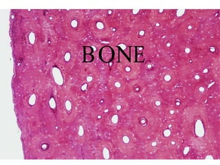
• Mature bone cells that sit in lacunae
• Gap junctions between osteocytes provide
nutrition (15 cells in a row)
• Maintain bony matrix; long lived cells
• Stimulated by calcitonin; inhibited by PTH
9. Osteocytes with Canaliculi
Photomicrograph of dried
bone ground very thin.
The lacunae and
canaliculi filled with air
deflect the light and
appear dark, showing the
communication between
these structures through
which nutrients derived
from blood vessels flow.
10. Osteoclasts
• Derived from monocytes; engulf bony material
• Active osteoblasts stimulate osteoclast activity
• L arge, branched, motile cells
• Secrete enzymes that digest matrix
14. On this image,
Remodeling
the deepest red
color is bone
while pink
represents
either
fibrocartilage
(i.e., collagen
within
cartilage) or
mineralized
cartilage. The
central clearing
represents the
invasion of bone into calcified cartilage. Osteoblast are laying down
new bone toward the left of the upper boundary of this
cavity. Ostoblasts are removing previously-formed bone .
17. E ndochondral Ossification
Photomicrograph of endochondral
ossification. In the upper region is
a row of osteoblasts with intense
cytoplasmic basophilia, a feature
to be expected in cells
synthesizing a glycoprotein
(collagen).
Note an osteoblast being captured
in the bone matrix (arrow).
Between the layer of osteoblasts
and the calcified bone matrix is a
pale region made of noncalcified
bone matrix called Osteoid.
18. Osteon
• L ong cylinder parallel
to long axis of
diaphysis
• Consists of:
– Haversian canal with
nerves, blood vessels;
lamellae with osteocytes
• Haversian canals
communicate with
marrow cavity,
periosteum, other canals
through V olkmann’ s
canals
26. Bone Tissue: Supportive Connective Tissue
E xtracellular Matrix
25% Water
25% Protein or organic matrix
95% Collagen Fibers
5% Chondroitin Sulfate
50% Crystalized Mineral Salts
Hydroxyapatite
(Calcium Phosphate)
Other substances: L ead, Gold,
Strontium, Plutonium, etc.
28. Compact Bone
• Compact bone is arranged in units called osteons or
Haversian systems.
• Osteons (Haversian canal) contain blood vessels,
lymphatic vessels, nerves
• Surrounding this canal are concentric rings of
osteocytes along with the calcified matrix.
• Osteons are aligned in the same direction along lines
of stress. These lines can slowly change as the stresses
on the bone changes.
29. Compact bone
- arranged into concentric rings called Haversian systems
- provides strength
- is external & solid
- Haversian system consists of:
lamella - concentric ring of matrix
lacuna - openings between lamellae for osteocytes
osteocytes - mature bone cell
Haversian canal - in center of lamella; houses vessels
Canaliculi - radiating channels between lacuna
and Haversian canal for nutrients and wastes
Volkmann canal - crosswise canals from Haversian canal
to exterior containing blood vessels and nerves
31. Histology of Compact Bone
• Osteon is concentric rings (lamellae) of calcified matrix
surrounding a vertically oriented blood vessel
• Osteocytes are found in spaces called lacunae
• Osteocytes communicate through canaliculi filled with
extracellular fluid that connect one cell to the next cell
• Interstitial lamellae represent older osteons that have been
partially removed during tissue remodeling
35. The Trabeculae of Spongy Bone
• L attice work of thin plates of bone called trabeculae oriented
along lines of stress
• Spaces in between these struts are filled with red marrow
where blood cells develop
• Found in ends of long bones and inside flat bones such as the
hipbones, sternum, sides of skull, and ribs.
No true Osteons.
36. Spongy Bone
• Spongy (cancellous) bone does
not contain osteons.
• It consists of trabeculae
surrounding many red marrow
filled spaces.
• It forms most of the structure of
short, flat, and irregular bones,
and the epiphyses of long bones.
• Spongy bone tissue is light and
supports and protects the red
bone marrow.
Spongy bone
- irregular lattice work of bone called trabecula
- spaces filled with red bone marrow
- osteocytes trapped within calcium matrix
37. BONE FORMATION
• All embryonic connective tissue begins as mesenchyme.
• Bone formation is termed osteogenesis or ossification and
begins when mesenchymal cells provide the template for
subsequent ossification.
• Two types of ossification occur.
– Intramembranous ossification is the formation of
bone directly from or within fibrous connective
tissue membranes.
– E ndochondrial ossification is the formation of bone
from hyaline cartilage models.
38. Two K inds of
Ossification
1. Intramembranous
Ossification
2. E ndochondral
Ossification
39. Intramembranous Ossification
Also called dermal ossification because it
normally occurs in the deeper layers of
connective tissue of the dermis of the skin.
• All roofing bones of the Skull
Frontal bone
Parietal bones
Occipital bone
Temporal bones
• Mandible
• Clavicle
42. E ndochondral Ossification
Developing bones are deposited as a hyaline
cartilage model and then this cartilage is
replaced by bone tissue.
All bones of the body except:
• All roofing bones of the Skull
• Mandible
• Clavicle
46. Growth at epiphyseal
plates
Zones of epiphyseal plates
Zone of Resting Cartilage
Zone of Proliferating Cartilage
Zone of Hypertrophic Cartilage
Zone of Calcified Cartilage
47. • Zone of resting cartilage Zones of Growth in
– anchors growth plate to bone E piphyseal Plate
• Zone of proliferating cartilage
– rapid cell division (stacked coins)
• Zone of hypertrophic cartilage
– cells enlarged & remain in
columns
• Zone of calcified cartilage
– thin zone, cells mostly dead since
matrix calcified
– osteoclasts removing matrix
– osteoblasts & capillaries move in
to create bone over calcified
cartilage
48. Growth at epiphyseal plates
Zones of epiphyseal plates
Zone of Resting Cartilage
Zone of Proliferating
Cartilage
Zone of Hypertrophic
Cartilage
Zone of Calcified Cartilage
49.
50.
51. Growth in Thickness
• Bone can grow in thickness or diameter only by
appositional growth.
• The steps in these process are:
– Periosteal cells differentiate into osteoblasts which
secrete collagen fibers and organic molecules to
form the matrix.
– Ridges fuse and the periosteum becomes the
endosteum.
– New concentric lamellae are formed.
– Osetoblasts under the peritsteum form new
circumferential lamellae.
52. Bone Growth in Width
• Only by appositional growth at the bone’s surface
• Periosteal cells differentiate into osteoblasts and form bony ridges
and then a tunnel around periosteal blood vessel.
• C oncentric lamellae fill in the tunnel to form an osteon.
54. Factors That Affect Bone Growth
1. Minerals
2. Vitamins
3. Hormones
4. E xercise
55. Factors That Affect Bone Growth
Minerals
Calcium Makes bone matrix hard
Hypocalcemia: low blood calcium levels.
Hypercalcemia: high blood calcium levels.
Phosphorus Makes bone matrix hard
Magnesium Deficiency inhibits osteoblasts
Boron May inhibit calcium loss,
increase levels of estrogens
Manganese Inhibits formation of new bone
tissue
56. Factors That Affect Bone Growth
Vitamins
Vitamin A Controls activity, distribution, and
coordination of osteoblasts/osteoclasts
Vitamin B12 May inhibit osteoblast activity
Vitamin C Helps maintain bone matrix,
deficiency leads to decreased collagen
production which inhibits bone
growth and repair
(scury) disorder due to a lack of Vitamin C
Vitamin D (Calcitriol) Helps build bone by
increasing calcium absorption.
Deficiencies result in “Rickets” in
children
57.
58. Factors That Affect Bone Growth
Hormones
Human Growth Hormone Promotes general growth of all
body tissue and normal growth in
children
Insulin-like Growth Factor Stimulates uptake of amino acids
and protein synthesis
Insulin Promotes normal bone growth and
maturity
Thyroid Hormones Promotes normal bone growth and
maturity
E strogen and Increases osteogenesis at puberty
Testosterone and is responsible for gender
differences of skeletons
62. Steps in Fracture Repair
1. Formation of a fracture hematoma
Immediately after the fracture, there is
a sharp fracture line with associated
soft tissue swelling. At the fracture
Site, there is abundant hematoma
with beginning fibroblastic penetration.
63. Steps in Fracture Repair
2. Fibrocartilaginous Callus
Formation
At 2 weeks there is much visible callus.
There is also bone resorption and
osteoporosis, both difficult to see in
this case because of the overlying callus.
There has been migration of chondroblasts
into the area and cartilage is beginning
to cover the ends of the fracture. New
osteous tissue is produced
enchondrally.