SlideShare une entreprise Scribd logo
1  sur  38
Protein Structure andProtein Structure and
FunctionFunction
ANGEL L SALAMAN, Ph.D.ANGEL L SALAMAN, Ph.D.
angelsalaman@yahoo.comangelsalaman@yahoo.com
1.1. Amino AcidsAmino Acids
2.2. Type of Amino AcidsType of Amino Acids
3.3. Primary StructurePrimary Structure
4.4. Secondary StructureSecondary Structure
5.5. Tertiary InteractionsTertiary Interactions
6.6. Quaternary structureQuaternary structure
Presentation OutlinePresentation Outline
Amino AcidsAmino Acids
 Amino acids are the basic structural unit ofAmino acids are the basic structural unit of
all proteins. A 'free' amino acid (a singleall proteins. A 'free' amino acid (a single
amino acid) always has:amino acid) always has:
• an amino group -NH2,an amino group -NH2,
• a carboxyl group -COOHa carboxyl group -COOH
• a hydrogen -Ha hydrogen -H
• a chemical group or side chain -"R".a chemical group or side chain -"R".
 These are all joined to a central carbonThese are all joined to a central carbon
atom, the a -carbon.atom, the a -carbon.
Since the amino acids (except glycine) have 4 different
groups attached to the a-carbon, they are optically
active stereoisomers although only L-isomers are found
in proteins.
Amino AcidsAmino Acids
 L-isomerL-isomer
In proteins, only the L-In proteins, only the L-
isomer is found normally.isomer is found normally.
 As you travel onwardAs you travel onward
(from the carbonyl carbon(from the carbonyl carbon
to the amino group), theto the amino group), the
R group of L-amino acidsR group of L-amino acids
will be on the left aswill be on the left as
shown in the molecularshown in the molecular
graphic on the rightgraphic on the right
 In the above diagrams, the "R" representsIn the above diagrams, the "R" represents
"a chemical group" and gives each amino"a chemical group" and gives each amino
acid its distinctive properties.acid its distinctive properties.
 With amino acids, this chemical group isWith amino acids, this chemical group is
called the "sidechain", and it specifies thecalled the "sidechain", and it specifies the
amino acid. There are only 20 differentamino acid. There are only 20 different
amino acids found in proteins, each withamino acids found in proteins, each with
it's own defining sidechain. Theseit's own defining sidechain. These
sidechains have a widespread range ofsidechains have a widespread range of
chemical (e.g.charge etc) and structuralchemical (e.g.charge etc) and structural
(e.g. shape etc) diversity.(e.g. shape etc) diversity.
Types of Amino AcidsTypes of Amino Acids
ALIPHATIC AMINO ACIDSALIPHATIC AMINO ACIDS
Aliphatic amino acids are hydrophobic. They do not like to be in contact with
water molecules in an aqueous solution. They are often located in the core of
the protein, surrounded by the rest of the protein, and "shielded" by them
from the aqueous surroundings.
•Glycine (Gly; G): is the simplest and smallest of all amino acids, and the
only one which is not optically active since it has a single hydrogen atom as
it's sidechain
•Alanine (Ala; A): has a methyl group as it's sidechain.
•Valine (Val; V): has a slightly longer sidechain again, and this time there is
a branch. As the aliphatic side chains get longer they are also more
hydrophobic
•Leucine (Leu; L): similar to Valine execept it has another methyl group.
•Isoleucine (Ile; I): is again similar to leucine and valine except that the
orientation of the atoms in the sidechain is slightly different. Isoleucine also
has two centers of asymmetry.
•Proline (Pro; P): the sidechain is bonded to both the a-carbon but also to
the amino group. This has marked effects on the architecture of the proteins.
And although aliphatic, it does not mind being in contact with water as much
as the others.
AROMATIC AMINO ACIDSAROMATIC AMINO ACIDS
These are the amino acids which contain an aromatic ring as partThese are the amino acids which contain an aromatic ring as part
of their sidechains. Because of the hydrophobic nature ofof their sidechains. Because of the hydrophobic nature of
aromatic rings these amino acids are highly hydrophobicaromatic rings these amino acids are highly hydrophobic
 PhenylalaninePhenylalanine (Phe; F)(Phe; F)
is the first of all the aromatic amino acids. It contains a phenylis the first of all the aromatic amino acids. It contains a phenyl
ring attached to a methylene group. Due to the phenyl ring it isring attached to a methylene group. Due to the phenyl ring it is
a hydrophobic amino acid.a hydrophobic amino acid.
 TyrosineTyrosine (Tyr; Y)(Tyr; Y)
contains a hydroxyl group at the end of the phenyl ring. Thiscontains a hydroxyl group at the end of the phenyl ring. This
makes tyrosine less hydrophobic than phenylalanine. It is alsomakes tyrosine less hydrophobic than phenylalanine. It is also
a reactive group, whereas the sidechains so far have all beena reactive group, whereas the sidechains so far have all been
unreactive.unreactive.
 TryptophanTryptophan (Trp; W)(Trp; W)
has a slightly different ring attached to the methylene group.has a slightly different ring attached to the methylene group.
This is an indole ring and it is highly hydrophobic.This is an indole ring and it is highly hydrophobic.
SULPHUR CONTAININGSULPHUR CONTAINING
AMINO ACIDSAMINO ACIDS
 There are two amino acids which contain a sulphurThere are two amino acids which contain a sulphur
atom. They are both very special but for completelyatom. They are both very special but for completely
different reasons.different reasons.
 CysteineCysteine (Cys; C)(Cys; C)
contains a sulphydryl group (-SH). This is extremelycontains a sulphydryl group (-SH). This is extremely
reactive, and can form hydrogen bonds. Cysteine isreactive, and can form hydrogen bonds. Cysteine is
very important because it can also form disulphidevery important because it can also form disulphide
bridges. Even though the -SH group of cysteine canbridges. Even though the -SH group of cysteine can
form hydrogen bonds the long aliphatic part of the sideform hydrogen bonds the long aliphatic part of the side
chain makes it quite hydrophobic.chain makes it quite hydrophobic.
 MethionineMethionine (Met; M)(Met; M)
is a very special amino acid in that it is the "start"is a very special amino acid in that it is the "start"
amino acid in the process of translation (proteinamino acid in the process of translation (protein
synthesis), and therefore, begins every single proteinsynthesis), and therefore, begins every single protein
made. It too has a sulphur atom, but this time it is in amade. It too has a sulphur atom, but this time it is in a
thioether linkage, and is relatively unreactive.thioether linkage, and is relatively unreactive.
Methionine has a highly hydrophobic sidechain.Methionine has a highly hydrophobic sidechain.
HYDROPHILLIC AMINO ACIDSHYDROPHILLIC AMINO ACIDS
 These amino acids can be split into aThese amino acids can be split into a
several groups. There are thoseseveral groups. There are those
which are neutral, those which arewhich are neutral, those which are
acidic, and those which are basic.acidic, and those which are basic.
ACIDIC AMINO ACIDSACIDIC AMINO ACIDS
 These amino acids are highly polar, and are nearlyThese amino acids are highly polar, and are nearly
always negatively charged at physiological pH.always negatively charged at physiological pH.
 AspartateAspartate (Asp; D)(Asp; D)
is really aspartic acid. It is called aspartate because it isis really aspartic acid. It is called aspartate because it is
usually negatively charged at physiological pH and so itusually negatively charged at physiological pH and so it
is named for the carboxylate anion. (Compare aceticis named for the carboxylate anion. (Compare acetic
acid and acetate.).acid and acetate.).
 GlutamateGlutamate (Glu; E)(Glu; E)
is also called glutamic acid. The side chain of glutamateis also called glutamic acid. The side chain of glutamate
also has a carboxylate group which has a negativealso has a carboxylate group which has a negative
charge at physiological pHcharge at physiological pH
NEUTRAL POLAR AMINO ACIDSNEUTRAL POLAR AMINO ACIDS
These amino acids are not charged at physiological pH. However they all haveThese amino acids are not charged at physiological pH. However they all have
groups on their side-chains which are polar and can form hydrogen bonds.groups on their side-chains which are polar and can form hydrogen bonds.
For this reason the amino acids are classed as hydrophillic.For this reason the amino acids are classed as hydrophillic.
 SerineSerine (Ser; S)(Ser; S)
contains an aliphatic chain with a hydroxyl group. It looks like thecontains an aliphatic chain with a hydroxyl group. It looks like the
hydroxylated version of alanine. The OH group make the amino acid highlyhydroxylated version of alanine. The OH group make the amino acid highly
reactive and hydrophillic as it readily forms hydrogen bonds.reactive and hydrophillic as it readily forms hydrogen bonds.
 ThreonineThreonine (Thr; T)(Thr; T)
is another neutral amino acid which has a highly reactive (and highlyis another neutral amino acid which has a highly reactive (and highly
hydrophillic) hydroxyl group. This is an odd amino acid in that it containshydrophillic) hydroxyl group. This is an odd amino acid in that it contains
two centers of asymmetry (two asymetric carbon atoms). This property istwo centers of asymmetry (two asymetric carbon atoms). This property is
shared only by isoleucine.shared only by isoleucine.
 AsparagineAsparagine (Asn; N)(Asn; N)
is the amide derivative of Aspartic acid. When the carboxylate sidechain isis the amide derivative of Aspartic acid. When the carboxylate sidechain is
amidated the resulting amide is uncharged. There is a terminal amideamidated the resulting amide is uncharged. There is a terminal amide
group as opposed to the carboxyl group on aspartate.group as opposed to the carboxyl group on aspartate.
 GlutamineGlutamine (Gln; Q)(Gln; Q)
is similar to Asparagine, with a terminal amide instead of a carboxyl groupis similar to Asparagine, with a terminal amide instead of a carboxyl group
as in glutamate. These two are called the amide derivatives of their parentas in glutamate. These two are called the amide derivatives of their parent
amino acids.amino acids.
Primary StructurePrimary Structure
 A protein has a completely defined order of aminoA protein has a completely defined order of amino
acids, called itsacids, called its sequencesequence..
 This defined sequence is called the primaryThis defined sequence is called the primary
structure of the protein. The sequence, orstructure of the protein. The sequence, or
primary structure is specified by the sequence ofprimary structure is specified by the sequence of
the piece of DNA containing a gene for thatthe piece of DNA containing a gene for that
protein, and the sequence is unique to theprotein, and the sequence is unique to the
individual protein.individual protein.
 All the information necessary to make a protein isAll the information necessary to make a protein is
contained in the primary structure. However, ascontained in the primary structure. However, as
we shall see soon, the primary structure is onlywe shall see soon, the primary structure is only
the first level of structural complexity of proteins.the first level of structural complexity of proteins.
Secondary structureSecondary structure
 Generally proteins have a core of hydrophobicGenerally proteins have a core of hydrophobic
residues surrounded by a shell of hydrophilicresidues surrounded by a shell of hydrophilic
residues. Since the peptide bonds themselves areresidues. Since the peptide bonds themselves are
polar they are neutralized by hydrogen bondingpolar they are neutralized by hydrogen bonding
with each other when in the hydrophobicwith each other when in the hydrophobic
environment. This gives rise to regions of theenvironment. This gives rise to regions of the
polypeptide that form regular 3D structuralpolypeptide that form regular 3D structural
patterns called 'patterns called 'secondary structuresecondary structure'. There are'. There are
two main types of secondary structure:two main types of secondary structure:
 α-helicesα-helices
 β-sheetβ-sheet
αα-Helices-Helices
 The a-helix is a rod like structure. ItThe a-helix is a rod like structure. It
is one peptide chain which has coiledis one peptide chain which has coiled
so that adjacent residues are 1.5Aso that adjacent residues are 1.5A
apart. There are H-bonds betweenapart. There are H-bonds between
the N-H and the C=O groups of thethe N-H and the C=O groups of the
backbone. These link the peptidebackbone. These link the peptide
bonds between every fourth aminobonds between every fourth amino
acid.acid.
 The sidechains all extend outwardsThe sidechains all extend outwards
from the helix.from the helix.
 Although the helix itself is quite a rigid structure,Although the helix itself is quite a rigid structure,
it can be curved or kinked, which lends someit can be curved or kinked, which lends some
flexibility to the protein structure.flexibility to the protein structure.
 Two or more polypeptides can entwine to formTwo or more polypeptides can entwine to form
very long, very stable, structures called a-helicalvery long, very stable, structures called a-helical
coiled coils. These are often found in keratincoiled coils. These are often found in keratin
(hair), myosin (muscle), epidermin (skin), and(hair), myosin (muscle), epidermin (skin), and
fibrin (blood clots). The coiled coils serve afibrin (blood clots). The coiled coils serve a
mechanical role for the proteins mentionedmechanical role for the proteins mentioned
above, which are all fibrous proteins.above, which are all fibrous proteins.
αα-Helices-Helices
αα-Helices-Helices
ββ-Sheets-Sheets
 This secondary structure differs considerably fromThis secondary structure differs considerably from
the a-helix. However b-sheets are classified withthe a-helix. However b-sheets are classified with
a-helices since they are a regular structurala-helices since they are a regular structural
element and are held together by hydrogenelement and are held together by hydrogen
bonds between the -N-H and C=O groups of thebonds between the -N-H and C=O groups of the
peptide bond.peptide bond.
 A polypeptide in a b-sheet is called a b-strand,A polypeptide in a b-sheet is called a b-strand,
and is almost fully extended (not coiled) so thereand is almost fully extended (not coiled) so there
is 3.5A between adjacent residues.is 3.5A between adjacent residues.
 The b-sheet is stabilized by H-bonds between theThe b-sheet is stabilized by H-bonds between the
N-H and the C=O groups of different peptides orN-H and the C=O groups of different peptides or
different parts of the same peptide. The b-strandsdifferent parts of the same peptide. The b-strands
in a sheet can either run in the same (parallel) orin a sheet can either run in the same (parallel) or
different (anti-parallel) direction.different (anti-parallel) direction.
ββ-Sheets-Sheets
MOTIFSMOTIFS
 Some simple combinations ofSome simple combinations of secondary structuresecondary structure elements haveelements have
been found to frequently occur inbeen found to frequently occur in protein structureprotein structure and areand are
referred to as 'super-secondary structure' orreferred to as 'super-secondary structure' or motifsmotifs..
 For example, the β-hairpin motif consists of two adjacentFor example, the β-hairpin motif consists of two adjacent
antiparallel β-strands joined by a small loop. It is present in mostantiparallel β-strands joined by a small loop. It is present in most
antiparallel β structures both as an isolated ribbon and as part ofantiparallel β structures both as an isolated ribbon and as part of
more complex β-sheets. Another common super-secondarymore complex β-sheets. Another common super-secondary
structure is the β-α-β motif, which is frequently used to connectstructure is the β-α-β motif, which is frequently used to connect
two parallel β-strands.two parallel β-strands.
 The central α-helix connects the C-termini of the first strand toThe central α-helix connects the C-termini of the first strand to
the N-termini of the second strand, packing its side chains againstthe N-termini of the second strand, packing its side chains against
the β-sheet and therefore shielding the hydrophobic residues ofthe β-sheet and therefore shielding the hydrophobic residues of
the β-strands from the surface.the β-strands from the surface.
DOMAINSDOMAINS
 Several motifs pack together to form compact, local, semi-Several motifs pack together to form compact, local, semi-
independent units calledindependent units called domainsdomains.. The overall 3DThe overall 3D
structure of the polypeptide chain is referred to as thestructure of the polypeptide chain is referred to as the
protein's 'protein's 'tertiary structuretertiary structure'.'.
 Domains are the fundamental units of tertiary structureDomains are the fundamental units of tertiary structure,,
each domain containing an individual hydrophobic core builteach domain containing an individual hydrophobic core built
from secondary structural units connected by loop regions.from secondary structural units connected by loop regions.
 The packing of the polypeptide is usually much tighter inThe packing of the polypeptide is usually much tighter in
the interior than the exterior of the domain producing athe interior than the exterior of the domain producing a
solid-like core and a fluid-like surface. In fact, core residuessolid-like core and a fluid-like surface. In fact, core residues
are often conserved in a protein family, whereas theare often conserved in a protein family, whereas the
residues in loops are less conserved, unless they areresidues in loops are less conserved, unless they are
involved in the protein's function.involved in the protein's function.
 Protein tertiary structure can be divided into four mainProtein tertiary structure can be divided into four main
classes based on the secondary structural content of theclasses based on the secondary structural content of the
domain.domain.
DOMAINSDOMAINS
 The concept of the domain was first proposed in 1973 byThe concept of the domain was first proposed in 1973 by
Wetlaufer after X-ray crystallographic studies of henWetlaufer after X-ray crystallographic studies of hen
lysozymelysozyme (Phillips, 1966), papain (Drenth et al., 1968) and(Phillips, 1966), papain (Drenth et al., 1968) and
by limited proteolysis studies ofby limited proteolysis studies of immunoglobulinsimmunoglobulins (Porter,(Porter,
1973; Edelman, 1973). Wetlaufer defined domains as1973; Edelman, 1973). Wetlaufer defined domains as
stable units ofstable units of protein structureprotein structure that could foldthat could fold
autonomously.autonomously.
 DefinitionDefinition:: a compact structural domain that is founda compact structural domain that is found
amongst diverse proteins is likely to fold independentlyamongst diverse proteins is likely to fold independently
within its structural environment.within its structural environment.
 In a multidomain protein, each domain may fulfill its ownIn a multidomain protein, each domain may fulfill its own
functionfunction independently, or in a concerted manner with itsindependently, or in a concerted manner with its
neighbors. Domains can either serve as modules forneighbors. Domains can either serve as modules for
building up large assemblies such as virus particles orbuilding up large assemblies such as virus particles or
muscle fibers, or can provide specific catalytic or bindingmuscle fibers, or can provide specific catalytic or binding
sites as found in enzymes or regulatory proteins.sites as found in enzymes or regulatory proteins.
DOMAINS (Classes)DOMAINS (Classes)
 AllAll α-domainsα-domains have a domain core built exclusively fromhave a domain core built exclusively from
α-helices. This class is dominated by small folds, many ofα-helices. This class is dominated by small folds, many of
which form a simple bundle with helices running up andwhich form a simple bundle with helices running up and
down.down.
 AllAll β-domainsβ-domains have a core comprising of antiparallel β-have a core comprising of antiparallel β-
sheets, usually two sheets packed against each other.sheets, usually two sheets packed against each other.
Various patterns can be identified in the arrangement ofVarious patterns can be identified in the arrangement of
the strands, often giving rise to the identification ofthe strands, often giving rise to the identification of
recurring motifs, for example the greek key motif.recurring motifs, for example the greek key motif.
 α+β domainsα+β domains are a mixture of all-α and all-β motifs.are a mixture of all-α and all-β motifs.
Classification of proteins into this class is difficult becauseClassification of proteins into this class is difficult because
of overlaps to the other three classes and therefore is notof overlaps to the other three classes and therefore is not
used in theused in the CATHCATH domain database.domain database.
 α/β domainsα/β domains are made from a combination of β-α-βare made from a combination of β-α-β
motifs that predominantly form a parallel β-sheetmotifs that predominantly form a parallel β-sheet
surrounded by amphipathic α-helices. The secondarysurrounded by amphipathic α-helices. The secondary
structures are arranged in layers or barrels.structures are arranged in layers or barrels.
Pyruvate kinase
•An appropriate example is pyruvate
kinase, a glycolytic enzyme that plays
an important role in regulating the
flux from fructose-1,6-biphosphate to
pyruvate. It contains an all-β
regulatory domain, an α/β-substrate
binding domain and an α/β-nucleotide
binding domain, connected by several
polypeptide linkers (George and
Heringa, 2002). Each domain in this
protein occurs in diverse sets of
protein families.
DOMAINSDOMAINS
Ribbon diagram of the SH2
domain from P56-Lck tyrosine
kinase
The Src homology 2 domain (or
SH2 domain) is a protein domain
of about 100 amino acid residues
first identified as a conserved
sequence region among the
oncoproteins Src and Fps.
SH2 domains typically bind a
phosphorylated tyrosine residue
Phosphorylation of tyrosine
residues in a protein occurs during
signal transduction and is carried
out by tyrosine kinases. In this
way, phosphorylation of a
substrate by tyrosine kinases acts
as a switch to trigger binding to an
SH2 domain-containing protein.
Pawson, T., Gish, G., Nash, P. (2001). SH2
domains, interaction modules and cellular wiring.
Trends in Cell Biology 11(12): 504-511
DOMAINSDOMAINS
This image (courtesy of P. J.
Bjorkman from Nature 329:506,
1987) is a schematic
representation of the extracellular
portion of HLA-A2, a human class I
histocompatibility molecule. It also
illustrates two common examples
of secondary structure: the
stretches of beta conformation
are represented by the broad green
arrows (pointing N -> C terminal);
regions of alpha helix are shown
as helical ribbons. The pairs of
purple spheres represent the
disulfide bridges.
•The Structural Classification of
Proteins: SCOP is to some a
magical hierarchical classification of
protein structures. As is simplified
greatly in the figure on the right -
SCOP classifies protein structures
at many levels from overall class (all
alpha helix, all beta strand etc.)
down to protein domain and species
level.
•The SCOP tree takes the protein
domain as the evolutionary unit and
represents evolutionary
relationships between proteins such
that anything classified within a
superfamily is evolutionarily related.
•Whereas protein domains within
the same class or fold are not
necessarily evolutionarily related.
Tertiary InteractionsTertiary Interactions
 This is the term given to the overall three dimensionalThis is the term given to the overall three dimensional
structure of the polypeptide, also written 3o structure. Thisstructure of the polypeptide, also written 3o structure. This
describes how the peptide chain folds so that sidechains aredescribes how the peptide chain folds so that sidechains are
packed and organized. This may bring two sidechains whichpacked and organized. This may bring two sidechains which
are distant, in terms of primary structure, close together. Itare distant, in terms of primary structure, close together. It
also creates binding sites and other functional regions onalso creates binding sites and other functional regions on
the surface of the protein.the surface of the protein.
 If we know the 3D structure of the protein, we can begin toIf we know the 3D structure of the protein, we can begin to
understand how the protein works. If the protein comesunderstand how the protein works. If the protein comes
from a bacterium or a virus, we can start to design drugsfrom a bacterium or a virus, we can start to design drugs
which affect the bacterial or viral form of the protein, andwhich affect the bacterial or viral form of the protein, and
not the host (eukaryotic) form. If an animal is making anot the host (eukaryotic) form. If an animal is making a
defective protein (as is the case in cystic fibrosis patients)defective protein (as is the case in cystic fibrosis patients)
then by using protein engineering and gene therapy, it maythen by using protein engineering and gene therapy, it may
be possible to get the animal to start making the functionalbe possible to get the animal to start making the functional
protein. All this leads to a new area of science calledprotein. All this leads to a new area of science called
"Structural Biology"."Structural Biology".
 There are 5 forces affecting the 3D shape of proteins. All ofThere are 5 forces affecting the 3D shape of proteins. All of
them build upon the framework started by the secondarythem build upon the framework started by the secondary
structures.structures.
Hydrophobic InteractionsHydrophobic Interactions
 Non-Polar/Hydrophobic/Aliphatic sidechains cannot hydrogenNon-Polar/Hydrophobic/Aliphatic sidechains cannot hydrogen
bond with water, hence the network of water is disrupted bybond with water, hence the network of water is disrupted by
them. This is avoided if the non-polar sidechains arethem. This is avoided if the non-polar sidechains are
removed or shielded from the water and buried in theremoved or shielded from the water and buried in the
interior of the protein. In this coiled coil structure note thatinterior of the protein. In this coiled coil structure note that
the interior of the structure is occupied solely bythe interior of the structure is occupied solely by
hydrophobic residues.hydrophobic residues.
 Hydrophobic interaction are a major driving force in proteinHydrophobic interaction are a major driving force in protein
folding. Hydrophobic interactions are also important in thefolding. Hydrophobic interactions are also important in the
formation of secondary structure elements such as a-helicesformation of secondary structure elements such as a-helices
and b-sheets. The peptide backbone is relatively hydrophillicand b-sheets. The peptide backbone is relatively hydrophillic
because of the C=O and N-H groups of each peptide bond.because of the C=O and N-H groups of each peptide bond.
However in the centre of the protein these tend to be in aHowever in the centre of the protein these tend to be in a
hydrophobic environment. One way to reduce thehydrophobic environment. One way to reduce the
hydrophilicity of the peptide backbone is to for hydrogenhydrophilicity of the peptide backbone is to for hydrogen
bonds between the peptide atoms. The a-helix and b-sheetbonds between the peptide atoms. The a-helix and b-sheet
are two structures which maximise the hydrogen bondingare two structures which maximise the hydrogen bonding
between the peptide bonds of the backbone and reduce itsbetween the peptide bonds of the backbone and reduce its
hydrophilicity.hydrophilicity.
Hydrogen BondsHydrogen Bonds
 When unfolded, all polar/hydrophillic sidechains canWhen unfolded, all polar/hydrophillic sidechains can
interact via H-bonds with water. When the protein folds,interact via H-bonds with water. When the protein folds,
they must H-bond to each other and exclude much of thethey must H-bond to each other and exclude much of the
water. All groups capable of forming a hydrogen bondwater. All groups capable of forming a hydrogen bond
MUST, hence H-bonding in the backbone (C=O to N-H) byMUST, hence H-bonding in the backbone (C=O to N-H) by
way of helices and sheets is an efficient way of ensuringway of helices and sheets is an efficient way of ensuring
maximum H-bonding.maximum H-bonding.
 Sidechains can either accept (as in C=O) or donate (as inSidechains can either accept (as in C=O) or donate (as in
N-H, or O-H) an H-bond.N-H, or O-H) an H-bond.
 The capacity of proteins to form hydrogen bonds is anThe capacity of proteins to form hydrogen bonds is an
important determinant of protein stability. Hydrogen bondsimportant determinant of protein stability. Hydrogen bonds
can be between backbone groups, as in helices and sheets;can be between backbone groups, as in helices and sheets;
between side chains, such as serine or threonine O-Hbetween side chains, such as serine or threonine O-H
groups and carbonyl carbons of side chains (-C=O); andgroups and carbonyl carbons of side chains (-C=O); and
between backbone groups and side chain groups.between backbone groups and side chain groups.
Ion PairsIon Pairs
 When amino acid sidechains of opposite charge are in closeWhen amino acid sidechains of opposite charge are in close
proximity, they can form an ion pair (also called a saltproximity, they can form an ion pair (also called a salt
bridge). But they are also capable of hydrogen bonding andbridge). But they are also capable of hydrogen bonding and
hence, are usually found on the surface of the protein. Ifhence, are usually found on the surface of the protein. If
they can form a salt bridge, they will usually be buried.they can form a salt bridge, they will usually be buried.
Since charge is affected by pH, so is the formation and theSince charge is affected by pH, so is the formation and the
breakage of these ion pairs.breakage of these ion pairs.
 Salt bridges also increase the stability of the tertiarySalt bridges also increase the stability of the tertiary
structure.structure.
NOTE:NOTE:Hydrophobic interactions, hydrogen bonds and saltHydrophobic interactions, hydrogen bonds and salt
bridges are all non-covalent interactionsbridges are all non-covalent interactions
 These are all relatively weak interactions but the largeThese are all relatively weak interactions but the large
number in a protein combine to give the overall stability ofnumber in a protein combine to give the overall stability of
the structure.the structure.
Disulphide BondsDisulphide Bonds
 If two cysteine sidechains are close to one another and the localIf two cysteine sidechains are close to one another and the local
environment is conducive to oxidation,then they can form aenvironment is conducive to oxidation,then they can form a
disulphide bond/bridge as shown. The residues can either be indisulphide bond/bridge as shown. The residues can either be in
the same peptide chain (forming a loop) or in different chains.the same peptide chain (forming a loop) or in different chains.
 As Disulphide bonds are covalent linkages they add considerablyAs Disulphide bonds are covalent linkages they add considerably
to the stability of proteins in which they occur. However theyto the stability of proteins in which they occur. However they
mainly occur in secreted proteins and in the parts of membranemainly occur in secreted proteins and in the parts of membrane
proteins which face the outside.proteins which face the outside.
Note that disuphide bridges only form after the protein has foldedNote that disuphide bridges only form after the protein has folded
into its tertiary structure. This folding is driven by the combinedinto its tertiary structure. This folding is driven by the combined
energy of all of the weak interactions described in the previousenergy of all of the weak interactions described in the previous
section above.section above.
Cysteine side chains in cytosolic proteins are usually found as -SHCysteine side chains in cytosolic proteins are usually found as -SH
groups as the cytosolic environmemt is sufficiently reducing togroups as the cytosolic environmemt is sufficiently reducing to
prevent disulphides from forming.prevent disulphides from forming.
Non-Covalent BondsNon-Covalent Bonds
 The tertiary structure of proteins is maintained byThe tertiary structure of proteins is maintained by
a host of weak, non-covalent interactions:a host of weak, non-covalent interactions:
 Hydrophobic InteractionsHydrophobic Interactions
 Hydrogen BondsHydrogen Bonds
 Salt BridgesSalt Bridges
 Protein stability is a function of many weakProtein stability is a function of many weak
interactions. If these interactions are maintainedinteractions. If these interactions are maintained
the protein keeps its tertiary ("native") stucture.the protein keeps its tertiary ("native") stucture.
However once the bonds maintaining theHowever once the bonds maintaining the
structure begin to be disrupted the tertiarystructure begin to be disrupted the tertiary
structure is destroyed and the protein is said tostructure is destroyed and the protein is said to
be "denatured"be "denatured"
Subunits & Quaternary StructureSubunits & Quaternary Structure
 Proteins containing more than one polypeptideProteins containing more than one polypeptide
chain exhibit an additional level of structuralchain exhibit an additional level of structural
organisation.organisation.
 In this case, each polypeptide chain is aIn this case, each polypeptide chain is a
"subunit". Quaternary (4o) structure describes"subunit". Quaternary (4o) structure describes
the way the subunits are arranged together andthe way the subunits are arranged together and
the nature of their contacts.the nature of their contacts.
 Subunits associate by non-covalent interactionsSubunits associate by non-covalent interactions
similar to those involved in tertiary structure:similar to those involved in tertiary structure:
 Hydrophobic InteractionsHydrophobic Interactions
 Hydrogen bondsHydrogen bonds
 Salt Bridges / Ion PairsSalt Bridges / Ion Pairs
DENATURATION
loss of 3-D conformation by heat, pH, organic solvents, detergents
RENATURATION - regaining of biological activity via self-
assembly
Factors Which Can Denature AFactors Which Can Denature A
ProteinProtein
 Heat: increase atomic vibrations, ruinsHeat: increase atomic vibrations, ruins
interactions, need heat of >50interactions, need heat of >50oo
C to denatureC to denature
most proteins.most proteins.
 pH: breaks the salt bridges.pH: breaks the salt bridges.
 Organic Solvents: disrupt hydrophobicOrganic Solvents: disrupt hydrophobic
interactionsinteractions
 Detergents: hydrophobic part pushes into proteinDetergents: hydrophobic part pushes into protein
corecore
 When a protein is denatured, it loses it's 3DWhen a protein is denatured, it loses it's 3D
shape. When a protein is denatured, it loses it'sshape. When a protein is denatured, it loses it's
biological activity. Therefore...biological activity. Therefore...
A PROTEINS TERTIARY STRUCTURE ISA PROTEINS TERTIARY STRUCTURE IS
ESSENTIAL TO IT'S BIOLOGICAL ACTIVITYESSENTIAL TO IT'S BIOLOGICAL ACTIVITY
REFERENCES
 Introduction to Protein Structure, C.Branden and J.Tooze, Garland,
New York (1991).
 A novel super-secondary structure of proteins and the relation
between the structure and amino acid sequence, A.F.Efimov, FEBS
Lett. 166, 33-38 (1984).
 Structure of alpha-alpha-hairpins with short connections,
A.V.Efimov, Protein Engin.4, 245-250 (1991).
 Structure of beta-beta-hairpins and beta-beta-corners, A.V.Efimov,
FEBS Lett., 284, 288-292 (1991).
 Structure and evolution of calcium modulated proteins,
R.H.Kretsinger, CRC Crit.Rev.Biochem. 8, 119-174 (1980).
 Comparison of the structures of cro and lambda repressor proteins
in bacteriophage lambda, D.H.Ohlendorf, W.F.Anderson, M.Lewis,
C.O.Pabo and B.W.Matthews, J.Molec.Biol. 169 757-769 (1983).
 Conformation of beta-hairpins in protein structures, B.L.Sibanda,
T.L.Blundell and J.M.Thornton, J.Molec.Biol. 206, 759-777 (1989).
 Beta-hairpin families in globular proteins, B.L.Sibanda and
J.M.Thornton, Nature 316, 170-174 (1985).

Contenu connexe

Tendances

Amino Acids: Structures, Physical and Chemical properties, Groups, Functions
Amino Acids: Structures, Physical and Chemical properties, Groups, FunctionsAmino Acids: Structures, Physical and Chemical properties, Groups, Functions
Amino Acids: Structures, Physical and Chemical properties, Groups, FunctionsSt Xaviers
 
Biological roles of proteins
Biological roles of proteinsBiological roles of proteins
Biological roles of proteinsrajeshkumar590473
 
Structural organisation of protiens
Structural organisation of protiensStructural organisation of protiens
Structural organisation of protiensVikas CJ Vikki
 
Protein structure: details
Protein structure: detailsProtein structure: details
Protein structure: detailsdamarisb
 
PROPERTIES OF AMINOACIDS.pptx
PROPERTIES OF AMINOACIDS.pptxPROPERTIES OF AMINOACIDS.pptx
PROPERTIES OF AMINOACIDS.pptxSuganyaPaulraj
 
Characteristics and Properties of Amino Acids (AA)
Characteristics and Properties of Amino Acids (AA)Characteristics and Properties of Amino Acids (AA)
Characteristics and Properties of Amino Acids (AA)Mohamed Hassanien
 
Structure of proteins
Structure of proteinsStructure of proteins
Structure of proteinsDevyani Joshi
 
Membrane Proteins & its types
Membrane Proteins & its typesMembrane Proteins & its types
Membrane Proteins & its typesSabahat Ali
 
Lipids: Structure and Functions
Lipids: Structure and FunctionsLipids: Structure and Functions
Lipids: Structure and Functionsvibhakhanna1
 
Amino acid structure classification and properties
Amino acid structure classification and propertiesAmino acid structure classification and properties
Amino acid structure classification and propertiesdeepalakshmi59
 
Globular proteins
Globular proteinsGlobular proteins
Globular proteinsSabahat Ali
 
Fibrous proteins
Fibrous proteinsFibrous proteins
Fibrous proteinsKinza Ayub
 
Tm of DNA - melting temperature of DNA
Tm of DNA - melting temperature of DNATm of DNA - melting temperature of DNA
Tm of DNA - melting temperature of DNAMandira bhosale
 
Cytoskeleton presentation (introduction structure & function)
Cytoskeleton presentation (introduction structure & function)Cytoskeleton presentation (introduction structure & function)
Cytoskeleton presentation (introduction structure & function)Dryogeshcsv
 
Proteins-Classification ,Structure of protein, properties and biological impo...
Proteins-Classification ,Structure of protein, properties and biological impo...Proteins-Classification ,Structure of protein, properties and biological impo...
Proteins-Classification ,Structure of protein, properties and biological impo...SoniaBajaj10
 

Tendances (20)

Amino Acids: Structures, Physical and Chemical properties, Groups, Functions
Amino Acids: Structures, Physical and Chemical properties, Groups, FunctionsAmino Acids: Structures, Physical and Chemical properties, Groups, Functions
Amino Acids: Structures, Physical and Chemical properties, Groups, Functions
 
Biological roles of proteins
Biological roles of proteinsBiological roles of proteins
Biological roles of proteins
 
Glycolipid ppt
Glycolipid pptGlycolipid ppt
Glycolipid ppt
 
Amino acid
Amino acid Amino acid
Amino acid
 
Structural organisation of protiens
Structural organisation of protiensStructural organisation of protiens
Structural organisation of protiens
 
Protein structure: details
Protein structure: detailsProtein structure: details
Protein structure: details
 
PROPERTIES OF AMINOACIDS.pptx
PROPERTIES OF AMINOACIDS.pptxPROPERTIES OF AMINOACIDS.pptx
PROPERTIES OF AMINOACIDS.pptx
 
Protein structure
Protein structureProtein structure
Protein structure
 
Characteristics and Properties of Amino Acids (AA)
Characteristics and Properties of Amino Acids (AA)Characteristics and Properties of Amino Acids (AA)
Characteristics and Properties of Amino Acids (AA)
 
Structure of proteins
Structure of proteinsStructure of proteins
Structure of proteins
 
Membrane Proteins & its types
Membrane Proteins & its typesMembrane Proteins & its types
Membrane Proteins & its types
 
Lipids: Structure and Functions
Lipids: Structure and FunctionsLipids: Structure and Functions
Lipids: Structure and Functions
 
Amino acid structure classification and properties
Amino acid structure classification and propertiesAmino acid structure classification and properties
Amino acid structure classification and properties
 
Globular proteins
Globular proteinsGlobular proteins
Globular proteins
 
Fibrous proteins
Fibrous proteinsFibrous proteins
Fibrous proteins
 
Tm of DNA - melting temperature of DNA
Tm of DNA - melting temperature of DNATm of DNA - melting temperature of DNA
Tm of DNA - melting temperature of DNA
 
Cytoskeleton presentation (introduction structure & function)
Cytoskeleton presentation (introduction structure & function)Cytoskeleton presentation (introduction structure & function)
Cytoskeleton presentation (introduction structure & function)
 
Protein folding
Protein foldingProtein folding
Protein folding
 
Proteins-Classification ,Structure of protein, properties and biological impo...
Proteins-Classification ,Structure of protein, properties and biological impo...Proteins-Classification ,Structure of protein, properties and biological impo...
Proteins-Classification ,Structure of protein, properties and biological impo...
 
Amino acid classification
Amino acid classificationAmino acid classification
Amino acid classification
 

Similaire à Protein Structure and Function

PROTEIN-AND-AMINO-ACIDS-PPT.p df
PROTEIN-AND-AMINO-ACIDS-PPT.p         dfPROTEIN-AND-AMINO-ACIDS-PPT.p         df
PROTEIN-AND-AMINO-ACIDS-PPT.p dfChyeOdchigue
 
Amino Acids for Dentistry
Amino Acids  for DentistryAmino Acids  for Dentistry
Amino Acids for DentistryAyman Abdo
 
Amino acids.pptx
Amino acids.pptxAmino acids.pptx
Amino acids.pptxshoba shoba
 
aminoacidbysridarshinichandra1-1-160424175247.pdf
aminoacidbysridarshinichandra1-1-160424175247.pdfaminoacidbysridarshinichandra1-1-160424175247.pdf
aminoacidbysridarshinichandra1-1-160424175247.pdfMariamMansour32
 
Properties of amino acid side chains report ni lady
Properties of amino acid side chains   report ni ladyProperties of amino acid side chains   report ni lady
Properties of amino acid side chains report ni ladyAya Chavez
 
Properties of amino acid side chains report ni lady
Properties of amino acid side chains   report ni ladyProperties of amino acid side chains   report ni lady
Properties of amino acid side chains report ni ladyAya Chavez
 
chemistryofaminoacidsproteins-140307154509-phpapp02.pdf
chemistryofaminoacidsproteins-140307154509-phpapp02.pdfchemistryofaminoacidsproteins-140307154509-phpapp02.pdf
chemistryofaminoacidsproteins-140307154509-phpapp02.pdfanju468752
 
biochemistry-secondary structure of proteins
biochemistry-secondary structure of proteinsbiochemistry-secondary structure of proteins
biochemistry-secondary structure of proteinsroyabasser
 
Lec2 aminoacid classification-aimec
Lec2 aminoacid classification-aimecLec2 aminoacid classification-aimec
Lec2 aminoacid classification-aimecShamim Akram
 
Lec2 amino.a.classification microsoft power
Lec2 amino.a.classification microsoft powerLec2 amino.a.classification microsoft power
Lec2 amino.a.classification microsoft powerDrShamimAkram
 
Amino acid
Amino acidAmino acid
Amino acidNimmiRoy
 
Lecture 1 - General Properties of Amino Acids(2) (1).pdf
Lecture 1 - General Properties of Amino Acids(2) (1).pdfLecture 1 - General Properties of Amino Acids(2) (1).pdf
Lecture 1 - General Properties of Amino Acids(2) (1).pdfKundaBwalya1
 

Similaire à Protein Structure and Function (20)

PROTEIN-AND-AMINO-ACIDS-PPT.p df
PROTEIN-AND-AMINO-ACIDS-PPT.p         dfPROTEIN-AND-AMINO-ACIDS-PPT.p         df
PROTEIN-AND-AMINO-ACIDS-PPT.p df
 
Amino Acids for Dentistry
Amino Acids  for DentistryAmino Acids  for Dentistry
Amino Acids for Dentistry
 
Amino acids structure
Amino acids structureAmino acids structure
Amino acids structure
 
Amino acids.pptx
Amino acids.pptxAmino acids.pptx
Amino acids.pptx
 
Amino acids
Amino acidsAmino acids
Amino acids
 
Amino acids
Amino acidsAmino acids
Amino acids
 
aminoacidbysridarshinichandra1-1-160424175247.pdf
aminoacidbysridarshinichandra1-1-160424175247.pdfaminoacidbysridarshinichandra1-1-160424175247.pdf
aminoacidbysridarshinichandra1-1-160424175247.pdf
 
Amino Acids.docx
Amino Acids.docxAmino Acids.docx
Amino Acids.docx
 
Properties of amino acid side chains report ni lady
Properties of amino acid side chains   report ni ladyProperties of amino acid side chains   report ni lady
Properties of amino acid side chains report ni lady
 
Properties of amino acid side chains report ni lady
Properties of amino acid side chains   report ni ladyProperties of amino acid side chains   report ni lady
Properties of amino acid side chains report ni lady
 
chemistryofaminoacidsproteins-140307154509-phpapp02.pdf
chemistryofaminoacidsproteins-140307154509-phpapp02.pdfchemistryofaminoacidsproteins-140307154509-phpapp02.pdf
chemistryofaminoacidsproteins-140307154509-phpapp02.pdf
 
Aminoacids.pptx
Aminoacids.pptxAminoacids.pptx
Aminoacids.pptx
 
Amino acids.ppt
Amino acids.pptAmino acids.ppt
Amino acids.ppt
 
biochemistry-secondary structure of proteins
biochemistry-secondary structure of proteinsbiochemistry-secondary structure of proteins
biochemistry-secondary structure of proteins
 
Lec2 aminoacid classification-aimec
Lec2 aminoacid classification-aimecLec2 aminoacid classification-aimec
Lec2 aminoacid classification-aimec
 
Lec2 amino.a.classification microsoft power
Lec2 amino.a.classification microsoft powerLec2 amino.a.classification microsoft power
Lec2 amino.a.classification microsoft power
 
Proteins
ProteinsProteins
Proteins
 
Amino acid
Amino acidAmino acid
Amino acid
 
Protein-An overview
Protein-An overviewProtein-An overview
Protein-An overview
 
Lecture 1 - General Properties of Amino Acids(2) (1).pdf
Lecture 1 - General Properties of Amino Acids(2) (1).pdfLecture 1 - General Properties of Amino Acids(2) (1).pdf
Lecture 1 - General Properties of Amino Acids(2) (1).pdf
 

Plus de angelsalaman

Basis Validation of Biomanufacturing Processes
Basis Validation of Biomanufacturing ProcessesBasis Validation of Biomanufacturing Processes
Basis Validation of Biomanufacturing Processesangelsalaman
 
Environmental Characterization of Controlled Rooms
Environmental Characterization of Controlled RoomsEnvironmental Characterization of Controlled Rooms
Environmental Characterization of Controlled Roomsangelsalaman
 
Isolation and Screening of Secondary Metabolites
Isolation and Screening of Secondary MetabolitesIsolation and Screening of Secondary Metabolites
Isolation and Screening of Secondary Metabolitesangelsalaman
 
Downstream Processing in Biopharmaceuticals
Downstream Processing in BiopharmaceuticalsDownstream Processing in Biopharmaceuticals
Downstream Processing in Biopharmaceuticalsangelsalaman
 
Protein Purification
Protein PurificationProtein Purification
Protein Purificationangelsalaman
 
Protein Detection Methods and Application
Protein Detection Methods and ApplicationProtein Detection Methods and Application
Protein Detection Methods and Applicationangelsalaman
 
Presentation Protein Synthesis
Presentation Protein SynthesisPresentation Protein Synthesis
Presentation Protein Synthesisangelsalaman
 
Laboratorio de Microbiologia Seguridad
Laboratorio de Microbiologia SeguridadLaboratorio de Microbiologia Seguridad
Laboratorio de Microbiologia Seguridadangelsalaman
 
Bioburden Validation Strategy for Cleaning Validation
Bioburden Validation Strategy for Cleaning ValidationBioburden Validation Strategy for Cleaning Validation
Bioburden Validation Strategy for Cleaning Validationangelsalaman
 

Plus de angelsalaman (10)

Basis Validation of Biomanufacturing Processes
Basis Validation of Biomanufacturing ProcessesBasis Validation of Biomanufacturing Processes
Basis Validation of Biomanufacturing Processes
 
Environmental Characterization of Controlled Rooms
Environmental Characterization of Controlled RoomsEnvironmental Characterization of Controlled Rooms
Environmental Characterization of Controlled Rooms
 
Isolation and Screening of Secondary Metabolites
Isolation and Screening of Secondary MetabolitesIsolation and Screening of Secondary Metabolites
Isolation and Screening of Secondary Metabolites
 
Downstream Processing in Biopharmaceuticals
Downstream Processing in BiopharmaceuticalsDownstream Processing in Biopharmaceuticals
Downstream Processing in Biopharmaceuticals
 
Bioreactor Basis
Bioreactor BasisBioreactor Basis
Bioreactor Basis
 
Protein Purification
Protein PurificationProtein Purification
Protein Purification
 
Protein Detection Methods and Application
Protein Detection Methods and ApplicationProtein Detection Methods and Application
Protein Detection Methods and Application
 
Presentation Protein Synthesis
Presentation Protein SynthesisPresentation Protein Synthesis
Presentation Protein Synthesis
 
Laboratorio de Microbiologia Seguridad
Laboratorio de Microbiologia SeguridadLaboratorio de Microbiologia Seguridad
Laboratorio de Microbiologia Seguridad
 
Bioburden Validation Strategy for Cleaning Validation
Bioburden Validation Strategy for Cleaning ValidationBioburden Validation Strategy for Cleaning Validation
Bioburden Validation Strategy for Cleaning Validation
 

Dernier

TEST BANK For Radiologic Science for Technologists, 12th Edition by Stewart C...
TEST BANK For Radiologic Science for Technologists, 12th Edition by Stewart C...TEST BANK For Radiologic Science for Technologists, 12th Edition by Stewart C...
TEST BANK For Radiologic Science for Technologists, 12th Edition by Stewart C...ssifa0344
 
PossibleEoarcheanRecordsoftheGeomagneticFieldPreservedintheIsuaSupracrustalBe...
PossibleEoarcheanRecordsoftheGeomagneticFieldPreservedintheIsuaSupracrustalBe...PossibleEoarcheanRecordsoftheGeomagneticFieldPreservedintheIsuaSupracrustalBe...
PossibleEoarcheanRecordsoftheGeomagneticFieldPreservedintheIsuaSupracrustalBe...Sérgio Sacani
 
Biopesticide (2).pptx .This slides helps to know the different types of biop...
Biopesticide (2).pptx  .This slides helps to know the different types of biop...Biopesticide (2).pptx  .This slides helps to know the different types of biop...
Biopesticide (2).pptx .This slides helps to know the different types of biop...RohitNehra6
 
Green chemistry and Sustainable development.pptx
Green chemistry  and Sustainable development.pptxGreen chemistry  and Sustainable development.pptx
Green chemistry and Sustainable development.pptxRajatChauhan518211
 
Forensic Biology & Its biological significance.pdf
Forensic Biology & Its biological significance.pdfForensic Biology & Its biological significance.pdf
Forensic Biology & Its biological significance.pdfrohankumarsinghrore1
 
Zoology 4th semester series (krishna).pdf
Zoology 4th semester series (krishna).pdfZoology 4th semester series (krishna).pdf
Zoology 4th semester series (krishna).pdfSumit Kumar yadav
 
9654467111 Call Girls In Raj Nagar Delhi Short 1500 Night 6000
9654467111 Call Girls In Raj Nagar Delhi Short 1500 Night 60009654467111 Call Girls In Raj Nagar Delhi Short 1500 Night 6000
9654467111 Call Girls In Raj Nagar Delhi Short 1500 Night 6000Sapana Sha
 
Presentation Vikram Lander by Vedansh Gupta.pptx
Presentation Vikram Lander by Vedansh Gupta.pptxPresentation Vikram Lander by Vedansh Gupta.pptx
Presentation Vikram Lander by Vedansh Gupta.pptxgindu3009
 
Botany 4th semester series (krishna).pdf
Botany 4th semester series (krishna).pdfBotany 4th semester series (krishna).pdf
Botany 4th semester series (krishna).pdfSumit Kumar yadav
 
CALL ON ➥8923113531 🔝Call Girls Kesar Bagh Lucknow best Night Fun service 🪡
CALL ON ➥8923113531 🔝Call Girls Kesar Bagh Lucknow best Night Fun service  🪡CALL ON ➥8923113531 🔝Call Girls Kesar Bagh Lucknow best Night Fun service  🪡
CALL ON ➥8923113531 🔝Call Girls Kesar Bagh Lucknow best Night Fun service 🪡anilsa9823
 
Animal Communication- Auditory and Visual.pptx
Animal Communication- Auditory and Visual.pptxAnimal Communication- Auditory and Visual.pptx
Animal Communication- Auditory and Visual.pptxUmerFayaz5
 
Nanoparticles synthesis and characterization​ ​
Nanoparticles synthesis and characterization​  ​Nanoparticles synthesis and characterization​  ​
Nanoparticles synthesis and characterization​ ​kaibalyasahoo82800
 
Asymmetry in the atmosphere of the ultra-hot Jupiter WASP-76 b
Asymmetry in the atmosphere of the ultra-hot Jupiter WASP-76 bAsymmetry in the atmosphere of the ultra-hot Jupiter WASP-76 b
Asymmetry in the atmosphere of the ultra-hot Jupiter WASP-76 bSérgio Sacani
 
Chemistry 4th semester series (krishna).pdf
Chemistry 4th semester series (krishna).pdfChemistry 4th semester series (krishna).pdf
Chemistry 4th semester series (krishna).pdfSumit Kumar yadav
 
Physiochemical properties of nanomaterials and its nanotoxicity.pptx
Physiochemical properties of nanomaterials and its nanotoxicity.pptxPhysiochemical properties of nanomaterials and its nanotoxicity.pptx
Physiochemical properties of nanomaterials and its nanotoxicity.pptxAArockiyaNisha
 
Natural Polymer Based Nanomaterials
Natural Polymer Based NanomaterialsNatural Polymer Based Nanomaterials
Natural Polymer Based NanomaterialsAArockiyaNisha
 
Stunning ➥8448380779▻ Call Girls In Panchshil Enclave Delhi NCR
Stunning ➥8448380779▻ Call Girls In Panchshil Enclave Delhi NCRStunning ➥8448380779▻ Call Girls In Panchshil Enclave Delhi NCR
Stunning ➥8448380779▻ Call Girls In Panchshil Enclave Delhi NCRDelhi Call girls
 
Biological Classification BioHack (3).pdf
Biological Classification BioHack (3).pdfBiological Classification BioHack (3).pdf
Biological Classification BioHack (3).pdfmuntazimhurra
 
Spermiogenesis or Spermateleosis or metamorphosis of spermatid
Spermiogenesis or Spermateleosis or metamorphosis of spermatidSpermiogenesis or Spermateleosis or metamorphosis of spermatid
Spermiogenesis or Spermateleosis or metamorphosis of spermatidSarthak Sekhar Mondal
 

Dernier (20)

TEST BANK For Radiologic Science for Technologists, 12th Edition by Stewart C...
TEST BANK For Radiologic Science for Technologists, 12th Edition by Stewart C...TEST BANK For Radiologic Science for Technologists, 12th Edition by Stewart C...
TEST BANK For Radiologic Science for Technologists, 12th Edition by Stewart C...
 
PossibleEoarcheanRecordsoftheGeomagneticFieldPreservedintheIsuaSupracrustalBe...
PossibleEoarcheanRecordsoftheGeomagneticFieldPreservedintheIsuaSupracrustalBe...PossibleEoarcheanRecordsoftheGeomagneticFieldPreservedintheIsuaSupracrustalBe...
PossibleEoarcheanRecordsoftheGeomagneticFieldPreservedintheIsuaSupracrustalBe...
 
Biopesticide (2).pptx .This slides helps to know the different types of biop...
Biopesticide (2).pptx  .This slides helps to know the different types of biop...Biopesticide (2).pptx  .This slides helps to know the different types of biop...
Biopesticide (2).pptx .This slides helps to know the different types of biop...
 
Green chemistry and Sustainable development.pptx
Green chemistry  and Sustainable development.pptxGreen chemistry  and Sustainable development.pptx
Green chemistry and Sustainable development.pptx
 
CELL -Structural and Functional unit of life.pdf
CELL -Structural and Functional unit of life.pdfCELL -Structural and Functional unit of life.pdf
CELL -Structural and Functional unit of life.pdf
 
Forensic Biology & Its biological significance.pdf
Forensic Biology & Its biological significance.pdfForensic Biology & Its biological significance.pdf
Forensic Biology & Its biological significance.pdf
 
Zoology 4th semester series (krishna).pdf
Zoology 4th semester series (krishna).pdfZoology 4th semester series (krishna).pdf
Zoology 4th semester series (krishna).pdf
 
9654467111 Call Girls In Raj Nagar Delhi Short 1500 Night 6000
9654467111 Call Girls In Raj Nagar Delhi Short 1500 Night 60009654467111 Call Girls In Raj Nagar Delhi Short 1500 Night 6000
9654467111 Call Girls In Raj Nagar Delhi Short 1500 Night 6000
 
Presentation Vikram Lander by Vedansh Gupta.pptx
Presentation Vikram Lander by Vedansh Gupta.pptxPresentation Vikram Lander by Vedansh Gupta.pptx
Presentation Vikram Lander by Vedansh Gupta.pptx
 
Botany 4th semester series (krishna).pdf
Botany 4th semester series (krishna).pdfBotany 4th semester series (krishna).pdf
Botany 4th semester series (krishna).pdf
 
CALL ON ➥8923113531 🔝Call Girls Kesar Bagh Lucknow best Night Fun service 🪡
CALL ON ➥8923113531 🔝Call Girls Kesar Bagh Lucknow best Night Fun service  🪡CALL ON ➥8923113531 🔝Call Girls Kesar Bagh Lucknow best Night Fun service  🪡
CALL ON ➥8923113531 🔝Call Girls Kesar Bagh Lucknow best Night Fun service 🪡
 
Animal Communication- Auditory and Visual.pptx
Animal Communication- Auditory and Visual.pptxAnimal Communication- Auditory and Visual.pptx
Animal Communication- Auditory and Visual.pptx
 
Nanoparticles synthesis and characterization​ ​
Nanoparticles synthesis and characterization​  ​Nanoparticles synthesis and characterization​  ​
Nanoparticles synthesis and characterization​ ​
 
Asymmetry in the atmosphere of the ultra-hot Jupiter WASP-76 b
Asymmetry in the atmosphere of the ultra-hot Jupiter WASP-76 bAsymmetry in the atmosphere of the ultra-hot Jupiter WASP-76 b
Asymmetry in the atmosphere of the ultra-hot Jupiter WASP-76 b
 
Chemistry 4th semester series (krishna).pdf
Chemistry 4th semester series (krishna).pdfChemistry 4th semester series (krishna).pdf
Chemistry 4th semester series (krishna).pdf
 
Physiochemical properties of nanomaterials and its nanotoxicity.pptx
Physiochemical properties of nanomaterials and its nanotoxicity.pptxPhysiochemical properties of nanomaterials and its nanotoxicity.pptx
Physiochemical properties of nanomaterials and its nanotoxicity.pptx
 
Natural Polymer Based Nanomaterials
Natural Polymer Based NanomaterialsNatural Polymer Based Nanomaterials
Natural Polymer Based Nanomaterials
 
Stunning ➥8448380779▻ Call Girls In Panchshil Enclave Delhi NCR
Stunning ➥8448380779▻ Call Girls In Panchshil Enclave Delhi NCRStunning ➥8448380779▻ Call Girls In Panchshil Enclave Delhi NCR
Stunning ➥8448380779▻ Call Girls In Panchshil Enclave Delhi NCR
 
Biological Classification BioHack (3).pdf
Biological Classification BioHack (3).pdfBiological Classification BioHack (3).pdf
Biological Classification BioHack (3).pdf
 
Spermiogenesis or Spermateleosis or metamorphosis of spermatid
Spermiogenesis or Spermateleosis or metamorphosis of spermatidSpermiogenesis or Spermateleosis or metamorphosis of spermatid
Spermiogenesis or Spermateleosis or metamorphosis of spermatid
 

Protein Structure and Function

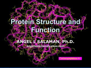
  • 1. Protein Structure andProtein Structure and FunctionFunction ANGEL L SALAMAN, Ph.D.ANGEL L SALAMAN, Ph.D. angelsalaman@yahoo.comangelsalaman@yahoo.com
  • 2. 1.1. Amino AcidsAmino Acids 2.2. Type of Amino AcidsType of Amino Acids 3.3. Primary StructurePrimary Structure 4.4. Secondary StructureSecondary Structure 5.5. Tertiary InteractionsTertiary Interactions 6.6. Quaternary structureQuaternary structure Presentation OutlinePresentation Outline
  • 3. Amino AcidsAmino Acids  Amino acids are the basic structural unit ofAmino acids are the basic structural unit of all proteins. A 'free' amino acid (a singleall proteins. A 'free' amino acid (a single amino acid) always has:amino acid) always has: • an amino group -NH2,an amino group -NH2, • a carboxyl group -COOHa carboxyl group -COOH • a hydrogen -Ha hydrogen -H • a chemical group or side chain -"R".a chemical group or side chain -"R".  These are all joined to a central carbonThese are all joined to a central carbon atom, the a -carbon.atom, the a -carbon.
  • 4. Since the amino acids (except glycine) have 4 different groups attached to the a-carbon, they are optically active stereoisomers although only L-isomers are found in proteins. Amino AcidsAmino Acids
  • 5. Amino AcidsAmino Acids  L-isomerL-isomer In proteins, only the L-In proteins, only the L- isomer is found normally.isomer is found normally.  As you travel onwardAs you travel onward (from the carbonyl carbon(from the carbonyl carbon to the amino group), theto the amino group), the R group of L-amino acidsR group of L-amino acids will be on the left aswill be on the left as shown in the molecularshown in the molecular graphic on the rightgraphic on the right
  • 6.  In the above diagrams, the "R" representsIn the above diagrams, the "R" represents "a chemical group" and gives each amino"a chemical group" and gives each amino acid its distinctive properties.acid its distinctive properties.  With amino acids, this chemical group isWith amino acids, this chemical group is called the "sidechain", and it specifies thecalled the "sidechain", and it specifies the amino acid. There are only 20 differentamino acid. There are only 20 different amino acids found in proteins, each withamino acids found in proteins, each with it's own defining sidechain. Theseit's own defining sidechain. These sidechains have a widespread range ofsidechains have a widespread range of chemical (e.g.charge etc) and structuralchemical (e.g.charge etc) and structural (e.g. shape etc) diversity.(e.g. shape etc) diversity. Types of Amino AcidsTypes of Amino Acids
  • 7. ALIPHATIC AMINO ACIDSALIPHATIC AMINO ACIDS Aliphatic amino acids are hydrophobic. They do not like to be in contact with water molecules in an aqueous solution. They are often located in the core of the protein, surrounded by the rest of the protein, and "shielded" by them from the aqueous surroundings. •Glycine (Gly; G): is the simplest and smallest of all amino acids, and the only one which is not optically active since it has a single hydrogen atom as it's sidechain •Alanine (Ala; A): has a methyl group as it's sidechain. •Valine (Val; V): has a slightly longer sidechain again, and this time there is a branch. As the aliphatic side chains get longer they are also more hydrophobic •Leucine (Leu; L): similar to Valine execept it has another methyl group. •Isoleucine (Ile; I): is again similar to leucine and valine except that the orientation of the atoms in the sidechain is slightly different. Isoleucine also has two centers of asymmetry. •Proline (Pro; P): the sidechain is bonded to both the a-carbon but also to the amino group. This has marked effects on the architecture of the proteins. And although aliphatic, it does not mind being in contact with water as much as the others.
  • 8. AROMATIC AMINO ACIDSAROMATIC AMINO ACIDS These are the amino acids which contain an aromatic ring as partThese are the amino acids which contain an aromatic ring as part of their sidechains. Because of the hydrophobic nature ofof their sidechains. Because of the hydrophobic nature of aromatic rings these amino acids are highly hydrophobicaromatic rings these amino acids are highly hydrophobic  PhenylalaninePhenylalanine (Phe; F)(Phe; F) is the first of all the aromatic amino acids. It contains a phenylis the first of all the aromatic amino acids. It contains a phenyl ring attached to a methylene group. Due to the phenyl ring it isring attached to a methylene group. Due to the phenyl ring it is a hydrophobic amino acid.a hydrophobic amino acid.  TyrosineTyrosine (Tyr; Y)(Tyr; Y) contains a hydroxyl group at the end of the phenyl ring. Thiscontains a hydroxyl group at the end of the phenyl ring. This makes tyrosine less hydrophobic than phenylalanine. It is alsomakes tyrosine less hydrophobic than phenylalanine. It is also a reactive group, whereas the sidechains so far have all beena reactive group, whereas the sidechains so far have all been unreactive.unreactive.  TryptophanTryptophan (Trp; W)(Trp; W) has a slightly different ring attached to the methylene group.has a slightly different ring attached to the methylene group. This is an indole ring and it is highly hydrophobic.This is an indole ring and it is highly hydrophobic.
  • 9. SULPHUR CONTAININGSULPHUR CONTAINING AMINO ACIDSAMINO ACIDS  There are two amino acids which contain a sulphurThere are two amino acids which contain a sulphur atom. They are both very special but for completelyatom. They are both very special but for completely different reasons.different reasons.  CysteineCysteine (Cys; C)(Cys; C) contains a sulphydryl group (-SH). This is extremelycontains a sulphydryl group (-SH). This is extremely reactive, and can form hydrogen bonds. Cysteine isreactive, and can form hydrogen bonds. Cysteine is very important because it can also form disulphidevery important because it can also form disulphide bridges. Even though the -SH group of cysteine canbridges. Even though the -SH group of cysteine can form hydrogen bonds the long aliphatic part of the sideform hydrogen bonds the long aliphatic part of the side chain makes it quite hydrophobic.chain makes it quite hydrophobic.  MethionineMethionine (Met; M)(Met; M) is a very special amino acid in that it is the "start"is a very special amino acid in that it is the "start" amino acid in the process of translation (proteinamino acid in the process of translation (protein synthesis), and therefore, begins every single proteinsynthesis), and therefore, begins every single protein made. It too has a sulphur atom, but this time it is in amade. It too has a sulphur atom, but this time it is in a thioether linkage, and is relatively unreactive.thioether linkage, and is relatively unreactive. Methionine has a highly hydrophobic sidechain.Methionine has a highly hydrophobic sidechain.
  • 10. HYDROPHILLIC AMINO ACIDSHYDROPHILLIC AMINO ACIDS  These amino acids can be split into aThese amino acids can be split into a several groups. There are thoseseveral groups. There are those which are neutral, those which arewhich are neutral, those which are acidic, and those which are basic.acidic, and those which are basic.
  • 11. ACIDIC AMINO ACIDSACIDIC AMINO ACIDS  These amino acids are highly polar, and are nearlyThese amino acids are highly polar, and are nearly always negatively charged at physiological pH.always negatively charged at physiological pH.  AspartateAspartate (Asp; D)(Asp; D) is really aspartic acid. It is called aspartate because it isis really aspartic acid. It is called aspartate because it is usually negatively charged at physiological pH and so itusually negatively charged at physiological pH and so it is named for the carboxylate anion. (Compare aceticis named for the carboxylate anion. (Compare acetic acid and acetate.).acid and acetate.).  GlutamateGlutamate (Glu; E)(Glu; E) is also called glutamic acid. The side chain of glutamateis also called glutamic acid. The side chain of glutamate also has a carboxylate group which has a negativealso has a carboxylate group which has a negative charge at physiological pHcharge at physiological pH
  • 12. NEUTRAL POLAR AMINO ACIDSNEUTRAL POLAR AMINO ACIDS These amino acids are not charged at physiological pH. However they all haveThese amino acids are not charged at physiological pH. However they all have groups on their side-chains which are polar and can form hydrogen bonds.groups on their side-chains which are polar and can form hydrogen bonds. For this reason the amino acids are classed as hydrophillic.For this reason the amino acids are classed as hydrophillic.  SerineSerine (Ser; S)(Ser; S) contains an aliphatic chain with a hydroxyl group. It looks like thecontains an aliphatic chain with a hydroxyl group. It looks like the hydroxylated version of alanine. The OH group make the amino acid highlyhydroxylated version of alanine. The OH group make the amino acid highly reactive and hydrophillic as it readily forms hydrogen bonds.reactive and hydrophillic as it readily forms hydrogen bonds.  ThreonineThreonine (Thr; T)(Thr; T) is another neutral amino acid which has a highly reactive (and highlyis another neutral amino acid which has a highly reactive (and highly hydrophillic) hydroxyl group. This is an odd amino acid in that it containshydrophillic) hydroxyl group. This is an odd amino acid in that it contains two centers of asymmetry (two asymetric carbon atoms). This property istwo centers of asymmetry (two asymetric carbon atoms). This property is shared only by isoleucine.shared only by isoleucine.  AsparagineAsparagine (Asn; N)(Asn; N) is the amide derivative of Aspartic acid. When the carboxylate sidechain isis the amide derivative of Aspartic acid. When the carboxylate sidechain is amidated the resulting amide is uncharged. There is a terminal amideamidated the resulting amide is uncharged. There is a terminal amide group as opposed to the carboxyl group on aspartate.group as opposed to the carboxyl group on aspartate.  GlutamineGlutamine (Gln; Q)(Gln; Q) is similar to Asparagine, with a terminal amide instead of a carboxyl groupis similar to Asparagine, with a terminal amide instead of a carboxyl group as in glutamate. These two are called the amide derivatives of their parentas in glutamate. These two are called the amide derivatives of their parent amino acids.amino acids.
  • 13.
  • 14. Primary StructurePrimary Structure  A protein has a completely defined order of aminoA protein has a completely defined order of amino acids, called itsacids, called its sequencesequence..  This defined sequence is called the primaryThis defined sequence is called the primary structure of the protein. The sequence, orstructure of the protein. The sequence, or primary structure is specified by the sequence ofprimary structure is specified by the sequence of the piece of DNA containing a gene for thatthe piece of DNA containing a gene for that protein, and the sequence is unique to theprotein, and the sequence is unique to the individual protein.individual protein.  All the information necessary to make a protein isAll the information necessary to make a protein is contained in the primary structure. However, ascontained in the primary structure. However, as we shall see soon, the primary structure is onlywe shall see soon, the primary structure is only the first level of structural complexity of proteins.the first level of structural complexity of proteins.
  • 15. Secondary structureSecondary structure  Generally proteins have a core of hydrophobicGenerally proteins have a core of hydrophobic residues surrounded by a shell of hydrophilicresidues surrounded by a shell of hydrophilic residues. Since the peptide bonds themselves areresidues. Since the peptide bonds themselves are polar they are neutralized by hydrogen bondingpolar they are neutralized by hydrogen bonding with each other when in the hydrophobicwith each other when in the hydrophobic environment. This gives rise to regions of theenvironment. This gives rise to regions of the polypeptide that form regular 3D structuralpolypeptide that form regular 3D structural patterns called 'patterns called 'secondary structuresecondary structure'. There are'. There are two main types of secondary structure:two main types of secondary structure:  α-helicesα-helices  β-sheetβ-sheet
  • 16. αα-Helices-Helices  The a-helix is a rod like structure. ItThe a-helix is a rod like structure. It is one peptide chain which has coiledis one peptide chain which has coiled so that adjacent residues are 1.5Aso that adjacent residues are 1.5A apart. There are H-bonds betweenapart. There are H-bonds between the N-H and the C=O groups of thethe N-H and the C=O groups of the backbone. These link the peptidebackbone. These link the peptide bonds between every fourth aminobonds between every fourth amino acid.acid.  The sidechains all extend outwardsThe sidechains all extend outwards from the helix.from the helix.
  • 17.  Although the helix itself is quite a rigid structure,Although the helix itself is quite a rigid structure, it can be curved or kinked, which lends someit can be curved or kinked, which lends some flexibility to the protein structure.flexibility to the protein structure.  Two or more polypeptides can entwine to formTwo or more polypeptides can entwine to form very long, very stable, structures called a-helicalvery long, very stable, structures called a-helical coiled coils. These are often found in keratincoiled coils. These are often found in keratin (hair), myosin (muscle), epidermin (skin), and(hair), myosin (muscle), epidermin (skin), and fibrin (blood clots). The coiled coils serve afibrin (blood clots). The coiled coils serve a mechanical role for the proteins mentionedmechanical role for the proteins mentioned above, which are all fibrous proteins.above, which are all fibrous proteins. αα-Helices-Helices
  • 19. ββ-Sheets-Sheets  This secondary structure differs considerably fromThis secondary structure differs considerably from the a-helix. However b-sheets are classified withthe a-helix. However b-sheets are classified with a-helices since they are a regular structurala-helices since they are a regular structural element and are held together by hydrogenelement and are held together by hydrogen bonds between the -N-H and C=O groups of thebonds between the -N-H and C=O groups of the peptide bond.peptide bond.  A polypeptide in a b-sheet is called a b-strand,A polypeptide in a b-sheet is called a b-strand, and is almost fully extended (not coiled) so thereand is almost fully extended (not coiled) so there is 3.5A between adjacent residues.is 3.5A between adjacent residues.  The b-sheet is stabilized by H-bonds between theThe b-sheet is stabilized by H-bonds between the N-H and the C=O groups of different peptides orN-H and the C=O groups of different peptides or different parts of the same peptide. The b-strandsdifferent parts of the same peptide. The b-strands in a sheet can either run in the same (parallel) orin a sheet can either run in the same (parallel) or different (anti-parallel) direction.different (anti-parallel) direction.
  • 21. MOTIFSMOTIFS  Some simple combinations ofSome simple combinations of secondary structuresecondary structure elements haveelements have been found to frequently occur inbeen found to frequently occur in protein structureprotein structure and areand are referred to as 'super-secondary structure' orreferred to as 'super-secondary structure' or motifsmotifs..  For example, the β-hairpin motif consists of two adjacentFor example, the β-hairpin motif consists of two adjacent antiparallel β-strands joined by a small loop. It is present in mostantiparallel β-strands joined by a small loop. It is present in most antiparallel β structures both as an isolated ribbon and as part ofantiparallel β structures both as an isolated ribbon and as part of more complex β-sheets. Another common super-secondarymore complex β-sheets. Another common super-secondary structure is the β-α-β motif, which is frequently used to connectstructure is the β-α-β motif, which is frequently used to connect two parallel β-strands.two parallel β-strands.  The central α-helix connects the C-termini of the first strand toThe central α-helix connects the C-termini of the first strand to the N-termini of the second strand, packing its side chains againstthe N-termini of the second strand, packing its side chains against the β-sheet and therefore shielding the hydrophobic residues ofthe β-sheet and therefore shielding the hydrophobic residues of the β-strands from the surface.the β-strands from the surface.
  • 22. DOMAINSDOMAINS  Several motifs pack together to form compact, local, semi-Several motifs pack together to form compact, local, semi- independent units calledindependent units called domainsdomains.. The overall 3DThe overall 3D structure of the polypeptide chain is referred to as thestructure of the polypeptide chain is referred to as the protein's 'protein's 'tertiary structuretertiary structure'.'.  Domains are the fundamental units of tertiary structureDomains are the fundamental units of tertiary structure,, each domain containing an individual hydrophobic core builteach domain containing an individual hydrophobic core built from secondary structural units connected by loop regions.from secondary structural units connected by loop regions.  The packing of the polypeptide is usually much tighter inThe packing of the polypeptide is usually much tighter in the interior than the exterior of the domain producing athe interior than the exterior of the domain producing a solid-like core and a fluid-like surface. In fact, core residuessolid-like core and a fluid-like surface. In fact, core residues are often conserved in a protein family, whereas theare often conserved in a protein family, whereas the residues in loops are less conserved, unless they areresidues in loops are less conserved, unless they are involved in the protein's function.involved in the protein's function.  Protein tertiary structure can be divided into four mainProtein tertiary structure can be divided into four main classes based on the secondary structural content of theclasses based on the secondary structural content of the domain.domain.
  • 23. DOMAINSDOMAINS  The concept of the domain was first proposed in 1973 byThe concept of the domain was first proposed in 1973 by Wetlaufer after X-ray crystallographic studies of henWetlaufer after X-ray crystallographic studies of hen lysozymelysozyme (Phillips, 1966), papain (Drenth et al., 1968) and(Phillips, 1966), papain (Drenth et al., 1968) and by limited proteolysis studies ofby limited proteolysis studies of immunoglobulinsimmunoglobulins (Porter,(Porter, 1973; Edelman, 1973). Wetlaufer defined domains as1973; Edelman, 1973). Wetlaufer defined domains as stable units ofstable units of protein structureprotein structure that could foldthat could fold autonomously.autonomously.  DefinitionDefinition:: a compact structural domain that is founda compact structural domain that is found amongst diverse proteins is likely to fold independentlyamongst diverse proteins is likely to fold independently within its structural environment.within its structural environment.  In a multidomain protein, each domain may fulfill its ownIn a multidomain protein, each domain may fulfill its own functionfunction independently, or in a concerted manner with itsindependently, or in a concerted manner with its neighbors. Domains can either serve as modules forneighbors. Domains can either serve as modules for building up large assemblies such as virus particles orbuilding up large assemblies such as virus particles or muscle fibers, or can provide specific catalytic or bindingmuscle fibers, or can provide specific catalytic or binding sites as found in enzymes or regulatory proteins.sites as found in enzymes or regulatory proteins.
  • 24. DOMAINS (Classes)DOMAINS (Classes)  AllAll α-domainsα-domains have a domain core built exclusively fromhave a domain core built exclusively from α-helices. This class is dominated by small folds, many ofα-helices. This class is dominated by small folds, many of which form a simple bundle with helices running up andwhich form a simple bundle with helices running up and down.down.  AllAll β-domainsβ-domains have a core comprising of antiparallel β-have a core comprising of antiparallel β- sheets, usually two sheets packed against each other.sheets, usually two sheets packed against each other. Various patterns can be identified in the arrangement ofVarious patterns can be identified in the arrangement of the strands, often giving rise to the identification ofthe strands, often giving rise to the identification of recurring motifs, for example the greek key motif.recurring motifs, for example the greek key motif.  α+β domainsα+β domains are a mixture of all-α and all-β motifs.are a mixture of all-α and all-β motifs. Classification of proteins into this class is difficult becauseClassification of proteins into this class is difficult because of overlaps to the other three classes and therefore is notof overlaps to the other three classes and therefore is not used in theused in the CATHCATH domain database.domain database.  α/β domainsα/β domains are made from a combination of β-α-βare made from a combination of β-α-β motifs that predominantly form a parallel β-sheetmotifs that predominantly form a parallel β-sheet surrounded by amphipathic α-helices. The secondarysurrounded by amphipathic α-helices. The secondary structures are arranged in layers or barrels.structures are arranged in layers or barrels.
  • 25. Pyruvate kinase •An appropriate example is pyruvate kinase, a glycolytic enzyme that plays an important role in regulating the flux from fructose-1,6-biphosphate to pyruvate. It contains an all-β regulatory domain, an α/β-substrate binding domain and an α/β-nucleotide binding domain, connected by several polypeptide linkers (George and Heringa, 2002). Each domain in this protein occurs in diverse sets of protein families. DOMAINSDOMAINS
  • 26. Ribbon diagram of the SH2 domain from P56-Lck tyrosine kinase The Src homology 2 domain (or SH2 domain) is a protein domain of about 100 amino acid residues first identified as a conserved sequence region among the oncoproteins Src and Fps. SH2 domains typically bind a phosphorylated tyrosine residue Phosphorylation of tyrosine residues in a protein occurs during signal transduction and is carried out by tyrosine kinases. In this way, phosphorylation of a substrate by tyrosine kinases acts as a switch to trigger binding to an SH2 domain-containing protein. Pawson, T., Gish, G., Nash, P. (2001). SH2 domains, interaction modules and cellular wiring. Trends in Cell Biology 11(12): 504-511 DOMAINSDOMAINS
  • 27. This image (courtesy of P. J. Bjorkman from Nature 329:506, 1987) is a schematic representation of the extracellular portion of HLA-A2, a human class I histocompatibility molecule. It also illustrates two common examples of secondary structure: the stretches of beta conformation are represented by the broad green arrows (pointing N -> C terminal); regions of alpha helix are shown as helical ribbons. The pairs of purple spheres represent the disulfide bridges.
  • 28. •The Structural Classification of Proteins: SCOP is to some a magical hierarchical classification of protein structures. As is simplified greatly in the figure on the right - SCOP classifies protein structures at many levels from overall class (all alpha helix, all beta strand etc.) down to protein domain and species level. •The SCOP tree takes the protein domain as the evolutionary unit and represents evolutionary relationships between proteins such that anything classified within a superfamily is evolutionarily related. •Whereas protein domains within the same class or fold are not necessarily evolutionarily related.
  • 29. Tertiary InteractionsTertiary Interactions  This is the term given to the overall three dimensionalThis is the term given to the overall three dimensional structure of the polypeptide, also written 3o structure. Thisstructure of the polypeptide, also written 3o structure. This describes how the peptide chain folds so that sidechains aredescribes how the peptide chain folds so that sidechains are packed and organized. This may bring two sidechains whichpacked and organized. This may bring two sidechains which are distant, in terms of primary structure, close together. Itare distant, in terms of primary structure, close together. It also creates binding sites and other functional regions onalso creates binding sites and other functional regions on the surface of the protein.the surface of the protein.  If we know the 3D structure of the protein, we can begin toIf we know the 3D structure of the protein, we can begin to understand how the protein works. If the protein comesunderstand how the protein works. If the protein comes from a bacterium or a virus, we can start to design drugsfrom a bacterium or a virus, we can start to design drugs which affect the bacterial or viral form of the protein, andwhich affect the bacterial or viral form of the protein, and not the host (eukaryotic) form. If an animal is making anot the host (eukaryotic) form. If an animal is making a defective protein (as is the case in cystic fibrosis patients)defective protein (as is the case in cystic fibrosis patients) then by using protein engineering and gene therapy, it maythen by using protein engineering and gene therapy, it may be possible to get the animal to start making the functionalbe possible to get the animal to start making the functional protein. All this leads to a new area of science calledprotein. All this leads to a new area of science called "Structural Biology"."Structural Biology".  There are 5 forces affecting the 3D shape of proteins. All ofThere are 5 forces affecting the 3D shape of proteins. All of them build upon the framework started by the secondarythem build upon the framework started by the secondary structures.structures.
  • 30. Hydrophobic InteractionsHydrophobic Interactions  Non-Polar/Hydrophobic/Aliphatic sidechains cannot hydrogenNon-Polar/Hydrophobic/Aliphatic sidechains cannot hydrogen bond with water, hence the network of water is disrupted bybond with water, hence the network of water is disrupted by them. This is avoided if the non-polar sidechains arethem. This is avoided if the non-polar sidechains are removed or shielded from the water and buried in theremoved or shielded from the water and buried in the interior of the protein. In this coiled coil structure note thatinterior of the protein. In this coiled coil structure note that the interior of the structure is occupied solely bythe interior of the structure is occupied solely by hydrophobic residues.hydrophobic residues.  Hydrophobic interaction are a major driving force in proteinHydrophobic interaction are a major driving force in protein folding. Hydrophobic interactions are also important in thefolding. Hydrophobic interactions are also important in the formation of secondary structure elements such as a-helicesformation of secondary structure elements such as a-helices and b-sheets. The peptide backbone is relatively hydrophillicand b-sheets. The peptide backbone is relatively hydrophillic because of the C=O and N-H groups of each peptide bond.because of the C=O and N-H groups of each peptide bond. However in the centre of the protein these tend to be in aHowever in the centre of the protein these tend to be in a hydrophobic environment. One way to reduce thehydrophobic environment. One way to reduce the hydrophilicity of the peptide backbone is to for hydrogenhydrophilicity of the peptide backbone is to for hydrogen bonds between the peptide atoms. The a-helix and b-sheetbonds between the peptide atoms. The a-helix and b-sheet are two structures which maximise the hydrogen bondingare two structures which maximise the hydrogen bonding between the peptide bonds of the backbone and reduce itsbetween the peptide bonds of the backbone and reduce its hydrophilicity.hydrophilicity.
  • 31. Hydrogen BondsHydrogen Bonds  When unfolded, all polar/hydrophillic sidechains canWhen unfolded, all polar/hydrophillic sidechains can interact via H-bonds with water. When the protein folds,interact via H-bonds with water. When the protein folds, they must H-bond to each other and exclude much of thethey must H-bond to each other and exclude much of the water. All groups capable of forming a hydrogen bondwater. All groups capable of forming a hydrogen bond MUST, hence H-bonding in the backbone (C=O to N-H) byMUST, hence H-bonding in the backbone (C=O to N-H) by way of helices and sheets is an efficient way of ensuringway of helices and sheets is an efficient way of ensuring maximum H-bonding.maximum H-bonding.  Sidechains can either accept (as in C=O) or donate (as inSidechains can either accept (as in C=O) or donate (as in N-H, or O-H) an H-bond.N-H, or O-H) an H-bond.  The capacity of proteins to form hydrogen bonds is anThe capacity of proteins to form hydrogen bonds is an important determinant of protein stability. Hydrogen bondsimportant determinant of protein stability. Hydrogen bonds can be between backbone groups, as in helices and sheets;can be between backbone groups, as in helices and sheets; between side chains, such as serine or threonine O-Hbetween side chains, such as serine or threonine O-H groups and carbonyl carbons of side chains (-C=O); andgroups and carbonyl carbons of side chains (-C=O); and between backbone groups and side chain groups.between backbone groups and side chain groups.
  • 32. Ion PairsIon Pairs  When amino acid sidechains of opposite charge are in closeWhen amino acid sidechains of opposite charge are in close proximity, they can form an ion pair (also called a saltproximity, they can form an ion pair (also called a salt bridge). But they are also capable of hydrogen bonding andbridge). But they are also capable of hydrogen bonding and hence, are usually found on the surface of the protein. Ifhence, are usually found on the surface of the protein. If they can form a salt bridge, they will usually be buried.they can form a salt bridge, they will usually be buried. Since charge is affected by pH, so is the formation and theSince charge is affected by pH, so is the formation and the breakage of these ion pairs.breakage of these ion pairs.  Salt bridges also increase the stability of the tertiarySalt bridges also increase the stability of the tertiary structure.structure. NOTE:NOTE:Hydrophobic interactions, hydrogen bonds and saltHydrophobic interactions, hydrogen bonds and salt bridges are all non-covalent interactionsbridges are all non-covalent interactions  These are all relatively weak interactions but the largeThese are all relatively weak interactions but the large number in a protein combine to give the overall stability ofnumber in a protein combine to give the overall stability of the structure.the structure.
  • 33. Disulphide BondsDisulphide Bonds  If two cysteine sidechains are close to one another and the localIf two cysteine sidechains are close to one another and the local environment is conducive to oxidation,then they can form aenvironment is conducive to oxidation,then they can form a disulphide bond/bridge as shown. The residues can either be indisulphide bond/bridge as shown. The residues can either be in the same peptide chain (forming a loop) or in different chains.the same peptide chain (forming a loop) or in different chains.  As Disulphide bonds are covalent linkages they add considerablyAs Disulphide bonds are covalent linkages they add considerably to the stability of proteins in which they occur. However theyto the stability of proteins in which they occur. However they mainly occur in secreted proteins and in the parts of membranemainly occur in secreted proteins and in the parts of membrane proteins which face the outside.proteins which face the outside. Note that disuphide bridges only form after the protein has foldedNote that disuphide bridges only form after the protein has folded into its tertiary structure. This folding is driven by the combinedinto its tertiary structure. This folding is driven by the combined energy of all of the weak interactions described in the previousenergy of all of the weak interactions described in the previous section above.section above. Cysteine side chains in cytosolic proteins are usually found as -SHCysteine side chains in cytosolic proteins are usually found as -SH groups as the cytosolic environmemt is sufficiently reducing togroups as the cytosolic environmemt is sufficiently reducing to prevent disulphides from forming.prevent disulphides from forming.
  • 34. Non-Covalent BondsNon-Covalent Bonds  The tertiary structure of proteins is maintained byThe tertiary structure of proteins is maintained by a host of weak, non-covalent interactions:a host of weak, non-covalent interactions:  Hydrophobic InteractionsHydrophobic Interactions  Hydrogen BondsHydrogen Bonds  Salt BridgesSalt Bridges  Protein stability is a function of many weakProtein stability is a function of many weak interactions. If these interactions are maintainedinteractions. If these interactions are maintained the protein keeps its tertiary ("native") stucture.the protein keeps its tertiary ("native") stucture. However once the bonds maintaining theHowever once the bonds maintaining the structure begin to be disrupted the tertiarystructure begin to be disrupted the tertiary structure is destroyed and the protein is said tostructure is destroyed and the protein is said to be "denatured"be "denatured"
  • 35. Subunits & Quaternary StructureSubunits & Quaternary Structure  Proteins containing more than one polypeptideProteins containing more than one polypeptide chain exhibit an additional level of structuralchain exhibit an additional level of structural organisation.organisation.  In this case, each polypeptide chain is aIn this case, each polypeptide chain is a "subunit". Quaternary (4o) structure describes"subunit". Quaternary (4o) structure describes the way the subunits are arranged together andthe way the subunits are arranged together and the nature of their contacts.the nature of their contacts.  Subunits associate by non-covalent interactionsSubunits associate by non-covalent interactions similar to those involved in tertiary structure:similar to those involved in tertiary structure:  Hydrophobic InteractionsHydrophobic Interactions  Hydrogen bondsHydrogen bonds  Salt Bridges / Ion PairsSalt Bridges / Ion Pairs
  • 36. DENATURATION loss of 3-D conformation by heat, pH, organic solvents, detergents RENATURATION - regaining of biological activity via self- assembly
  • 37. Factors Which Can Denature AFactors Which Can Denature A ProteinProtein  Heat: increase atomic vibrations, ruinsHeat: increase atomic vibrations, ruins interactions, need heat of >50interactions, need heat of >50oo C to denatureC to denature most proteins.most proteins.  pH: breaks the salt bridges.pH: breaks the salt bridges.  Organic Solvents: disrupt hydrophobicOrganic Solvents: disrupt hydrophobic interactionsinteractions  Detergents: hydrophobic part pushes into proteinDetergents: hydrophobic part pushes into protein corecore  When a protein is denatured, it loses it's 3DWhen a protein is denatured, it loses it's 3D shape. When a protein is denatured, it loses it'sshape. When a protein is denatured, it loses it's biological activity. Therefore...biological activity. Therefore... A PROTEINS TERTIARY STRUCTURE ISA PROTEINS TERTIARY STRUCTURE IS ESSENTIAL TO IT'S BIOLOGICAL ACTIVITYESSENTIAL TO IT'S BIOLOGICAL ACTIVITY
  • 38. REFERENCES  Introduction to Protein Structure, C.Branden and J.Tooze, Garland, New York (1991).  A novel super-secondary structure of proteins and the relation between the structure and amino acid sequence, A.F.Efimov, FEBS Lett. 166, 33-38 (1984).  Structure of alpha-alpha-hairpins with short connections, A.V.Efimov, Protein Engin.4, 245-250 (1991).  Structure of beta-beta-hairpins and beta-beta-corners, A.V.Efimov, FEBS Lett., 284, 288-292 (1991).  Structure and evolution of calcium modulated proteins, R.H.Kretsinger, CRC Crit.Rev.Biochem. 8, 119-174 (1980).  Comparison of the structures of cro and lambda repressor proteins in bacteriophage lambda, D.H.Ohlendorf, W.F.Anderson, M.Lewis, C.O.Pabo and B.W.Matthews, J.Molec.Biol. 169 757-769 (1983).  Conformation of beta-hairpins in protein structures, B.L.Sibanda, T.L.Blundell and J.M.Thornton, J.Molec.Biol. 206, 759-777 (1989).  Beta-hairpin families in globular proteins, B.L.Sibanda and J.M.Thornton, Nature 316, 170-174 (1985).