Inflammation....
•Télécharger en tant que PPTX, PDF•
59 j'aime•2,715 vues

INTRODUCTION HISTORY CAUSES OF INFLAMMATION CLASSIFICATION ACUTE INFLAMMATION CHEMICAL MEDIATORS OF INFLAMMATION OUTCOMES OF ACUTE INFLAMMATION CHRONIC INFLAMMATION INFLAMMATORY DISEASES REFERENCES

Recommandé
Contenu connexe
Tendances
Tendances (20)
Similaire à Inflammation....
Similaire à Inflammation.... (20)
Plus de ANIL KUMAR
Plus de ANIL KUMAR (11)
Dernier
Dernier (20)
Inflammation....
- 1. INFLAMMATION
- 2. CONTENTS INTRODUCTION HISTORY CAUSES OF INFLAMMATION CLASSIFICATION ACUTE INFLAMMATION CHEMICAL MEDIATORS OF INFLAMMATION OUTCOMES OF ACUTE INFLAMMATION CHRONIC INFLAMMATION INFLAMMATORY DISEASES REFERENCES
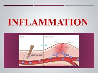
- 3. INTRODUCTION Inflammation is defined as a protective response involving host cells, blood vessels, and proteins and other mediators that is intended to eliminate the initial cause of cell injury, as well as the necrotic cells and tissues resulting from the original insult, and to initiate the process of repair (ROBBINS). Term inflammation is derived from Latin word ‘inflammare’ = ‘to burn’. Our Principal safeguard against injury Fundamentally designed to localize and eliminate the causative agents & limit tissue injury. Majority of lesions are named by adding suffix – itis= belonging to.
- 4. HISTORY Egyptians first described in 3000 B.C Celsus (1st century AD) − 4 cardinal signs of inflammation Virchow- fifth sign functio laesa (loss of function) John Hunter (1973) − inflammation is not a disease but a nonspecific response that has a salutary effect on its host. Julius Conheim (1839 to 1884) described the process of inflammation. In 1924 Lewis explained role of chemical mediators in inflammation.
- 5. CAUSES OF INFLAMMATION Physical agents - mechanical injuries, alteration in temperatures and pressure, radiation injuries. Chemical agents- including the ever increasing lists of drugs and toxins. Biologic agents (infectious)-bacteria,viruses,fungi, parasites Immunologic disorders- hypersensitivity reactions, autoimmunity,immunodeficiency states etc Genetic/metabolic disorders- examples gout, diabetes mellitus etc.
- 6. HOW IS INFLAMMATION DIFFERENT FROM INFECTION? Infection is invasion into the body by harmful microbes and their resultant ill effects by toxins. Protective mechanism of body against various etiological agents.
- 7. CLASSIFICATION Based on duration of the lesion and histologic appearances CHRONIC ACTIVE INFLAMMATION is the type of chronic inflammation in which during the course of disease there are acute exacerbations of activity.
- 8. Steps of Inflammation 5R’s 1. Recognition of injurious agent 2. Recruitment of leukocytes 3. Removal of agent 4. Regulation of the response 5. Resolution SEQUENCE OF EVENTS
- 9. ACUTE INFLAMMATION Acute inflammatory response by the host to any agent is a continuous process involving ACUTE INFLAMMATION VASCULAR EVENTS HAEMODYNAMIC CHANGES ALTERED VASCULAR PERMEABILITY CELLULAR EVENTS EXUDATION OF LEUKOCYTES PHAGOCYTOSIS
- 10. I.VASCULAR EVENTS A. HAEMODYNAMIC CHANGES: 1. TRANSIENT VASOCONSTRICTION 2. VASODILATATION (arterioles, venules and capillaries) obvious within half an hour of injury Increase blood volume in microvascular bed Redness and warmth 3. Elevation of HYDROSTATIC PRESSURE Results in transudation of fluid in the extracellular space swelling
- 11. Increased vascular permeability Increased concentration of RBCs Raised blood viscosity Slower blood flow slowing followed by 5.LEUCOYTE MARGINATION (neutrophils mainly) to the vascular endothelium Leukocytes then move and migrate through gaps between the endothelial cells in the extravascular space. 4. Slowing or stasis
- 12. LEWIS EXPERIMENT: Lewis induced the changes in the skin of the inner aspect of forearm by firm stroking with a blunt point. The reaction so elicited is known as TRIPLE RESPONSE or RED LINE RESPONSE consisting of : RED LINE- local vasodilatation of capillaries and venules FLARE - vasodilatation of adjacent arterioles WHEAL - transudation of fluid into the extravascular space
- 13. B. ALTERED VASCULAR PERMEABILITY In normal circumstances fluid balance is maintained by 2 opposing set of forces: 1. Forces that cause OUTWARD MOVEMENT of fluid from microcirculation are intravascular hydrostatic pressure and osmotic pressure of interstitial fluid. 2. Forces that cause INWARD MOVEMENT of interstitial fluid into circulation are intravascular osmotic pressure and hydrostatic pressure of interstitial fluid. STARLING’S HYPOTHESIS
- 14. Normally whatever little fluid is left in the interstitial compartment is drained by the lymphatics and thus no oedema results. In inflamed tissues, the endothelial lining becomes leaky. Consequently, the intravascular osmotic pressure decreases and osmotic pressure of the interstitial fluid increases resulting in excessive outward flow of fluid into the interstitial compartment. EXUDATIVE INFLAMMATORY OEDEMA
- 15. MECHANISMS OF INCREASED VASCULAR PERMEABILITY
- 16. A. EXUDATION OF LEUCOCYTES Most important feature of inflammatory response. The escape of leucocytes from lumen of microvasculature to the interstitial tissue. In acute inflammation, polymorphonuclear neutrophils comprise the first line of defense, followed later by the monocytes and macrophages. II.CELLULAR EVENTS CHANGES LEADING TO MIGRATION 1. CHANGES IN THE FORMED ELEMENTS OF BLOOD The normal axial flow consists of central stream of cells comprised by leucocytes and RBCs and peripheral cell-free layer of plasma close to vessel wall
- 17. VASODILATATION subsequently, SLOWING of BLOOD STREAM The central stream of cells widens and peripheral plasma zone becomes narrower because of loss of plasma by exudation. MARGINATION The neutrophils of the central column come close to the vessel wall PAVEMENTING
- 18. 2. ROLLING AND ADHESION: Peripherally marginated and pavemented neutrophils slowly roll over the endothelial cells lining the vessel wall. ROLLING PHASE Transient bond between the leucocytes and the endothelial cells becoming firmer. ADHESION PHASE
- 19. SELECTINS : E-selectin (cytokine-activated Endothelial cells) P-selectin (Preformed and stored in endothelial cells) L-selectin (expressed on surface of Lymphocytes and neutrophils) INTEGRINS: Activated during the process of loose and transient adhesions between the endothelial cells and leucocytes. IMMUNOGLOBULIN GENE SUPER FAMILY ADHESION MOLECULE: ICAM-1,VCAM-1. ADHESION MOLECULES:
- 20. 3. EMIGRATION Neutrophils move till a suitable site is reached CYTOPLASMIC PSEUDOPODS Subsequently, crosses the basement membrane by damaging it locally with secreted collagenases and escape out into the extravascular space. EMIGRATION
- 21. Simultaneously escape of RBCs takes place through the gaps between the endothelial cells. DIAPEDESIS Diapedesis gives Hemorrhagic appearance to the inflammatory exudate. DIAPEDESIS
- 22. The chemotactic factor mediated transmigration of leucocytes after crossing several barriers to reach the interstitial tissues is called CHEMOTAXIS. Well illustrated by BOYDEN’S CHAMBER EXPERIMENT. In this, a millipore filter separates the suspension of leucocytes from the test solution in tissue culture chamber. If the test solution contains chemotactic agent, the leucocytes migrate through the pores of filter towards the chemotactic agent. 4. CHEMOTAXIS
- 23. The following agents act as potent chemotactic substances for neutrophils: i) Leukotriene B4 (LT-B4). ii) Components of complement system (C5a and C3a in particular). iii) Cytokines (Interleukins, in particular IL-8) iv) Soluble bacterial products (such as formylated peptides).
- 24. B. PHAGOCYTOSIS ENGULFMENT of solid particulate material by the cells (cell eating). The cells performing this function are PHAGOCYTES. There are two main types of cells: 1. PMNs which appear early in acute inflammatory response and are called as MICROPHAGES. 2. Circulating monocytes and fixed tissue mononuclear phagocytes called as MACROPHAGES. The process of phagocytosis is same for both polymorphs and macrophages and involves 3 steps:
- 25. RECOGNITION AND ATTACHMENT OF PARTICLE ENGULFMENT WITH FORMATION OF PHAGOCYTIC VESICLE KILLING & DEGRADATION STAGE
- 26. CHEMICAL MEDIATORS OF INFLAMMATION Also called as PERMEABILITY factors or ENDOGENOUS factors. The substances acting as chemical mediators of inflammation may be released from the cells, the plasma, or damaged tissue itself.
- 27. VASOACTIVE AMINES HISTAMINE SOURCE : 1) Mast cells in C.T adjacent to blood vessels 2) Blood basophils 3) Platelets STIMULI : Injury Immune reactions Fragments of complement -C3a , C5a Neuropeptides such as substance P Cytokines IL – 1 , 8 SEROTONIN SOURCE : 1) Platelets 2) Enterochromaffin cells 3) nervous tissue 4) mast cells STIMULI : Platelet aggregation after contact with collagen , thrombin Action: vasodilatation, increased vascular permeability, itching and pain
- 28. ARACHIDONIC ACID METABOLITES A fatty acid with 2 main sources : Directly through diet Through conversion of essential fatty acid, linoleic acid to arachidonic acid ARACHIDONIC ACID ARACHIDONIC ACID METABOLITES Cyclo-oxygenase pathway Lipo-oxygenase Pathway CYCLO – OXY- GENASE PATHWAY • Prostaglandin - PGD2 , E2 , F2. • Thromboxane A2 • Prostacyclin LIPO – OXY – GENASE PATHWAY 5 – HETE Leukotrienes
- 30. LYSOSOMAL COMPONENTS GRANULES OF NEUTROPHILS: PRIMARY • MPO • ACID HYDROLASES • ACID PHOSPHATASE • PHOSPHOLIPASE • PROTEASES SECONDARY • COLLAGENASE • ALKALINE PHOSPHATASE • LACTOFERRIN • LYSOZYME GRANULES OF MONOCYTES AND TISSUE MACROPHAGES: ACID PROTEASE COLLAGENASE ELASTASE PLASMINOGEN ACTIVATOR
- 31. First described in early 1970’s by Jacques Benvinste Source: Platelets Basophils Mast cells Neutrophils Macrophages Endothelial Cells Platelet aggregation Vasoconstriction and Bronchoconstriction At extreme low concentration cause vasodilation, increased vescular permeability Increased leukocyte adhesion Chemotaxis Platelet Activating Factor Functions:
- 32. CYTOKINES/CHEMOKINES proteins produced mainly by- activated lymphocytes & macrophages , also from endothelium, epithelium & connective tissue cells. Molecularly defined cytokines are called Interleukins Produced rapidly in response to noxious stimuli as part of innate immunity Includes IL-1, IL-6, IL-8, Tumour necrosis factor (TNF- a nd b), Interferon (IFN)-gamma Other chemokines (IL-8, MCP-1, eotaxin, PF-4)
- 33. CYTOKINE CELL SOURCE MAIN ACTION IL-1 Monocytes/macrophages. • Emigration of neutrophils and macrophages • Role in fever and shock IL-6 Monocytes/macrophages. • Hepatic production of acute phase protein • Differentiation and growth of T and B cells IL-8 Monocytes/macrophages, T cells, neutrophils. • Induces migration of neutrophils, macrophages and T cells • Stimulates release of histamine from basophils • Stimulates angiogenesis TNF-α Monocytes/macrophages, mast cells/ basophils. • Hepatic production of acute phase protein • Systemic features (fever, shock, anorexia) • Enhanced leucocyte cytotoxicity • Induction of pro-inflammatory cytokines IFN- g T cells, NK cells All cells • Activation of macrophages and NK cells • Stimulates secretion of Igs by B cells • Differentiation of T helper cells MCP-1 Fibroblasts, smooth muscle cells. • Chemoattractant for monocytes, T cells and NK cells • Stimulates release of histamine from basophils PF-4 Platelets, megakaryocytes Fibroblasts, endothelial cells • Chemoattractant for fibroblasts • Inhibitory to haematopoietic precursors and endothelial cell proliferation Major cytokines in inflammation
- 34. Chemokines: Chemokines are a family of small proteins that act primarily as chemoattractants for specific type of leukocytes. GROUP TYPE OF CHEMOKINE FUNCTION C – X – C IL-8 Activation and chemotaxis of Neutrophils C – C MCP-1 Eotaxin MIP-1 RANTES Attracts Monocytes, eosinophil, basophils and lymphocytes but NOT Neutrophils C Lympholactin Specific for lymphocytes C – X3 – C Fractalkine Chemoattractant for monocytes and T cells
- 35. OXYGEN METABOLITES Released by activated neutrophils and macrophages. Superoxide oxygen, hydrogen peroxide , hydroxl ion. ACTION : Endothelial cell damage and thereby increasing vascular permeability Damage to cells and tissue matrix by activating protease and inactivating anti protease
- 36. NITRIC OXIDE is formed by activated macrophages during the oxidation of arginine by the action of enzyme, NO synthase. NO plays the following roles in mediating inflammation: a) mediates in vascular dilation b) Anti-platelet activating agent c) Possibly microbicidal action
- 37. PLASMA DERIVED MEDIATORS (factor XII) KININ SYSTEM COMPLEMEN T SYSTEM CLOTTING SYSTEM FIBRINOLYTI C SYSTEM
- 38. 1. THE KININ SYSTEM Vasoactive peptides derived from plasma proteins called kininogens by action of proteases called kallikreins. Braykinin increases vascular permeability; contraction of smooth muscle; dilation of blood vessels; pain when injected into skin
- 39. 2.THE CLOTTING SYSTEM The actions of fibrinopeptides in inflammation are: increased vascular permeability chemotaxis for leucocyte anticoagulant activity.
- 40. 3.FIBRINOLYTIC SYSTEM Kallikrein along with plasminogen activator converts plasminogen to plasmin. Plasmin acts on fibrin & forms fibrin split products. Plasmin also activates C3 to C3a
- 41. Complement activation elaborates several cleavage products which mediate in inflammatory response Critical step in activation is the proteolysis of C3. This can occur in 3 pathways: Classic pathway Alternate pathway Lectin pathway 4.Complement System
- 42. Role of Mediators in Different Reactions of Inflammation
- 43. OUTCOMES OF ACUTE INFLAMMATION Resolution, Healing by scarring (fibrosis), Chronic inflammation
- 44. CHRONIC INFLAMMATION Definition: Chronic inflammation can be defined as a prolonged inflammatory process(weeks or months) where an active inflammation, tissue destruction and attempts at repair are proceeding simultaneously. CAUSES: 1.Persistent infections Certain microorganisms associated with intracellular infection such as tuberculosis, leprosy, certain fungi etc. 2.Prolonged exposure to nondegradable but partially toxic substances either endogenous lipid components which result in atherosclerosis or exogenous substances such as silica, asbestos.
- 45. 3. Progression from acute inflammation: Acute inflammation almost always progresses to chronic inflammation 4.Autoimmunity: Autoimmune diseases such as rheumatoid arthritis and systemic lupus erythematosis are chronic inflammations from the outset.
- 46. Chronic inflammation is of two types Specific - when the injurious agent causes a characteristic histologic tissue response e.g. tuberculosis, leprosy, syphilis Non-specific - when the irritant substance produces a nonspecific chronic inflammatory reaction with formation of granulation tissue and healing by fibrosis e.g. chronic osteomyelitis, chronic ulcer.
- 47. CELLS OF CHRONIC INFLAMMATION Monocytes and Macrophages are the primary cells. These cells constitute the mononuclear- phagocytic system. Macrophages are scavenger cells of the body. T-Lymphocytes are primarily involved in cellular immunity B-lymphocytes and Plasma cells produce antibodies. Mast cells and eosinophils appear predominantly in response to parasitic infestations & allergic reactions.
- 48. FEATURES OF CHRONIC INFLAMMATION 1.Mononuclear cell infiltration – phagocytes and lymphoid cells macrophages- most important cells 2. Tissue destruction or necrosis proteases, elastases, cytokines etc released by activated macrophages 3. Proliferative changes- blood vessels and fibroblasts proliferate forming granulation tissue. thereby healing by fibrosis and collagen laying.
- 49. GRANULOMATOUS INFLAMMATION A granuloma is basically a collection of epithelioid cells, it also usually contains multinucleated giant cell & is usually surrounded by a cuff of lymphocytes and occasional plasma cells. The word ‘granuloma’ is derived from granule meaning circumscribed granule-like lesion, and -oma which is a suffix commonly used for true tumours.
- 51. INFLAMMATORY DISEASES Gingivitis Periodontitis Osteomyelitis Cellulitis Pericoronitis Sialadenitis Glossitis.
- 52. GINGIVITIS Inflammation of gingiva is termed as gingivitis. Bleeding on probing – earliest sign of gingivitis Gingivitis is classified into four stages 1. Initial lesion 2. Early lesion 3. Established lesion 4.Advanced lesion
- 54. CLINICAL FEATURES Bleeding on probing is present Color : red to bluish red Consistency : soft, friable Texture : loss of stippling is seen Size : Swollen or ballooning of interdental papilla and/or gingival margin. Shape and contour : Blunts the marginal and papillary tissues
- 55. Elimination of the etiologic factors: plaque, calculus by scaling and root planning Correction of plaque retentive factors such as over contoured crowns, overhanging margins, narrow embrasure spaces, open contacts, ill fitting fixed or removable partial dentures, tooth malposition. MANAGEMENT
- 56. PERIODONTITIS It is “an inflammatory d/s of supporting tissues of teeth caused by specific microorganisams,resulting in progressive destruction of periodontal ligament &alveolar bone with pocket formation,recession or both”. Onset - any age; most common in adults Plaque initiates condition Subgingival calculus common finding Slow-moderate progression; periods of rapid progression possible Modified by local factors/systemic factors/stress/smoking
- 57. Extent: • Localized: <30% of sites affected • Generalized: > 30% of sites affected Severity: entire dentition or individual teeth/site • Slight = 1-2 mm CAL(clinical attachment loss) • Moderate = 3-4 mm CAL • Severe = 5 mm CAL
- 58. Gingiva moderately swollen Deep red to bluish-red tissues Blunted and rolled gingival margin Bleeding and/or suppuration Plaque/calculus deposits Variable pocket depths Loss of periodontal attachment Horizontal/vertical bone loss Tooth mobility CLINICAL FEATURES
- 59. The treatment consists of – 1. Non-surgical procedures • Scaling • Root planing • Curettage 2. Surgical procedure • Pocket reduction surgery • Resective • Regenerative • Correction of morphological / anatomic defects MANAGEMENT
- 60. OSTEOMYELITIS Osteomyelitis may be defined as “an inflammatory condition of bone, that begins as an infection of medullary cavity and haversian systems of the cortex & extends to involve the periosteum of the affected area.” PREDISPOSING FACTORS • Fractures due to trauma and road traffic accidents • Gun shot wounds • Radiation damage • Paget`s disease • Osteoporosis • Systemic disease : Malnutrition, Acute Leukemia, Uncontrolled diabetes, sickle cell anemia, Chronic alcoholism
- 62. It is a diffuse inflammation of soft tissues which tends to spread through tissue spaces and along the facial planes Streptococci – hyaluronidase – destroys hyaluronic acid Prevotella and porphyromonas species destroy collagen It occurs as a sequela of an apical abscess or osteomyelitis or periodontal infection. Other causes : infection following tooth extraction, injection(infected needle or through infected area) or following jaw fracture. CELLULITIS
- 63. Patient is moderately ill, elevated temperature and leucocytosis Painful swelling of the soft tissues involved that are firm and brawny. Involvement of superficial tissue spaces results in inflamed skin with orange peel appearance. Regional lymphadenitis CLINICAL FEATURES
- 64. Maxilla – perforates outer cortical layer of bone above buccinator attachment – swelling of upper half of the face. Extension towards eye may result in cavernous sinus thrombosis Mandible – perforates outer cortical layer of bone below buccinator attachment – swelling of lower half of the face. Extension to cervical area results in respiratory discomfort
- 65. Administration of antibiotics including anti anaerobics Removal of cause of infection Patient is advised not to massage the area with any medication Intravenous fluids to correct the dehydration. If it does not respond to conservative treatment, surgical intervention is recommended. TREATMENT
- 66. Pericoronitis is inflammation of the soft tissue associated with the crown of a partially erupted tooth. Most commonly seen in relation to the mandibular third molar. Common symptoms and signs are pain, swelling, trismus, halitosis, bad taste, inflammation of pericoronal flap and pus discharge from underneath it. PERICORONITIS
- 67. Most common cause - entrapment of plaque and food debris between crown of tooth and overlying gingival flap or operculum. This is an ideal area for the growth of bacteria and It is difficult to keep clean. Acute pericoronitis is characterized by a red, swollen, suppurating lesion which is tender, with severe throbbing pain radiating to the ear, throat, floor of the mouth, temporomandibular joint and posterior submandibular region.
- 68. Patient also complains of pain during swallowing (dysphagia), halitosis, a foul taste and an inability to close the jaws. Chronic pericoronitis is characterized by a dull pain with mild discomfort lasts for a day or two, with remission lasting for many months. Patient may also complain of a bad taste.
- 69. Acute phase - debridement of plaque and food debris, drainage of pus, irrigation with sterile saline, chlorhexidine or hydrogen peroxide, elimination of occlusal trauma and prophylactic antibiotic along with analgesics. Surgical intervention will be made after acute phase subsided. An extraction of partially or completely impacted third molar should be done or operculectomy. TREATMENT
- 70. Sialadenitis is the inflammation of the salivary glands Causes range from simple infection to autoimmune etiologies. Sialadenitis is usually caused by bacterial or viral infection Other causes: Trauma Radiation and Allergic reaction SIALADENITIS
- 71. CONCLUSION Without inflammation, infections would go unchecked. wounds would never heal, and injured organs may remain as permanent decaying lesions. In our day to day live we come across many cases starting from gingivitis to oral cancer wherein inflammation exerts a direct or indirect effect. So understanding inflammation helps us to know the various vascular and cellular changes, mediators involved and therefore help us to evaluate the significance of various antibiotics and anti-inflammatory drugs that we do prescribe, for controlling the same.
- 72. 1. Harsh mohan Textbook of pathology. 6th edition. Jaypee publication. 2. Kumar, abbas, aster Robbin’s textbook of pathology. 9th edition. Elsevier publication. 3. Rajendran R, Sivapathasundharam B Shafer’s textbook of oral pathology. 5th edition. Elsevier publication. 4. Newmann, Takei, Carranza textbook of clinical periodontology. 9th edition. W.B. saunders company publication. 5. Color Atlas of Pathology Pathologic Principles · Associated DiseaseUrsus-Nikolaus Riedes. 6. Fundamentals of Inflammation Charles N. Serhan. REFERENCES