retina.ppt
•Télécharger en tant que PPT, PDF•
0 j'aime•174 vues

The document discusses the anatomy and layers of the retina. It notes that the retina has 10 layers and extends from the optic disc to the ora serrata. The posterior pole, which includes the optic disc and macula lutea, contains the thickest areas of the retina. The 10 layers of the retina are described in detail, including the retinal pigment epithelium, rods and cones layer, and the various nuclear, plexiform, ganglion and nerve fiber layers. Specific structures like the fovea centralis and its avascular zone are also explained.

Recommandé
Contenu connexe
Tendances
Tendances (20)
Similaire à retina.ppt
Similaire à retina.ppt (20)
Plus de Al-Shifa College of Paramedical Science,Perinthalmanna
Plus de Al-Shifa College of Paramedical Science,Perinthalmanna (20)
Dernier
Dernier (20)
retina.ppt
- 1. OPTOM ASKAR.PK Mr. Askar.P. K, HOD & Associate Professor Of Optometry
- 2. OPTOM ASKAR.PK ANATOMY • Inner most layer • Thin, delicate & transparent membrane • Appears purplish red due to underlying visual purple of the rod & vascular choroid
- 3. OPTOM ASKAR.PK • After death –white opaque • Retina extends from optic disc to ora serrata • Grossly divided in to two • Posterior pole & peripheral retina
- 4. OPTOM ASKAR.PK • Posterior pole & peripheral retina separated by retinal equator • Retinal equator is the imaginary line
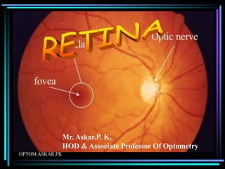
- 5. OPTOM ASKAR.PK POSTERIOR POLE • Posterior to the retinal equator • It includes two areas optic disc & macula lutea • It is best examined by slit lamp indirect biomicroscopy & direct ophthalmoscopy
- 6. OPTOM ASKAR.PK • Thickness –posterior pole: • Periphery- .56mm • Equator- .18- .2mm • Orra serrata- .1mm
- 7. OPTOM ASKAR.PK OPTIC DISC - It is pink coloured - Well defined circular area of 1.5mm in dia
- 8. OPTOM ASKAR.PK - All retinal layers terminate except nerve fibres - Nerve fibres pass through lamina cribrosa in to optic nerve
- 9. OPTOM ASKAR.PK • Compare to retina appears white opaque due to lamina cribrosa • A depression seen in the disc called physiological cup • Central retinal artery & vein emerge through the centre of this cup
- 10. OPTOM ASKAR.PK MACULA LUTEA • It is also called yellow spot
- 11. OPTOM ASKAR.PK • It is deeper red • Situated in posterior pole temporal to optic disc • It is 5.5mm in dia
- 12. OPTOM ASKAR.PK • Fovea centralis is the central depressed part of the macula • Fovea centralis 1.85mm in dia • Most sensitive part of the retina
- 13. OPTOM ASKAR.PK • In its centre shinning pit called foveola • Foveola dia 0.35mm • Not contain any retinal capillaries
- 14. OPTOM ASKAR.PK • Also called foveal avascular zone • Surrounding fovea are parafoveal & perifoveal areas • It is 0.5mm & 1.5mm in dia resp.
- 15. OPTOM ASKAR.PK FOVEA CENTRALIS • No rods , cones are packed tightly
- 16. OPTOM ASKAR.PK • Foveola largely consists of cones & their nuclei covered by thin internal limiting membrane • Other retinal layers absent in this region • Cone axons arranged obliquely henle’s layer to reach the margin of the fovea
- 17. OPTOM ASKAR.PK • PERIPHERAL RETINA • Area bounded posteriorly by retinal equator & anteriorly by ora serrata
- 18. OPTOM ASKAR.PK • Best examined by indirect ophthalmoscopy
- 19. OPTOM ASKAR.PK • ORA SERRATA • It is the peripheral margin where retina ends & ciliary body start • Here retina attached to vitreous & choroid • Pars plana extends anteriorly from ora
- 20. OPTOM ASKAR.PK
- 21. OPTOM ASKAR.PK
- 22. OPTOM ASKAR.PK • 1. pigment epithelium • 2.Layer of rods & cones • 3.external limiting membrane • 4.outer nuclear layer • 5.outer plexiform layer
- 23. OPTOM ASKAR.PK • 6.inner nuclear layer • 7.inner plexiform layer • 8.ganglion cell layer • 9.nerve fibre layer • 10.internal limiting membrane
- 24. OPTOM ASKAR.PK RETINAL PIGMENT EPITHELIUM • Outer most layer • Consists of single layer
- 25. OPTOM ASKAR.PK • Hexagonal shaped cells containing pigment • Firmly attached to underlying bruch’s membrane (choroid layer) • Loosely attached to the layer of rods & cones
- 26. OPTOM ASKAR.PK • Space btw RPE & sensory retina is called subretinal space • Fluid btw two layers is called subretinal fluid • Separation of RPE from the sensory retinal called RD
- 27. OPTOM ASKAR.PK • Play imp role in vit. A recycling & photoreceptor renewal • Maintain subretinal integrity by forming blood retinal barrier • Give mechanical support to photoreceptor
- 28. OPTOM ASKAR.PK LAYER OF RODS & CONES
- 29. OPTOM ASKAR.PK • Rods & cones are end organs of vision • Also called photoreceptors
- 30. OPTOM ASKAR.PK • Rods contain photosensitive substance (rhodopsin) • Rods subserve the peripheral vision & scotopic vision
- 31. OPTOM ASKAR.PK • Cones also contain photosensitive substance
- 32. OPTOM ASKAR.PK • Cones responsible for central vision (photopic) & colour vision • 120 million rods & 6.5 million cones • Highest density of cones in fovea
- 33. OPTOM ASKAR.PK • Cone density 40 – 45 % greater on nasal side than temporal side • Rods are absent at the fovea • Maximum below the optic disc • Reduces towards periphery • More rods in nasal than in the temporal retina
- 34. OPTOM ASKAR.PK RODS CELL • Each rod 40-60 m long • Outer segment is cylindrical • Inner segment thick & consist of two region –ellipsoid & myoid
- 35. OPTOM ASKAR.PK CONE CELL • Each cone 40-80 micron long • Largest at fovea • Shortest at periphery • Outer segment conical shape contain iodopsin
- 36. OPTOM ASKAR.PK EXTERNAL LIMITING MEMBRANE • Fenestrated membrane • Extending from ora serrata to the edge of optic disc • ELM is formed by the junction btw the cell membrane of photoreceptors & Muller's cells
- 37. OPTOM ASKAR.PK OUTER NUCLEAR LAYER • Formed by nuclei of rods & cones • Cone nuclei larger than rod nuclei • Lie in a single layer next to ELM • Num of rows of nuclei & thickness varies from region to region
- 38. OPTOM ASKAR.PK OUTER PLEXIFORM LAYER • It consist of connections of rods spherules and cone pedicles with the dendrites of bipolar cells and horizontal cells. • Thickest at macula-51micro m
- 39. OPTOM ASKAR.PK INNER NUCLEAR LAYER • It consist of cell bodies of bipolar cells • Consist of cell bodies of horizontal, amacrine and Muller's cells.
- 40. OPTOM ASKAR.PK • Consist of capillaries of central arteries of retina. • It is very thin • This layer is disappear in the fovea
- 41. OPTOM ASKAR.PK INNER PLEXIFORM LAYER • It consist of connection b/w the axons of bipolar cells, dendrites of ganglion cells and the process of amacrine cells • This layer is absent in foveola
- 42. OPTOM ASKAR.PK GANGLION CELL LAYER • It contains the cell bodies & the nuclei of ganglion cells.
- 43. OPTOM ASKAR.PK • Two types 1.midget ganglion cells 2.polysynaptic ganglion cells • Midget ganglion cell is present in macular region. • Polysynaptic ganglion cell is present in the peripheral retina.
- 44. OPTOM ASKAR.PK Contin…….. • Ganglion cell layer is absent in foveola
- 45. OPTOM ASKAR.PK NERVE FIBRE LAYER • Consist of axons of ganglion cells • It passes through the lamina cribrosa and become ensheathed by myelin to the optic nerve
- 46. OPTOM ASKAR.PK • Retinal vessels lie in the nerve fibre layer. • Nerve fibres varies in thickness from 0.5-2 microns • Nerve fibre layers are not myelinated.
- 47. OPTOM ASKAR.PK INTERNAL LIMITTING MEMBRAIN • Inner most • It separates retina from vitreous. • It is formed by union of terminal expansions of the mullers fibre.
- 48. OPTOM ASKAR.PK • Consist of collagen fibrils, prosteoglycans of the vitreous, basement membrane & plasma membrane of the muler’s cells
- 49. OPTOM ASKAR.PK Structure of fovea • No rods & cones • Other layers are very thin • Foveola consist of cones & their nuclei covered by a thin internal limiting membrane
- 50. OPTOM ASKAR.PK • All other layers are absent fovoela • In the fovea cone axons are arranged obliquely
- 51. OPTOM ASKAR.PK
- 52. OPTOM ASKAR.PK • Outer four layers of retina pigment epithelium layer of rods & cone external limiting membrane outer nuclear layer get nutrition from from - choriocapillaris
- 53. OPTOM ASKAR.PK • Six inner layers -outer plexiform layer -inner nuclear layer -inner plexiform layer -layer of ganglion cells -nerve fibre layer -internal limiting membrane get supply from central retinal artery
- 54. OPTOM ASKAR.PK • Outer plexiform layer get supply from central retinal artery partly & partly from choriocapillaris • Fovea supply by choriocapillaris • Macular region get supply from superior & inferior temporal branches of central retinal artery
- 55. OPTOM ASKAR.PK