Similaire à An automated 3D cup planning in total hip arthroplasty from a standard X‑ray radiograph using atlas-based 2D-3D pelvis shape reconstruction
PERFORMANCE EVALUATION OF TUMOR DETECTION TECHNIQUES ijcsa
Similaire à An automated 3D cup planning in total hip arthroplasty from a standard X‑ray radiograph using atlas-based 2D-3D pelvis shape reconstruction (20)
[2024]Digital Global Overview Report 2024 Meltwater.pdf
An automated 3D cup planning in total hip arthroplasty from a standard X‑ray radiograph using atlas-based 2D-3D pelvis shape reconstruction
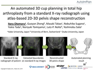
1. An automated 3D cup planning in total hip arthroplasty from a standard X‑ray radiograph using atlas-based 2D-3D pelvis shape reconstruction Itaru Otomarua, GuoyanZhengb, Masaki Takaoc, Nobuhiko Suganoc, Yukio Tadaa, Noriyuki Tomiyamac, Lutz-P. Nolteb, Yoshinobu Satoc a Kobe University, Japan b University of Bern, Switzerland c Osaka University, Japan Standard X-rayradiograph of patient Extracted boundaries on standard X-ray images Reconstructed3D pelvis shape Automated planning result
3. Background Automated planning [Otomaru et al., MICCAI 2009] Automatedsegmentation [Yokota et al., MICCAI 2009] 3D-CTimages 3D shape of pelvis and femurs Anatomical landmarks 3Dsurgical plan To stabilize quality of preoperative planning in THA, we have been developing a 3D-CT based automated planning system. (Otomaru et al., CAOS 2009, MICCAI 2008, 2009) Although this system is useful because acquisition of 3D-CT images is common in Japan, the usefulness may be limited in western countries.
4. Background Pelvic implant (cup) Automated planning [Otomaru et al., MICCAI 2009] Automatedsegmentation [Yokota et al., MICCAI 2009] Standard X-rayradiograph 3D-CTimages 3D shape of pelvis and femurs Anatomical landmarks 3Dsurgical plan Instead of 3D-CT, we use a single standard X-ray radiograph by estimating 3D shape using a 2D-3D reconstruction method [Zheng et al., MICCAI 2009]. In this study, we target pelvic implant (cup).
6. Materials | Standard X-ray radiograph We used actual patient X-ray radiograph for this study. Acquisition of the data was done at Osaka University Hospital. An only calibration needed in the 2D-3D reconstruction [Zheng., MICCAI 2009] was input of an scaling parameter. Any other calibration was unnecessary. In this study, we assumed the scaling parameter as known(which was calculated using 3D pelvis shape reconstructed from 3D-CT images). Spec of standard X-ray radiograph: Source-image distance was 1,200 mm. Pixel size was 0.1 x 0.1 mm2. Example of a standard X-ray image
7. Materials | Training datasets of statistical atlases Statistical shape atlas of pelvis for 2D-3D reconstruction [Zheng, MICCAI:2009] … 0mm 5.0 mm 10.0 mm (Thick) (Thin) Training datasets Statistical atlas of cup planning criteria for automated planning. [Otomaru et al., MICCAI:2009] We constructed statistical atlases from 34 training datasets and applied the proposed method to six test cases. Test cases were excluded from training datasets.
8. Processing flow of the proposed method Standard X-rayradiograph of patient Extracted boundaries on standard X-ray images Automated planning result Reconstructed3D pelvis shape Given the X-ray radiograph, pelvis’s boundaries are semi-automatically detected using a live-wire method. The 3D pelvic shape is automatically reconstructed from the extracted boundaries. Cup diameter and position are automatically determined from the 3D pelvic shape.
10. Experimental method We used only mildly diseased cases whose Crowe’s classification was one. (In our datasets, about 70 % of patients were mildly diseased. ) Mildly diseased case(Crowe I) Severely diseased case (Crowe IV)
11. Experimental method We used only mildly diseased cases whose Crowe’s classification was one. (In our datasets, about 70 % of patients were mildly diseased. ) Gold standard were defined as follows: 3D pelvis shape model reconstructed from 3D-CT images for validation of 2D-3D reconstruction. Cup diameter and position determined on 3D-CT images prepared by an experienced surgeon for validation of automated planning. Planning errors were defined as follows: Error of cup diameter and position between automated planning results and experienced surgeon’s plan.
12. Results | Pelvis shape reconstruction Case 2 Case 1 -5.0 5.0 mm 0.0 Average reconstruction error of six cases was 1.8 mm. Generally, reconstruction results was good both in whole pelvis and acetabular regions as shown in case 1. On the other hand, in some cases, there were shape errors in acetabular regions as shown in case 2.
13. Results | Automated cup planning X-ray: 1.7 +/- 1.5 mm X-ray: 4.4 +/- 0.7 mm 3D-CT: 1.0 +/- 1.1 mm 3D-CT: 3.6 +/- 1.8 mm Standard X-rayradiograph 3D-CTimages Standard X-rayradiograph 3D-CTimages Error of cup diameter [mm] Error of cup position [mm] There were no significant difference between errors of X-ray radiograph and 3D-CT images both in diameter and cup position.
14. Results | Examples of planning result 46 mm 50 mm 50 mm 48 mm 48 mm 50 mm Standard X-ray radiograph 3D-CT images Surgeon Standard X-ray radiograph 3D-CT images Surgeon Case 2 Case 1 As shown in case 1, errors of cup diameter were within 2 mm for planning of X-ray radiograph in five cases out of six cases. On the other hand, as shown in case 2, when reconstruction error was large, cup size error was also large.
16. Conclusion We constructed an automated 3D cup planning method for single standard X-ray radiograph. In mildly diseased cases, the cup planning errors were little large than the results of 3D-CT images (no significant difference). In this study, we assumed that the scaling parameter was known. According to our preliminary experiment, cup size did not change when scaling parameter changed 5% larger and smaller from the correct value. As future work, more validations are necessary because we only targeted mildly diseased cases whose Crowe’s classification was class one.
17. Thank you very much for your attention! For more details of our project … Web : http://www.image.med.osaka-u.ac.jp/AutoImPlan/ : http://www.youtube.com/user/autoimplan : Search “AutoImPlan” in Facebook.