Anatomyof female genital tract
•Télécharger en tant que PPTX, PDF•
125 j'aime•115,925 vues

anatomy of female genital tract

Recommandé
Contenu connexe
Tendances
Tendances (20)
Similaire à Anatomyof female genital tract
Similaire à Anatomyof female genital tract (20)
Plus de Ayman Shehata
Dernier
Dernier (20)
Anatomyof female genital tract
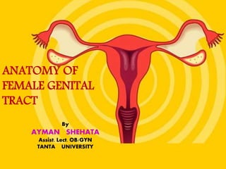
- 1. ANATOMY OF FEMALE GENITAL TRACT By AYMAN SHEHATA Assist. Lect. OB/GYN TANTA UNIVERSITY
- 2. • External genitalia • Internal genitalia • Accessory reproductive organs
- 4. Mons veneris Labia majora • Post commisure Labia minora • prepuce • Frenulum • Fourchette • Fossa navicularis clitoris • Glans • Body • 2 crura vestibule • 6 openings • bartholin’s abscess • Hymen External genitalia (vulva, pudendum)
- 6. External Genital Organs(vulva) • Mons pubis • Labia majora • Labia minora • Clitoris • Vestibule of the vagina External urethral orifice Vestibule glands paraurethral glands (Skene’s glands) Bartholin's gland vagina opening hymen External genitalia of adult female (parous)
- 7. MONS PUBIS The triangular mound of fatty tissue that covers the pubic bone It protects the pubic symphysis During adolescence sex hormones trigger the growth of pubic hair on the mons pubis Hair varies in coarseness curliness, amount, color and thickness
- 8. LABIA MAJORA Referred to as the outer lips They have a darker pigmentation The Labia Majora: Protect the introitus and urethral openings Are covered with hair and sebaceous glands Tend to be smooth, moist, and hairless Become flaccid with age and after childbirth
- 9. LABIA MINORA Referred to as the “inner lips” Made up of erectile, connective tissue that darkens and swells during sexual arousal Located inside the labia majora They are more sensitive and responsive to touch than the labia majora The labia minora tightens during intercourse
- 10. CLITORIS • Highly sensitive organ composed of nerves, blood vessels, and erectile tissue • Located under the prepuce • It is made up of a shaft and a glans • Becomes engorged with blood during sexual stimulation • Key to sexual pleasure for most women • Urethral opening is located directly below clitoris Clitoris
- 11. VAGINAL OPENING INTROITUS Opening may be covered by a thin sheath called the hymen Using the presence of an intact hymen for determining virginity is erroneous Some women are born without hymens The hymen can be perforated by many different events UNIT 3: FEMALE REPRODUCTIVE SYSTEM 11
- 16. • Blood supply – Arteries • Br. of internal pudendal artery – Labial A., – Transverse perineal A., – A to vestibular bulb, – deep & dorsal arteries to clitoris • Br. of Femoral A – Superficial & deep external pudendal A – Veins • Internal pudendal vein • Vesical/ vaginal venous plexuses • Long saphenous vein
- 17. • Nerve supply – Ant-sup part • Ilio inguinal & genital br. of genitofemoral N (L1,L2) – Post-inf part • Pudendal br. of post.cut. N of thigh (S123) – In between • Labial & perineal br.of Pudendal N (S234) • Lymphatics – Superf. Inguinal nodes – Gland of Cloquet – Ext. & Int. Iliac nodes
- 18. PERINEUM • The muscle and tissue located between the vaginal opening and anal canal • It supports and surrounds the lower parts of the urinary and digestive tracts • The perinium contains an abundance of nerve endings that make it sensitive to touch • An episiotomy is an incision of the perinium used during childbirth for widening the vaginal opening UNIT 3: FEMALE REPRODUCTIVE SYSTEM 18
- 21. Vagina • Fibromuscular membranous sheath • Excretory channel • Organ of copulation • Birth canal of parturition • 45° horizontal • 2.5cm diameter • distensibility
- 22. VAGINA • The vagina connects the cervix to the external genitals • It is located between the bladder and rectum Functions : • As a passageway for the menstrual flow • For uterine secretions to pass down through the introitus • As the birth canal during labor • With the help of two Bartholin’s glands becomes lubricated during SI
- 23. • Walls – Ant : 7.5 cm – Post : 9cm – 2 lateral walls • Fornices : – Ant : shallow – 2 lateral – Post : deep
- 28. Pelvic floor superior view
- 29. Pelvic floor inferior view
- 30. Pelvic floor side view
- 31. levator ani muscle is largest and strongest muscle in the pelvis Ischiococcygeus Iliococcygeus Pubococcygeus (Master sphincter) (weight bearing) (weight bearing)
- 32. Urogenital diaphragm • Braces the vagina and urethra (PUL) • Provides the stage for sexual quartet •Stabilizes the perineal body
- 33. The fetal head can damage the pelvic floor
- 34. Sexual quartet 1-Clitoris (Krause corpuscles) 2-Vestibular bulb (Erectile tissue) 3-Bartholin gland (Alkaline mucous) 4-Vagina (Copulatory canal)
- 35. • Structure – mucous coat – Submucous layer of loose areolar vascular tissues – Muscular layer – Fibrous coat (from endopelvic fascia) :highly vascular • Vaginal secretion – Doderlein’s bacilli : lactic acid from glycogen
- 36. • Blood Supply – Arteries • Cervicovaginal br. Of uterine A • Vaginal A • Middle rectal A • Internal pudendal • Anastomose---form 2 azygos arteries – Veins drain into: • Internal iliac V • Internal pudendal V • Lymphatics drain into • Upper 1/3rd : internal iliac nodes • Middle 1/3rd: external iliac nodes • Lower 1/3rd (below hymen) : superficial inguinal gp. • Nerve supply • Parasympathetic : S234 • Sympathetic: hypogastric plexus • Lower end : pudendal N (sensory )
- 37. Uterus • Hollow, pyriform muscular organ • Position: anteversion & anteflexion uterus dextrorotated • Measurements – 7.5cm long – 5cm wide – 3cm thick – Weight : 50-80gm
- 38. Parts of uterus
- 40. • Structure – Perimetrium : serous coat – Myometrium: thick bundles of sm. M • During pregnancy: – Outer long. – Middle interlacing – Inner circular – Endometrium • Surface epithelium • Lamina propria – Stromal cells – Endometrial glands – Vessels & nerves • Decidua in pregnancy • Secretion: scanty & watery
- 41. • Relation –Anterior • Above int.os : uterovesical pouch • Below int.os: separated from UB by loose areolar tissue –Posterior • Pouch of Douglas with coils on intestine –Lateral • Broad ligament • Mackenrodt’s ligament • Uterine A & ureter
- 42. • Ligaments of uterus – Uterosacral ligament – Transverse cervical/ Mackenrodt’s lig – Pubovesicocervical lig – Round ligament – Broad ligament • Mesovarium • Mesosalpinx • mesometrium • Suspensory lig of ovary/ infundibulopelvic ligament
- 43. Water flows under a bridge ureter Uterine artery
- 44. • Blood supply – Arteries: • Uterine A • Ovarian & Vaginal As. – Veins drain into • Internal iliac veins • Lymphatics • Nerves – Sympathetic • Motor : T5 & T6 • Sensory : T10 – L1 – Parasympathetic • Pelvic N ( S2,3,4) : both motor & sensory : ends in ganglia of Frankenhauser
- 45. 1 2 2 33 4 4 5 5 6 Figure: internal organs in female pelvis. 1. uterus, 2. ovaries, 3. fallopian tubes, 4. round ligaments, 5. utersacral ligaments, 6. rectouterine pouch (pouch of Douglas) , 7.broad ligaments. 7
- 47. Arterial system 1. Ovarian artery: the chief source of the blood for ovaries. 2. Uterine artery: corpus branch cervical-vaginal branch 3. Vaginal artery: main source of the blood for the middle part of vagina 4. Internal pudendal artery: supply for superficial perineum, labia majora, labia minora, lower part of the vagina, clitoris Blood supply for female genitalia
- 48. Fallopian tube (uterine tube, oviduct) • 10-14 cm • Lies within the superior border of broad ligament • 2 openings – Medially into cornua – Laterally into abdominal cavity
- 49. Parts of fallopian tube
- 51. Structure : -3 layers
- 52. • Blood Supply – Arteries • Uterine A • Ovarian A – Veins • Through pampiniforn plexus into ovarian veins • Lymphatics • Para-aortic nodes • Nerve supply – Uterine & ovarian nerves
- 53. The Ovary • Paired, situated on either side of uterus • Close to lateral pelvic wall • In ovarian fossa of Waldeyer • Size: 4*3*2 cm • Only intra-abdominal structure not covered by peritoneum • Medial pole: attached to uterine cornua by ovarian ligament • Laterally to the pelvic wall by infundibulopelvic ligament • Fimbrial end of oviduct close to ovary & attached to it via fimbria ovarica
- 55. • Structure : 2 parts • Lined single layer of germinal epithelium of Waldeyer (cuboidal epi.) • Tunica albuginea : stromal cells thickened beneath germinal epithelium • Contain primordial follicles • Corpus albicans/ atretic follicles Cortex • Loose connective tissues, blood vessels, nerves, muscles • Hilus cells : homologous to interstitial cells of testes Medulla
- 57. • Blood Supply – Arterial : Ovarian A – Veins : • Through pampiniform plexus ovarian V. Lt. Renal Vein IVC • Lymphatics • Para-aortic nodes • Nerve supply • Sympathetic supply from T10 along ovarian A.
- 58. Pelvic vessels
- 59. Pelvic vessels
- 60. • Lymphatic drainage: – External genital organ lymph group: 1. superficial inguinal lymph nodes. 2. deep inguinal lymph nodes – Pelvic lymph group: 1. iliac lymph group. 2. presacral lymph group. 3. lumbar lymph group.
- 62. The nerve of external genitalia • Pudendal nerve – Obstetric local anesthesia • Divided into 3 branches beside tuberosity – Inferior hemorrhoid nerve – Dorsal nerve – Perineal nerve
- 63. The nerve of internal genitalia • Sympathetic and parasympathetic nerves from lumbar and sacral spinal cord • Sympathetic nerve are derived from Plexus – Sacral plexus – Ovarian plexus
- 64. Pelvic innervation Frankenhauser’s plexus Pudendal nerve Hypogastric nerve (s) Superior hypogastric plexus Inferior mesentric plexus Celiac plexus Inferior hypogastric plexus PSN LUNA
- 66. BREASTS • Organs of sexual arousal • Contain mammary glands • Consist of connective tissue that serves as support • Each breast contain 15-25 clusters called lobes • Each lobule is connected by ducts that open into the nipples • The nipples are made up of erectile tissue • The pigmented around the nipples are called the areola • Breast size is determined primarily by heredity • Size also depends on the existing fat and glandular tissue • Breasts may exhibit cyclical changes, including increased swelling and tenderness prior to menstruation • Benign breast changes refer to fibrocystic disease • Lumps or masses that are noncancerous
- 67. UNIT 3: FEMALE REPRODUCTIVE SYSTEM 67