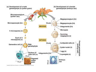
5. Pollen development and maturation
The end of meiosis in the microsporocyte or microspore
mother cell marks a turning point in microsporogenesis .
Results in the production of 4 microspores, each with its
own callose envelope.
A candidate gene for separation of microspores from
the tetrad in Arabidopsis anthers is designated as
QUARTET (QRT).
Outcome of microsporogenesis affected by this
mutation is release of microspore in tetrads
Failure of microspore separation in qrt mutants
Traced to the fusion of exine layer of adjacent
microspores Failure of protein degradation
6. Pollen development and maturation
The end of meiosis in the microsporocyte or microspore
mother cell marks a turning point in microsporogenesis .
Results in the production of 4 microspores, each with its
own callose envelope.
A candidate gene for separation of microspores from
the tetrad in Arabidopsis anthers is designated as
QUARTET (QRT).
Outcome of microsporogenesis affected by this
mutation is release of microspore in tetrads
Failure of microspore separation in qrt mutants
Traced to the fusion of exine layer of adjacent
microspores Failure of protein degradation
7. Pectin is absent in primary wall of wild type
microspores at the time of release from tetrad
Pectin remains as integral part of the
microspore wall of mutant
QRT gene functions in degradation of
pectin in order to separate the microspore
from tetrad.
Model of primary cell wall
8. Pollen grain has two cells: from first mitotic division
Vegetative cell- develops into pollen tube.
Contains most cytoplasmic organelles
Generative cell- small, produces the sperm
-cytoplasm partitioned unequally during
mitotic division of microspore
-lacks mitochondria and chloroplast
-at some point in pollen dev., divides by
mitosis, each daughter cell differentiates into sperm
cells, will lack also chloroplasts and mitochondria.
This is the basis of for the maternal inheritance of
chloroplast and mitochondrial genomes which
occurs in ca 90% of all angiosperm species.
9.
10.
11.
12.
13.
14.
15. Parts of ovule
1.nucellus- central body with vegetative cells enclosing the
Sporogenous cells
2. 1 or 2 integuments (unitegmic or bitegmic) enclosing the
Nucellus
3. funiculus-stalk connecting ovule with the placenta.
4. chalaza-where nucellus, integuments and the funiculus
merge
16. X-section of an ovary Ovary contains a cavity
lined with an epidermal
layer. Ovules develop
from the epidermal
cells and are contained
within the cavity of the
ovary, attached to its
inner surface by a
short stalk- funiculus
Ovary bears
ovules on a
ridge on the
ovary wall
called placenta
17. Developing ovules of Lilium. Ovule emerges from the
placenta as conical protuberance with the first
sporogenous cell,called archesporial cell.
Integuments formed by periclinal div. of epidermis
19. A, D. (bel mutant)
exposed nucellus
and a single
integument
F. Larger mutant ovule
Outer integument has
many cells.
20. Megaspore mother cell
differentiates from
surrounding nucellar tissue
and undergoes meiosis.
Inner integ.
Outer integ
Begin as
ridges of
tissue early in
Ovule dev.
C, E,F differential growth of
ovule causes them to curve
so micropyle is bent around
funiculus and placenta (C).
21. Development of embryo sac and female gamete (in
an anatropous ovule)
A hypodermal cell of the nucellus enlarges and
becomes differentiated into a megaspore mother cell
or megasporocyte. This diploid megaspore mother
cell increases in size and undergoes meiosis to form
a linear tetrad of 4 haploid megaspores, 3 of which
degenerate and the 4th becomes the functional
megaspore in monosporic types, all 4 become
functional in tetrasporic types
Female Gametophyte
The nucleus of the megaspore undergoes three
successive mitotic divisions forming eight nuclei. The
megaspore enlarges into an oval shaped structure
called the embryo sac. The eight nuclei of the
embryo sac arrange themselves in 3 groups.
22.
23. Micropyle
Inner integument
Outer integument
placenta
funuculus
Outer and inner integument completely overgrow the nucellus
Except for the micropyle.
--Begins with elongation of the functional megaspore,
usually at chalazal end.
-- initially megaspore is non-vacuolate but later small
vacuoles appear which may fuse to form large vacuole.
Development of
embryo sac
24. A. First megaspore mitosis yields binucleate embryo sac.
Spindle of first nuclear div oriented along the long axis of
the cell. Wall formation Does Not follow the nuclear
division. Both nuclei divide 2x, forming 4 in B then 8 in C
B. Large vacuole appears between the two daughter nuclei.
As cell expands, nuclei are pushed toward opposite poles
of the cell. Both nuclei from each pole divide twice
25. D. The 8 nuclei arrange themselves in two clusters of 4 nuclei
one at each opposite ends. One nucleus from each end
migrates towards the middle, called polar nuclei (named for
where they came from, not where they end up).
C.8-nucleate state . All
8 nuclei are present in
a common cytoplasm,
they move around
probably from
remnants of spindle
fibers from earlier
divisions.
26. Chalazal trio called antipodals
( Latin “against the foot”) at
opposite end of the egg and
antipodals
Egg apparatus consists of larger
egg flanked by two smaller cells
called synergids (greek for
“helpers” or cooperators
The large binucleate
29. Megaspore mother cell devs. from surrounding nucellar
tissue and undergoes meiotic division to form megaspore.
Nucellus considered as a megasporangium
funiculus
nucellus
chalaza- region where integuments fuse with funiculus
34. Mutants in ovule determination
1. bell (bel1)- ovule lacks inner integument
2. Aberrant testa shape (ats)- no clear distinction between
inner and outer integument
3. Extreme types of integument mutations:
aintegumenta (ant)
huellenhos (hll)
Do not develop integument and
embryo is disrupted.
36. Embryo sac cells
1.Egg- highly vacuolate, strongly polarized. In Arabidopsis,
a large vacuole aligned toward micropylar end and an
aggregation of cytoplasmic organelles and nucleus at
chalazal end. Ultrastructural simplicity of cytoplasm
characterize egg cells.
amount of cytoplasm is limited
cytoplasm spread as a thin layer surrounding vacuole
cytoplasm contains very little ER, limited no. of plastids
mitochondria, dictyosomes but high ribosomes which
are randomly distributed rather than aggregated as
polyribosomes
cell wall does not extend over the entire cell but wall
shows various attenuation toward chalazal pole
37. 2 Synergids- limited life span, wilt after fertilization.
Probably involved in nutrition of egg.
has extensive wall ingrowth at micropylar region
called filiform apparatus
metabolically active
3 Antipodals-transient existence , minimal cytoplasmic
organelle show nuclear abnormalities like
endoreplication
2 polar nuclei-metabolically active, extensive ER,
numerous plastids, mitochondria, dictyosomes and
polysomes, has large quantities of starch, proteins and
lipids
39. Germination of pollen tube
Pollen tubes
extend up to sev.
cm to reach
embryo sac. Cell
wall lacks
cellulose but has
another
polysaccharide-
callose- , a glucan
Callose –synthesized by Golgi and transported
to the extreme tip of pollen tube by Golgi-derived
vesicles .Fusion of vesicles with plasma
membrane expand the cell membrane of
elongating tube
Content of vesicles expand the wall of elongating tube
40. In angiosperms, to effect fertilization, the pollen grains
germinate on the stigma by putting forth tubes
(pollen tubes) which grow thru the style and find their way
into the ovules where they discharge the sperms in the
vicinity of the egg.
41. Page 114
Tube enters at the apex of the filiform apparatus and after
growing thru it arrives in the cytoplasm of the synergid. The
penetrated synergid starts degenerating before the arrival of
the Pollen tube, but after pollination. The process of
discharge takes place in seconds.
42. In cotton, the contents of the tube are discharged thru a
subterminal pore which faces the chalaza.
43. Pollen tube discharge: includes 2 sperms, the veg, nucleus
and a fair amount of cytoplasm. A portion of cytoplasm is
retained in the pollen tube.
No mixing between cytoplasm released by the pollen tube and
that of the synergid. They remain as two separate entities.