Nano particles
•Télécharger en tant que PPTX, PDF•
8 j'aime•825 vues

Use of nano particles in drug delivery.

Recommandé
Contenu connexe
Tendances
Tendances (20)
En vedette
En vedette (18)
Similaire à Nano particles
Similaire à Nano particles (20)
Plus de Bikash Singh
Plus de Bikash Singh (10)
Dernier
Dernier (20)
Nano particles
- 1. Use of Nanoparticles for drug Delivery
- 2. Nanoparticle – any particle that is sized between 1 and 100 nanometers (in terms of diameter) The use of nanoparticles allows one to change the pharmacokinetic properties of the drug without changing the active compound
- 3. Nanomedicine is the medical application of nanotechnology.
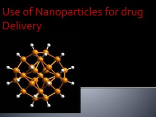
- 4. Liposomes Polymeric nanoparticles Dendrimers Fullerenes Quantum dots Metal nanoparticles Magnetic nanoparticles
- 6. In recent years, biodegradable polymeric nanoparticles have attracted considerable attention as potential drug delivery devices in view of their applications in drug targeting to particular organs/tissues, as carriers of DNA in gene therapy, and in their ability to deliver proteins, peptides and genes through a per oral route of administration
- 7. Approved by the FDA in 1996 for the treatment of multiple sclerosis (T-cell therapy) Synthetic polymer of amino amino acids (Cop1 composed of L-Ala, L-Lys, L-Glu, and L-Tyr) Administered subcutaneously Marketed byTEVA pharmaceuticals
- 8. Cop 1 polymer related to myelin binding protein (MBP) Binding to MHC leads to the activation ofT- suppressor cells Competes with several myelin-associated antigens to bind to MHC class II molecules Low toxicity; however, copaxone can only slow the progression of the disease and reduce the relapse rate
- 9. Gold nanoparticles Non-metals used : Carbon60
- 10. Gold nanoparticles can be novel nanocarriers of peptides and proteins of interest. Cationic tetraalkylammonium-functionalized GNPs recognize the surface of an anionic protein through complementary electrostatic interaction and inhibit its activity (Verma et al., 2004). The activity was recovered due to the release of free protein by treating the protein–particle complex with glutathione, showing GNPs as potential protein transporters. Pokharkar and coworkers have demonstrated functionalized gold nanoparticles as the carriers of insulin (Bhumkar and Joshi, 2007). Chitosan-coated particles strongly adsorb insulin on their surface, and are effective for transmucosal delivery of insulin.
- 11. Recent work has shown that the combination of phototherapy with conventional gene therapy offers a high possibility to improve the efficiency of gene delivery into cells (Mariko et al., 2005). For example, Niidome et al., (2006) have investigated the release of plasmid DNA from spherical gold nanoparticles after exposure to pulsed laser irradiation.
- 12. Interference of gene expression can also be performed using gold nanorods. Lee et al. have demonstrated the idea of a remote optical switch for localized and selective control of gene interference. Thiol-modified sense oligonucleotides were conjugated with gold nanorods.Thereafter, the antisense oligonucleotides were hybridized to the sense oligonucleotides.
- 13. After laser irradiation, the double strands of the oligonucleotides were denatured and the antisense oligonucleotides were released from the complex structure. These antisense oligonucleotides can bind to the corresponding mRNA while the sense oligonucleotides were still attached to gold nanorods through thiol bonds. Once the mRNA/oligodeoxynucleotide heteroduplex is formed, its structure will be recognized and degraded by RNaseH enzymes inside the cell, thereby inhibiting the normal genetic function of the mRNA (Lee et al., 2009).
- 14. Electroporation is another external stimulus that can be used to release genes from a gold nanoparticle. Kawano et al. (2006) have investigated gene delivery in vivo using gold nanoparticles excited with electrical pulses. In this study, gold nanoparticles were modified with mPEG-SH5000 and conjugated with plasmid DNA.
- 15. These were then injected into anesthetized mice. After a suitable delay to allow the conjugates to spread in the mouse, electrical pulses were then applied to the left lobe of its liver. The result was that gene expression was detected in the major mouse organs. In contrast, the injection of naked DNA resulted in a 10-fold lower level of detection. The degradation of DNA in blood, which occurs in times as short as 5 min, is evidently the reason for the inefficient transfection in the latter case.This study illustrates yet another interesting approach to improve gene delivery using gold nanoparticles.
- 17. Carbon nanotubes (CNTs), discovered by Japanese scientist Iijima in 1991 [1], are now considered to be a top class subject in academic researches as well as in various industrial areas
- 18. CNTs can be used as drug carriers to treat tumors .The efficacy of anticancer drugs used alone is restrained not only by their systemic toxicity and narrow therapeutic window but also by drug resistance and limited cellular penetration. Because CNTs can easily across the cytoplasmic membrane and nuclear membrane, anticancer drug transported by this vehicle will be liberated in situ with intact concentration and consequently, its action in the tumor cell will be higher than that administered alone by traditional therapy. Thus, the development of efficient delivery systems with the ability to enhance cellular uptake of existing potent drugs is needed.The high aspect ratio of CNTs offers great advantages over the existing delivery vectors, because the high surface area provides multiple attachment
- 19. Many anticancer drugs have been conjugated with functionalizedCNTs and successfully tested in vitro and in vivo such as epirubicin, doxorubicin, cisplatin, methotrexate, quercetin, and paclitaxel .
- 20. For drug delivery, most of the sites are accessible through either microcirculation by blood capillaries or pores present at various surfaces and membranes. Most of the apertures, openings, and gates at cellular or subcellular levels are of nanometer size Hence, nanoparticles are the most suited to reach the subcellular level.
- 21. For any moiety to remain in the vasculature, it needs to have its one dimension narrower than the cross-sectional diameter of the narrowest capillaries, which is about 2000 nm. Actually, for efficient transport the nanoparticle should be smaller than 300 nm. But, just moving in the vessels does not serve the drug delivery purpose. The delivery system must reach the site at the destination level. This requires crossing of the blood capillary wall to reach the extracellular fluid of the tissue and then again crossing of other cells, if they are in the way, and entering the target cell.These are the major barriers in the transit
- 22. A nanoparticle has to do a lot during this sojourn of the carrier through the vessels (capillaries) and across the barriers. There are two routes for crossing the blood capillaries and other cell layers, i.e., transcellular and paracellular. In the transcellular route, the particulate system has to enter the cell from one side and exit the cell from the other side to reach the tissue. The particulate system has to survive the intracellular environment to reach the target tissue. The other route is paracellular. In this, the particlulate system is not required to enter the cell; instead, it moves between the cells. These intercellular areas are known as the junctions. Passing through the junctions would obviate destruction of the carrier by the cell system.
- 23. Paracellular movement of moieties including ions, larger molecules, and leukocytes is controlled by the cytoskeletal association of tight junctions and the adherence junctions called apical junction complex. While tight junctions act as a regulated barrier, the adherence junctions are responsible for the development and stabilization of the tight junctions. Different epithelial and endothelial barriers have different permeabilities mainly because of the differences in the structure and the presence of tight junctions. While epithelia and brain capillary endothelium exhibit a high degree of barrier function, the vascular endothelium in other tissues has greater permeability. The tight junctions control the paracellular transport. For example, diffusion of large molecules may not be feasible, but migration of white cells is allowed
- 24. As the nanoparticle based drug delivery is achieved by particle transport, it is important to understand the blood flow rates and volumes of various organs and tissues. Considering the body’s distribution network, the blood vascular system, the body could be divided into several compartments based on the distributional sequencing and differentiation by the blood vascular system.