
Recommandé
Contenu connexe
Tendances
Tendances (18)
Similaire à Skin barrier models-molluscum
Similaire à Skin barrier models-molluscum (20)
Skin barrier models-molluscum
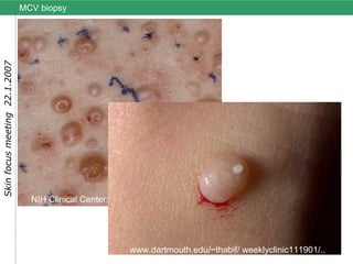
- 1. MCV biopsy NIH Clinical Center: Bugert and Turner, 1993 www.dartmouth.edu/~thabif/ weeklyclinic111901/..
- 2. MCV histopathology Lobular hyperplasia of epidermis into the dermis resulting in a cup-shaped lesion. The basal layers appear normal, but the keratinocytes of stratum malpighii become enlarged and acquire eosinophilic inclusion bodies - Henderson-Patterson bodies that contain virus particles. From biopsy; University of Heidelberg: Bugert, 1988
- 3. MCV in the host cell SEM Shelley and Burmeister, 1986 MCV can be purified in large quantities from skin biopsies
- 4. MCV virus preparation ELMI: 220- 450 nm filter MCV-HD-18-yellow-Cardiff 5 350 nm MCV-HD-10-red-Cardiff 1 Attempt to grow in cells in culture Abb. 1.1 , Schemazeichnung eines Pockenpartikels Tubuli Nukleosom Lateralkörper Aussenmembran Hülle Core Membran 100nm
- 5. MCV infection - biological features 1. only human host / no cell culture system 2. epidermal tissue tropism (gene acquisition-adaptation) 3. epidermal dedifferentiation 4. epidermal hyperproliferation-benign tumors 5. immuneevasion : lack of cellular immune response 6. cell receptor overexpression (EGFR and transferrinR) 7. HPV, EBV (analogy to other human epithelial viruses) EGF receptor 100nm
- 7. Minucell ® dual perfusion chamber cell culture system Minucell ® -Loop 3 sterile filter medium reservoir peristaltic pump 37 °C heater plate sterile filter Gas expander module sterile filter perfusion chamber sterile filter 3 way valve (injection port) microscopy
- 8. Emma Smith: Minucell system in operation (Rm. 115) 9/2004 injection / sample port