SlideShare une entreprise Scribd logo
1  sur  13
Télécharger pour lire hors ligne
13
Chapter IV
PATHOPHYSIOLOGY OF DIABETIC FOOT
 DEVELOPMENT OF SENSORY, MOTOR AND AUTONOMIC NEUROPATHY
 ASSOCIATIONS CONTRIBUTORY TO SENSORY NEUROPATHY IN DEVELOPING THE ULCERS
 MOTOR NEUROPATHY AND MECHANISMS OF INJURY
 AUTONOMIC NEUROPATHY IN DIABETIC FOOT
 PERIPHERAL VASCULAR DISEASE IN DIABETIC FOOT
 INFECTIONS AND RELATED ISSUES
 PAINFUL NEURO-RADICULOPATHY IN DIABETES
For a person with diabetes to develop a diabetic foot one or more of the following
three components are essential neuropathy, vasculopathy and infection. The fourth
extremely important risk factor is increased plantar pressure leading to ulceration.
Neuropathy and peripheral vasculopathy can be independent risk factors or could be
responsible in combination.12 Following is a description of what each of these
components contributes to the development of diabetic foot and its pathophysiologic
mechanism.
Diabetic Polyneuropathy:
The major components of diabetic polyneuropathy are:
1. Sensory neuropathy
2. Motor neuropathy and
3. Autonomic Neuropathy
1. Sensory Neuropathy:
14
Hyperglycemia is 'the' trigger that starts the cascade of neuropathy. It is well known
that the subsequent progressive unrelenting deterioration of sensorimotor and
autonomic function is a phenomenon related to the diabetic state per say and even
the rigorous control of blood glucose after the neuropathy sets in does not often help.
Given this, every effort at tight control which may include early aggressive life style
modification, drugs, diet and insulinization from the stage of diagnosis may go a
long way.
The sensory neuropathy can cause loss of a variety of sensation like touch, pressure,
temperature, vibration, position and pain. When the sensation of pain is lost it gives
rise to an insensate foot. The loss of pain sensation makes the foot vulnerable to
trivial trauma as it is not sensed. This causes a break in the skin and even if it is
inconspicuous and miniscule, it can become a portal of entry for the bacteria to enter.
As many as 81% amputations have been found to be due to initial minor trauma.14
Several mechanisms detailed below finally lead to the development of an ulcer.
Trauma, whether trivial or more substantial, needs to be of the repetitive nature to
become responsible for ulceration. Because the foot is insensate, the same type of
trauma can occur without the patient realizing that it is recurring. That leads to a
break in the skin and will lead to a noticeable ulcer that causes further facilitation to
the entry of bacteria and for infection to set in.
Distal symmetric sensory motor polyneuropathy is the most common form of
neuropathy in diabetes resulting either in pain, parasthesias, muscle atrophy or loss
of variety of sensations mentioned above. Radiculopathy due to thrombosis of vasa
nervorum leads to unilateral leg pain and weakness. Entrapment syndromes are
common in persons with diabetes like tarsal tunnel syndrome causing sensory
impairment of plantar skin and the weakness of the intrinsic pedal musculature.12
An insensate foot also gets pressure necrosis. Because of loss of sensation a patient
tends to keep his feet in the same position whether he stands for a long time or while
he is lying in the hospital bed or at home. This leads to the necrosis of the foot skin
under pressure at the pressure point. This happens particularly to heels in patients
lying in. The necrosis causes the skin to break down and an ulcer forms.
Glycosilation and the change in the shape of foot: The glycosilation in the ligaments
changes its properties. This results in restricted movements of joints. This Limited
Joint Mobility (LJM) affects the cycle of walking. At the take off stage in the cycle of
walking the normal extension of 1st MTP joint is restricted. The subcutaneous pad of
fat is thinner and it slips forward and does not cover the joint. Therefore the joint
bears extra pressure. This repetitive trauma of walking starts the process of
inflammation as during every cycle of walking there is anoxia that does not recover
fully in diabetes due to metabolic and vascular factors. This causes warmth and
erythema.
Other associations of sensory neuropathy:
Persons with diabetes loose proprioception, i.e., the sense of foot position with loss of
pain. This does not allow the patient to know how he places his feet. That results in
abnormal foot positions. An unevenly placed foot in varying or in the similar kind of
15
position, repetitively, develops unequal pressure areas on the plantar surface of the
foot. The part of the foot that gets excessive pressure and unnoticed repetitive
trauma can develop an ulcer. There are other causes of such unequal pressures also.
If the loss of pain sensation, numbness and loss of proprioception are present, there
will also be pressure unawareness. These patients do not tend to move their feet
from any one position. This causes continuous pressure.
Loss of temperature sensation:
The heat and cold sensations are carried by C fibers. Small Fibers sub-serve warm,
cold and pain sensation also. The rate of conduction for these sensations is slower,
have a period of latency of about 500 milliseconds before the impulse gets initiated.
Consequently there is a compounded delay in the sensation reaching the cortical
areas, its registration of the sensation and response to it. The rate of rise or fall of
temperature in degrees as well as time is also a determinant of cognition of these
sensations.
Heat and Cold Pain: In addition to the sensation of heat and cold there is the concept
of heat pain and cold pain that should be understood. These thresholds are higher
and lower than heat and cold sensation thresholds respectively. C Fibers are
unmyelinated, without specialized nerve endings, lying naked in tissues and are
distinct from A delta fibers that are responsible for deep aches and pain.
Abnormalities of C fibers are supposedly responsible for early occurrence of
symptoms. Other somatic sensations of pressure, touch, vibration, proprioception are
likely as not, may not be affected at this stage.29 Some authors have considered C
fibers to be the first to be affected in diabetic neuropathy.30
Symptomatic progression: Various symptoms occur initially when there is on going
damage to the nerves. Later relief of symptoms to a pain free state signifies further
damage and not amelioration. Amelioration of pain appears due to depletion of the
neuropeptides, substance P that are needed for the pain sensations as well as the
decrease in Nerve Growth Factors that maintain the C fibers. Adequacy of insulin,
either endogenous or exogenously administered in diabetic state maintains the
integrity of the nerve fibers.
Vasomotion: Diabetes affects rhythmic vasomotion of small arterioles due to
sympathetic damage early in disease. Loss of warm thermal threshold also occurs
early with C fiber damage and correlates significantly to reduced vasomotion.31
Exact cause effect or association between vasomotion and C fiber damage as a time
relationship is not established.31 Skin biopsies in persons with Diabetes show
uniform depletion of substance P, CGRP and the cytoplasmic proteins, for small fiber
specificity.32 Glabrous skin of the foot is far more affected by Diabetes than that of
hand.
Since these deficits may occur as a part of sensory neuropathy a patient is not likely
to be aware if his feet come in contact with a hot object. This causes necrosis of the
skin leading to ulcers. Some of the examples of this kind of thermal injuries cited by
Indian workers are the contact of feet on the hot exhausts of motor-bikes or the cover
of the engine in the driver's cabin in public transport system, where people tend to
rest their feet after removing whatever foot wear they have. Another common cause
16
of thermal injury is that Indians to go for hot fomentations or immersing the feet in
hot water. There are numerous examples of people with diabetes developing ulcers
walking on hot tiles barefoot within temple complexes, as religious reasons forbid
use of footwear. (Fig 8)
The nature of repetitive trauma:
Vigorous massage with counter irritants causes sliding of skin over fascia. This
possibly happens because of loss of subcutaneous fat tissue due to lipolysis. This
factor damages the underlying blood vessels. This in turn causes areas of patchy
gangrene in the skin. (Fig 9)
Foreign bodies in shoes are not sensed by the diabetic foot and can remain in the
shoe causing repeated trauma giving rise to high pressure at certain points. It may
cause small puncture wounds. (Fig 10)
Walking is the most frequent example of repetitive stress and trauma. In normal
foot, the pressure on the forefoot at its peak while walking is 600 kilo Pascals. To get
an idea of the kilopascal, we should know that the systolic pressure of 120mm of
mercury is an equivalent of 15 kilopascals. So it is easy to imagine how easily the
forefoot gets ischemic during walking, repeatedly. Absence of recovery from anoxia
and ischemia leads to inflammation that manifests as warmth and erythema. This has
important clinical implication. If the patient monitors the temperature of the foot by
back of the hand at 1st, 3rd and 5th metatarsal heads daily, especially at the end of
the day, he will immediately notice the increased warmth. At this point of time if
patient restricts the activity then the inflammation resolves. If patient continues the
same level of activity, then the exudate collects and the blister forms which breaks
down resulting in to a classical diabetic foot ulcer, commonly on 1st MTP joint.
Therefore it is necessary to remember that majority of the foot ulcer in diabetes are
from within out and not from outside in. Thus majority of these can be averted by
simply educating the patient in basic foot care.
Other pathogenetic mechanisms:
A-V shunting: These cycles of anoxia are enhanced by arteriovenous shunting if it is
present. The mechanism of A-V shunting is described elsewhere. The anoxia will
lead to inflammation in the deeper tissues but there is no pain and patients keep
walking. The inflammatory process then leads to tissue necrosis that leads to the
formation of ulcer.
Shear Forces: In addition to this, the feet are subjected to shear forces that are
comparable to walking on the rollers. Shear forces are perpendicular to the vertical
pressure exerted on the feet by patient's weight. There are anteroposterior and
mediolateral shears that are roughly a sixth of the vertical pressure forces. The longer
the stride, the higher are the shear forces. The faster one walks or longer, the greater
will be the shear forces. These forces are also quite damaging to the feet. (Fig 11)
The other causes of developing pressure points are found primarily in the motor
component of neuropathy. We will elaborate on foot pressures and its correlations in
biomechanics later.
17
Injuries of other kinds:
The various types of the injuries which act like trigger factors in the presence of
concomitant neuropathy and vasculopathy are home surgery, insect bites, chemical
injuries, footwear injury, pressure injury and thermal injury. The insect bites occur in
insensitive foot because the foot is exposed. The rodents are attracted by the smell of
the callous of the exposed foot and nibble at it, as the foot is insensate. The simple
precaution of routine use of socks can prevent this type of injury to an extent. Use of
strong chemicals and counter irritants to relive pain could be damaging.
2. Motor Neuropathy:
Motor neuropathy and some components of autonomic neuropathy cause change in
the shape of the foot. Motor neuropathy leads to the weakness and / or atrophy of
the interosseous muscles of the foot, resulting in the loss of ability of the small or the
intrinsic muscles to hold the foot in normal shape and place it in the correct position.
This leads to the deformity of foot. Once a foot is deformed then the pressure
distribution is unequal and some points develop excessive pressure. Such a foot then
becomes maladapted to the shoes one wears and the abnormal point of contact
becomes a pressure area. The repetitive pressure leads to keratosis and callus
formation. (Fig 12) The keratosis is said to develop more inappropriately in the
presence of sensory neuropathy that is almost always present. Development of a
callous is a good indicator of severity of neuropathy. The high pressure at the callus
results in the damage to the tissues of the foot and can start the formation of ulcer
below the callus that later on, breaks through. The pressure at callus point is nearly
20 times higher than the surrounding skin. Allowing the patient to walk with callus
is akin to patient walking with pebble in his shoes.
It is expressed by authorities like AJM Boulton that motor neuropathy and some
deformity as a consequence thereof can precede the development of sensory
neuropathy. It means that motor neuropathic abnormalities also would be high in
prevalence like sensory neuropathy which is known to be frequent.
The common deformities due to motor neuropathy:
Crowding of toes: The crowded tows tend to get above or below the adjacent toes or
come to lie in an oblique direction, while in the same plain. These tows may then
hurt the adjacent toes with their nails. (Fig 13) Crowding of toes in the presence of
sensory loss and poor foot hygiene can cause extensive web space fungal infection
that leads to gangrene. Teaching the patient to separate the toes with cotton wool,
foam pads or commercially available toe separators can avert this.
Cock up toes: The toes, especially the great toe could cock up and is not level with
the other toes of the foot. This makes them susceptible to trauma if shoes are
habitually worn. They come in repetitive traumatic contact with the upper of the
shoes and develop ulceration. There are other reasons for developing ulcers if there
are cock up toes. In these cases the fibro-fatty pad of the metatarsal head moves
forward and exposes metatarsal head to direct pressure and trauma. (Fig 14)
Hallux valgus is a great toe not in alignment with the next and causes deformity.
(Fig 15)
18
Hallux rigidus does not move with the movement of the foot does not bend or
extend hence becomes the site for repetitive injury and ulcer. (Fig 16)
Clawing of toes: Due to an imbalance in the muscle tone of the intrinsic muscles of
the foot the toes bend down. (Fig. 25) This brings the tips of the toes in contact with
the surface and bear pressure. The tips do not have the fatty insulation the toes in
normal position have. There is little between the skin and the bone and the pressure
thus tends to ulcerate the tips of the toes.
The cock up toes, clawed toes, hallux valgus or hallux rigidus develop an ulcer by
one more mechanism. The diabetic state is known to give rise to the glycation of long
lasting collagen fibres of the ligaments that hold various joints. The subsequent
inflammatory changes leading to advanced glycated end products make the
ligaments inflexible resulting in various abnormal postures of feet and toes making
them susceptible to ulceration. Persons with diabetes tend to choose tighter shoes
because then only they can feel the presence of it on their foot. This, along with
deformities causes pressure points, bunions and ulcerations. (Fig 17) The hammer
toes (Fig 11) cause the tip of the Hallux to bear the pressure as the toe flexes. At this
point the bone is directly in contact with the ground with no cushion of fat in
between. This leads to ulceration.
3. The pathophysiologic contribution of autonomic neuropathy:
Autonomic neuropathy results in two distinct mechanisms that contribute to diabetic
foot ulceration, i.e. sudomotor dysfunction and arterio-venous shunting in the foot.
Sudomotor dysfunction:
It is the dysfunction of the sympathetic nerves supplying the sweat glands in the
foot. This results in the reduction or absence of sweating which normally keeps the
feet moist. Feet become dry and the skin cracks more easily.12 It is said that feet that
sweat do not ulcerate.
Autonomic Neuropathy: Decrease in the flair reaction to a noxious stimulus is also
due to autonomic neuropathy resulting in reducing blood flow to the wound or
infected area. 13
Arteriovenous shunting:
It has two aspects - The mechanism by which it develops and the disastrous effects it
causes in the development of deformities leading to the development of diabetic foot.
Development of AV shunts:
Arteriovenous shunts connecting small arterioles to small veins and small venules
normally exist. They are preferentially either closed or are regulated under rigid
sympathetic control so that excess blood does not flow through them but is
preferentially directed to arterioles and from there to veins through capillary bed.
(Fig18.) Arterioles are less richly innervated by sympathetic nerves than are the
arteriovenous shunts. When sympathetic degeneration takes place the arteriovenous
shunts open much more, the rigid control closing them down normally, is now gone.
19
But the arterioles do not dilate to the similar extent causing greater resistance to
blood flow than the wider open AV shunt offers. That is how the blood gets shunted
through AV shunts.
The plantar circulation is organized with the small subcutaneous arteries penetrating
the dermis and anastomizing in the cutaneous arteriolar plexus. The arterioles arise
from these plexus and ascend to form sub-papillary plexuses. If AV shunting is
present they are likely to be deprived of the normal blood supply causing ischemia to
the skin and subcutaneous areas.
The effects of AV shunting on diabetic foot:
Since the blood is not perfused through the capillary bed via the arteriole but gets
shunted off earlier to that, at the level of larger arteriole, the tissues are left hypoxic,
the dermal capillary plexuses do not get perfused with blood resulting in lower
transcutaneous oxygen tension and lower skin temperature. The veins become
turgid, venous oxygen tension is high and in an operative setting the foot that is
hypoxic, paradoxically bleeds more profusely.
Hypoxia affects the bones of the feet. There is resorption of the bone that leads to
osteopenia, i.e. decrease in calcium content of the bones. These changes lead to
increase in frequency of fractures of the small bones. Since it is an insensate foot, the
patient continues to walk despite the fractures, as there is no pain. These two factors
can lead to considerable joint deformities, subluxation of talonavicular joint and
lateral migration of toes. These changes lead to more deformities and greater
susceptibility to ulceration as described above.
This is the setting of Charcot's joint. (Fig 19, 20)
The two classic deformities that finally lead to the Charcot's arthropathy are:
The rocker bottom deformity in which there is sub-luxation and displacement of
tarsal bones downwards.
The medial convexity that results from the displacement of talonavicular joint or
from tarso-metatarsal dislocations, results in toes overlapping each other. Such toes
can cause injury to the adjacent toes by their nails and this can happen repeatedly
leading to small puncture wound and ulceration. Alternatively, if the shoe size is
small then a bunion may form on the medial side of 1st metatarsophalangeal joint
that can then get injured and infection may start. (Fig 21)
Nails and diabetic foot ulcers:
Hypertrophied nails are possibly an effect of neuropathy. They can cause sub-
ungual ulcerations, or could be in growing to cause ulceration there. The irregularly
grown nails can and do injure adjacent toes to cause ulcers. (Fig 22)
A word on painless / painful foot and leg:
Persons with diabetes may have feet that are numb and insensate but get severe
nocturnal pains in their lower limbs. Deep calf pains are characteristic at night and
can be extremely severe. Prevalence of pain has been reported to be as high as 33 %
20
in diabetic feet.10 Sudden increase of pain in a painless chronic ulcer is indicative of
worsening of infection or reduced vascularity. If there is global pain of sudden onset
with cold feet and is of severe nature it could indicate an acute embolization or
thrombosis in a large artery. All persons with diabetes who have painful legs and
feet do not have diabetic polyneuropathy. In one series of 117 patients, 65.5 % had
peripheral neuropathy of possibly diabetic origin of the distal symmetric type. 7%
had femoral neuropathy 11% had peripheral vascular disease, 4% had tarsal tunnel
syndrome, 4% had lumbar canal stenosis, 3% had reflex sympathetic dystrophies and
6% had other miscellaneous causes. Hence it is essential to keep other causes in
mind.10
4. Peripheral (macro)vascular disease causing diabetic foot
ulceration:
The most significant lesion in peripheral vessel that contributes to the development
of ulceration is the atherosclerotic disease of tibial and peroneal vessels that leads to
decreased flow of blood through the foot, resulting in decreased delivery of oxygen,
nutrients and antibiotics to the foot further hampering the chances of healing,
although it does not affect the plantar arch.
Some of the characteristics of the diabetic peripheral (macro)vascular disease as
described in the international consensus of the diabetic foot are, 16 more common
(than normal population), affects younger individuals. There is clear evidence from
studies in India that the diabetes detection age is lowered by a decade now. There is
no sex difference among persons with diabetes.15 Women with diabetes had more
PAD than non-diabetic women in Framingham study with greater claudication as
well as higher cardiac disease.17 There is faster progress, the affliction is multi-
sequential, i.e. tandem lesions, is more distal, (more typically the infra-popliteal,
tibial, peroneal segments)15 Vascular surgery is more common on persons with
diabetes.18 Smoking complicates the PVD, doubles the risk in both sexes and is dose
related. The impact was discernible in advanced age.19,20,12 Smoking is atherogenic,
decreases the blood flow through vasospasm and increases blood viscosity and
clotting factors. Low HDL-cholesterol in Indians is now well-documented as is
hypertriglyceridemia. This pattern is considered atherogenic.
Understanding macrovascular disease in diabetes:
While diffuse atherosclerotic tandem lesions in diabetes can occur and do occur in
the proximal femoropopliteal arteries the characteristic arteries involved in diabetes
are below the trifurcation of the popliteal arteries tibial and peroneal.33
Reconstructive surgery is most frequently done for these vessels. The autonomic
neuropathy leads to the decreased flair reaction. Microangiopathy is not believed to
be the cause of foot ulceration.15 Monckeberg sclerosis affects the vascular media by
calcification is considered to leave the lumen intact and therefore does not jeopardize
the foot from the ischemic point of view, though in 15% of cases with diabetes where
ankle pressure is measured medial sclerosis gives an abnormally high ankle pressure
due to the non compressibility of the vessel.35
The smaller arteries: It is not uncommon to have ischemia in the arteries of toe as
evident from toe pressure measurements while the ankle pressure may be normal.34
21
Cholesterol emboli can affect the smaller vessels and convert them in end arteries
resulting in blue toe syndrome that will lead to gangrene. Hypertension is well
known to be associated with atherosclerosis.16a
Microangiopathy: It is not a significant factor in the development of foot ischemia. In
many instances there will be enough patent vessels below the ankle to allow for
reconstructive surgery on tibioperoneal vessels. Another protective factor is the
really rich collateral circulation within the plantar and dorsal arches and between the
two arches, as well as smaller unnamed communications all over the foot tissues
usually allows major operations and make bypasses useful. 34
Infections and compromise of the foot vessels:
Puncture or penetrating wounds of the plantar region or the web space infections
may go up in the central non expansible plantar space. The inflammatory exudate
that collects causes pressure on the small arteries in the tissues and will lead to
thrombosis or obliteration. This will lead to gangrene.
In diabetes the choice of treatment is important. ACE inhibitors are preferred. The
role of a small dose of diuretics has been recognized in therapy. Despite the
beneficial effects of beta-blockers to the cardiac outcome within the subgroup of
diabetes it will be very contentious to use beta blockers in significant peripheral
arterial disease. If it is used it will have to have adequate justification.21,12
Pathogenetic contribution of vasculopathy to the development of
neuropathy:
50% of diabetic patients who have had ulceration / amputation have (in the western
countries at least) co-existing neuropathy and peripheral vascular disease.1,2
Peripheral ischemia of lower limb can lead to ischemic nerve conduction failure.3 The
lower endoneurial hypoxia is the possible mechanism in human beings.4 The
presence of peripheral vascular disease co-relates with the peripheral neuropathy.5
Neuropathy is also caused by the lower transcutaneous oxygen tension but is
probably due to shunting and not due to peripheral vascular disease.6 The abnormal
vasa nervorum causing local ischemia have been proposed to be responsible for this
since vasodilators and vasoconstrictor PGI2 antagonists have been shown to produce
improvement.7,8,9 As discussed earlier - the recurrent ischemia of the forefoot on
walking cannot be compensated fully and immediately in the presence of peripheral
vascular disease and results in chronic hypoxia, inflammation etc. Similarly use of
beta blockers in significant peripheral arterial disease will have to have adequate
justification, due its propensity to vasoconstriction, despite its beneficial effects in
cardiac disease, although selective beta blockers like metoprolol or bisoprolol do not
seem to be contraindicated.21, 12
5. The infection and related issues:
The source of infection is usually the contamination of the break in the skin, which
may be imperceptible like cracks or fissures, puncture wounds or a major wound in a
neuropathic foot due to trauma of any cause. Staphylococcus aureus and beta
haemolytic streptococci rapidly colonise the break in the skin. The other sources
would the web space fungal infections and paronychia, viz, tinea pedis,
22
Onychomycosis. The devastating developments subsequent to an infected ulcer that
lead to the development of gangrene, necrotising fascitis and life threatening
situations like multi-organ failure should be guarded against. The pathophysiology
of these events can be constructed in the following sequence:
In persons with diabetes, infection results in microthrombi formation in the smaller
vessels unlike persons without diabetes where it results in vasodilatation. This
impairs blood flow in diabetes, converting the small arteries of the toes into end
arteries resulting in gangrene of the toes.
Osteomyelitis can be difficult to diagnose and remains a focus of uneradicated
infection and fails to indicate to the physician the need for longer antibiotic regimen.
The diagnosis of Osteomyelitis was missed in as many as two thirds of bone culture
proven cases.22 Excessive reliance on plain X rays by primary care physicians does
not help.23 Simple probing the bone can make a diagnosis of Osteomyelitis, while
scanning techniques are not always successful, some like Tc99 lack specificity, but
MRI is proving helpful.24, 25, 26
Clinically the trophic ulcers in diabetic neuropathy occur in the area of
metatarsophalangeal joints. The flexor tendons of the corresponding joint are
involved. The infected tendon carries the infection proximally in the leg, may become
devasularized and lead to loss of the sling support of the arch. This can enhances the
chances of the collapse of the arch. This is more likely to happen in case of ulcers
over the 1st MTP joints. Therefore eradication of the infection and salvage of the foot
is at the cost of the collapse of the arch. It is known that surgical procedures on the
foot in a person with diabetes can accelerate the process developing
neuroarthropathy.
Diabetes mellitus as an immunocompromized state
Failure of persons with diabetes to control the spread
Poor granuloma formation and inability to localise the infection is probably due to
the immunocompromized state, along with impaired wound healing and persistence
of abscesses.29 As a result of that an ulcer spreading deeper can contaminate a
tendon causing tenosinovitis. Tendons are avascular and are enclosed in loose facial
sheaths. They do not offer resistance to upward spread of the infection towards mid
leg. The soft tissue, which could be hypoxic either because of AV shunting or
peripheral vascular disease, does not offer resistance to infection.
There is a concept of plantar spaces like the palmer spaces. These are spaces that
contain the nerves and blood vessels and communicate with each other. Usually it is
the medial or lateral plantar space that gets infected and forms an abscess
corresponding to the original ulcer through which bacteria have entered. Infections
along the Flexor Hallucis Longus can go up to the level of middle of the leg.
Pathogenetically it is important to remember that the ulcers may look small or
benign but may indicate deeper abscesses. They are like a tip of the iceberg.
Necrotising fascitis, one of the dreaded complications is an aftermath of this
upwardly spreading infection.
23
Hyperglycaemia is responsible for alternations of immune functions of the white
cells.27
Some of the commonly mentioned abnormalities are as follows:
Bacterial endotoxemia, in presence of hyperglycaemia causes slower response in the
number of polymorphonuclear cells and also the speed of response. In one study
where granulocyte colony stimulating factor was used to augment the granulocyte
response in diabetic infected foot lesions suggested that along with increase in the
number of polymorphs, the study indicated that G-CSF may be deficient or
functionally sub-optimal in diabetic infection.11 Cruciani has indicated that use of G-
CSF may not reduce the duration of healing but significantly reduces the need for
both surgical intervention and amputation on a need to treat basis. There is no
mention about costs involved.36
Diabetes or uncontrolled hyperglycaemia cause decreased diapedesis towards the
site of infection, decreased chemotaxis, lower adherence that is normalised by
correction of hyperglycaemia.
The phagocytosis is supposedly poor, especially in diabetic ketoacidosis (an infected
foot is likely to present with DKA).28 Killing after phagocytosis is also poor due to
affectation possibly of superoxide bursts. Natural killer cells have a reduced capacity
to kill in presence of hyperglycaemia that gets restored by normoglycaemia. CD4+
lymphocytes and antibody dependant cellular cytotoxicity is supposed to decrease in
the presence of hyperglycaemia. The monocytes have decreased lectin like receptors
with decreased affinity.
An overview of the pathophysiology of diabetic foot:
The diabetic state per se will result in peripheral neuropathy, peripheral
vasculopathy also and autonomic neuropathy leading to deformities, abnormal
pressure dynamics and ulcer. The ischemic-immunocompromized state in the face of
anatomical peculiarities of foot will cause spread of infection.
The development of gangrene:
It is always the end result of the vascular closure to a part of the body. In spreading
infection, either vasculitis, or microthrombi or pressure of an inflammatory exudate
in plantar spaces could lead to vascular closure.
References:
1. Edmonds ME, Blundell MP, Morris ME, et al. Improved survival of the
diabetic foot: The role of the specialist foot clinic. QJ Med 1986; 232:763-771.
2. Thomson FJ, VevesA, Ashe H,et al. A team approach to diabetic foot care: The
Manchester experience. Foot1991; 2:75-82.
3. Low PA Recent advances in the pathogenesis of diabetic neuropathy. Muscle nerve
1987; 10:121-128.
24
4. Newrick PG, Wilson AJ, Jakubowski J, et al. Sural nerve oxygen tension in persons
with diabetes. BMJ1986; 293:1053-1054.
5. Ram Z, Sadeh M, Walden R, et al. Vascular insufficiency quantitatively aggravates
diabetic neuropathy. Arch Neurol 1991; 48:1239-1242.
6. Young MJ, Veves A, Walker MG, et al. Correlations between nerve function and
tissue oxygenation in diabetic patients: Further clues to diabetic neuropthy?
Diabetologia1992; 35:1146-1150.
7. Auwerx J, Bouillion R, Collen D, et al. Tissue type plasminogen activator antigen and
plasminogen activator inhibitor in diabetes mellitus. Arterisclerosis 1988; 8:68-72.
8. Beach KW, Strandness DE, Arteriosclerosis obliterans and associated risk factors in
insulin dependant and non-insulin dependant diabetes. Diabetes 1980; 29:882-888.
9. Fontboonne AM, et al. Insulin and cardiovascular disease: a Paris prospective study.
Diabetes Care 1991; 6:461-469.
10. Marvin E Levin. Overview of the diabetic foot: Pathogenesis, management and
prevention of Lesions, In International J of Diabetes in Developing countries.
11. Andrew Gogh, Mary Clapperton, Nancy Rolando, Alethea V M Foster, John Philpott-
Howard, Michael Edmunds, randomised Placebo Controlled Trial of Granulocyte
Colony Stimulating Factor in Diabetic Foot Infection. The Lancet, Vol 350,
September 30, 1997 pp 855 - 859.
12. Bowker, John H and Pfeifer, Michael A (2001) Levin and O'Neal's The Diabetic
Foot. 6th Edition,. Mosby St.Louis, pp - 219.
13. Parkhouse N, LeQuesne PM: Impaired neurogenic vascular response inpatients with
diabetes and neurogenic foot lesions. N Engl J Med 318:1306, 1988.
14. Pecoraro RE, Rieber GE, Burgess EM: Pathways to diabetic limb amputation: basis
for prevention. Diabetes Care 16:1187-1189, 1993.
15. LoGerfo FW, Coffman JD: Vascular and microvascular disease of the foot in
diabetes. N Engl J Med 311:1516, 1984.
16. International Consensus on the Diabetic foot by the International Working Group on
the Diabetic Foot 1999.
16a. Wilson PWF, Kannel WB, Epidemiology of hyperglycemia and atherosclerosis. In
Ruderman N, Williamson J, Brownlee M, (eds): Hyperglycemia, Diabetes and
Vascular Disease, New York, Oxford University Press, 1992.
17. Malone JM, Snyder M, Anderson G et al: Prevention of amputation by diabetic
education Am J Surg, 158:520, 1989.
18. Delbridge L, Appleberg M, Reeve TS: Factors associated with the development of
foot lesions in the diabetic. Surgery 93:78, 1983.
19. Beach KW, Strandness DE Jr: Arteriosclerosis obliterans and associated risk factors
in insulin dependent diabetes. Diabetes 29:882, 1980.
25
20. Bowker, John H and Pfeifer, Michael A (2001) Levin and O'Neal's The Diabetic
Foot. 6th Edition. Mosby, St.Louis, pp 225
21. Newman LG, Walker J, Palestro CJ, Unsuspected Osteomyelitis in diabetic foot
ulcers. JAMA, 266:1246-1251, 1991.
22. Edelman D, Matchar DB: Clinical and radiographic findings that lead to intervention
in Diabetic patients with foot ulcers: A nationwide survey of primary care physicians.
Diabetes Care 19:755-757, 1996.
23. Grayson ML, Gibbons GW, Balogh K, et al: probing to bone in infected pedal ulcers:
A clinical sign of underlying Osteomyelitis in diabetic patients. JAMA 273:712-728,
1995.
24. Littenberg B, Mushalin AI: The diagnostic technology assessment consortium:
Technetium bone scanning in the diagnosis of Osteomyelitis: A meta analysis of the
test performance. J Gen Intern Med 7:158-163, 1992.
25. Durham JR: The role of magnetic resonance imaging in the management of foot
abscess in the diabetic patients. In Yao JST, Pearce WH (eds): The ishcemic
extremity: Advances in treatment. East Norwalk, CT: Appleton & Lange, 1995.
26. Mac Mahon MM, Bistrian BR: Host defenses and susceptibility to infection in
patients with diabetes mellitus. Infect Dis Clin North Am 9:1-10, 1995.
27. Sentochnik DE, Eliopoulos GM: Infection and Diabetes. In Kahn CR, Weir GC,
(eds): Joslin's Diabetes Mellitus, 13th ed. Philadelphia: Lea and Febiger, 1994, pp
867-888.
28. Lipsky BA, Pecoraro RE, Larson SA et al: Outpatient management of uncomplicated
lower extremity infections in diabetic patients. Arch Intern Med 150:790-797, 1990.
29. Aron Vinik Exp Clin Endocrinol Diabetes, 109 (2001) Suppl 2).
30. Jamal et al, 1987, Dyck, 1988, Hanson et al, 1992, as quoted by Vinik vide above.
31. (Vinik ibid).
32. Levy et al, 1992,Wallargren et al, 1995, Lauria et al, 1998, and others.
33. Levin ME, Sicard GA: Evaluating and treating diabetic peripheral vascular disease:
Part 1. Clin Diabetes 5:62, 1987.
34. Chapter 9 pp 223 as quoted by Bowker and Pfeifer in The Diabetic Foot, Sixth
edition.
35. LoGerfo FW, Coffman JD: Vascular and microvascular disease of the foot in
diabetes. N Engl J Med 311:1516, 1984.
36. Cruciany et al, Diabetes Care 28:454-460, 2005.

Contenu connexe

Tendances

Tendinopathies of wrist and hand
Tendinopathies of wrist and handTendinopathies of wrist and hand
Tendinopathies of wrist and handdinesh sharma
 
Diabetic foot Wilson N. M.D. MBBS
Diabetic foot    Wilson N.  M.D. MBBSDiabetic foot    Wilson N.  M.D. MBBS
Diabetic foot Wilson N. M.D. MBBSM.D. Njeru
 
Prof sobh renal osteodystrophy
Prof sobh renal osteodystrophyProf sobh renal osteodystrophy
Prof sobh renal osteodystrophymostafa hegazy
 
common congenital deformities of hand
common congenital deformities of handcommon congenital deformities of hand
common congenital deformities of handSumer Yadav
 
Fat embolism syndrome
Fat embolism syndromeFat embolism syndrome
Fat embolism syndromeAftab Hussain
 
P M S 2 Fractures And Healing
P M S 2  Fractures And  HealingP M S 2  Fractures And  Healing
P M S 2 Fractures And HealingAriff Anuar
 
Diabetic foot lecture 2010
Diabetic foot lecture 2010Diabetic foot lecture 2010
Diabetic foot lecture 2010Donald Pelto
 
Avascular necrosis femoral head by DR RAJAT MALOT (MS,DNB, MNAMS,FELLLOWSHIP ...
Avascular necrosis femoral head by DR RAJAT MALOT (MS,DNB, MNAMS,FELLLOWSHIP ...Avascular necrosis femoral head by DR RAJAT MALOT (MS,DNB, MNAMS,FELLLOWSHIP ...
Avascular necrosis femoral head by DR RAJAT MALOT (MS,DNB, MNAMS,FELLLOWSHIP ...chitrapandey
 
Rickets and osteomalacia
Rickets and osteomalacia Rickets and osteomalacia
Rickets and osteomalacia Sankalp Bansal
 
Paediatric Septic Arthritis
Paediatric Septic ArthritisPaediatric Septic Arthritis
Paediatric Septic ArthritisJasmial Nand
 
OSTEOMYELITIS Introduction, Clinical features
OSTEOMYELITIS Introduction, Clinical featuresOSTEOMYELITIS Introduction, Clinical features
OSTEOMYELITIS Introduction, Clinical featuresBALAKRISHNA341
 
fractures of hand bones
fractures of hand bonesfractures of hand bones
fractures of hand bonesSumer Yadav
 
Giant cell tumors of bone
Giant cell tumors of boneGiant cell tumors of bone
Giant cell tumors of boneDrAbdulSuhail
 

Tendances (20)

Tendinopathies of wrist and hand
Tendinopathies of wrist and handTendinopathies of wrist and hand
Tendinopathies of wrist and hand
 
Dupuytrens contracture presentation
Dupuytrens contracture presentationDupuytrens contracture presentation
Dupuytrens contracture presentation
 
Diabetic neuropathy
Diabetic neuropathyDiabetic neuropathy
Diabetic neuropathy
 
Renal osteodystrophy
Renal osteodystrophyRenal osteodystrophy
Renal osteodystrophy
 
Diabetic foot Wilson N. M.D. MBBS
Diabetic foot    Wilson N.  M.D. MBBSDiabetic foot    Wilson N.  M.D. MBBS
Diabetic foot Wilson N. M.D. MBBS
 
AVN
AVNAVN
AVN
 
Prof sobh renal osteodystrophy
Prof sobh renal osteodystrophyProf sobh renal osteodystrophy
Prof sobh renal osteodystrophy
 
Knee
KneeKnee
Knee
 
Musculoskele
MusculoskeleMusculoskele
Musculoskele
 
common congenital deformities of hand
common congenital deformities of handcommon congenital deformities of hand
common congenital deformities of hand
 
Fat embolism syndrome
Fat embolism syndromeFat embolism syndrome
Fat embolism syndrome
 
P M S 2 Fractures And Healing
P M S 2  Fractures And  HealingP M S 2  Fractures And  Healing
P M S 2 Fractures And Healing
 
Diabetic foot lecture 2010
Diabetic foot lecture 2010Diabetic foot lecture 2010
Diabetic foot lecture 2010
 
Osteomyelitis
OsteomyelitisOsteomyelitis
Osteomyelitis
 
Avascular necrosis femoral head by DR RAJAT MALOT (MS,DNB, MNAMS,FELLLOWSHIP ...
Avascular necrosis femoral head by DR RAJAT MALOT (MS,DNB, MNAMS,FELLLOWSHIP ...Avascular necrosis femoral head by DR RAJAT MALOT (MS,DNB, MNAMS,FELLLOWSHIP ...
Avascular necrosis femoral head by DR RAJAT MALOT (MS,DNB, MNAMS,FELLLOWSHIP ...
 
Rickets and osteomalacia
Rickets and osteomalacia Rickets and osteomalacia
Rickets and osteomalacia
 
Paediatric Septic Arthritis
Paediatric Septic ArthritisPaediatric Septic Arthritis
Paediatric Septic Arthritis
 
OSTEOMYELITIS Introduction, Clinical features
OSTEOMYELITIS Introduction, Clinical featuresOSTEOMYELITIS Introduction, Clinical features
OSTEOMYELITIS Introduction, Clinical features
 
fractures of hand bones
fractures of hand bonesfractures of hand bones
fractures of hand bones
 
Giant cell tumors of bone
Giant cell tumors of boneGiant cell tumors of bone
Giant cell tumors of bone
 

En vedette

1362748586 2 chapter2
1362748586 2 chapter21362748586 2 chapter2
1362748586 2 chapter2dfsimedia
 
1362465385 managinig neuropathic ulcer skke (1)
1362465385 managinig neuropathic ulcer skke (1)1362465385 managinig neuropathic ulcer skke (1)
1362465385 managinig neuropathic ulcer skke (1)dfsimedia
 
1362748605 3 chapter3
1362748605 3 chapter31362748605 3 chapter3
1362748605 3 chapter3dfsimedia
 
1362748638 5 chapter5
1362748638 5 chapter51362748638 5 chapter5
1362748638 5 chapter5dfsimedia
 
1363267070 13 chapter13
1363267070 13 chapter131363267070 13 chapter13
1363267070 13 chapter13dfsimedia
 
1362748535 1 chapter1
1362748535 1 chapter11362748535 1 chapter1
1362748535 1 chapter1dfsimedia
 

En vedette (6)

1362748586 2 chapter2
1362748586 2 chapter21362748586 2 chapter2
1362748586 2 chapter2
 
1362465385 managinig neuropathic ulcer skke (1)
1362465385 managinig neuropathic ulcer skke (1)1362465385 managinig neuropathic ulcer skke (1)
1362465385 managinig neuropathic ulcer skke (1)
 
1362748605 3 chapter3
1362748605 3 chapter31362748605 3 chapter3
1362748605 3 chapter3
 
1362748638 5 chapter5
1362748638 5 chapter51362748638 5 chapter5
1362748638 5 chapter5
 
1363267070 13 chapter13
1363267070 13 chapter131363267070 13 chapter13
1363267070 13 chapter13
 
1362748535 1 chapter1
1362748535 1 chapter11362748535 1 chapter1
1362748535 1 chapter1
 

Similaire à Pathophysiology of Diabetic Foot Neuropathy

Diabetic foot vinay 1
Diabetic foot vinay 1Diabetic foot vinay 1
Diabetic foot vinay 1Vinay Jain
 
bio mechanics of diabetic foot ulcer
bio mechanics of diabetic foot ulcerbio mechanics of diabetic foot ulcer
bio mechanics of diabetic foot ulcersenphysio
 
Diabetes, Peripheral Neuropathy & How to Conduct
Diabetes, Peripheral Neuropathy & How to ConductDiabetes, Peripheral Neuropathy & How to Conduct
Diabetes, Peripheral Neuropathy & How to ConductTrevor Perkes
 
Musculo skeletal complication of diabetes mellitus
Musculo skeletal complication of diabetes mellitusMusculo skeletal complication of diabetes mellitus
Musculo skeletal complication of diabetes mellitusAminuArzet
 
Diabetic Foot Ulcer.pptx
Diabetic Foot Ulcer.pptxDiabetic Foot Ulcer.pptx
Diabetic Foot Ulcer.pptxGaniDwiCahya2
 
rheumatological manifestations of systemic diseases- diabetes
 rheumatological manifestations of systemic diseases- diabetes rheumatological manifestations of systemic diseases- diabetes
rheumatological manifestations of systemic diseases- diabetesvinmmcri
 
Diabetes Mellitus and it's orthotic management
Diabetes Mellitus and it's orthotic managementDiabetes Mellitus and it's orthotic management
Diabetes Mellitus and it's orthotic managementankitajhaa729
 
Diabetic Foot slide show vascular surgery
Diabetic Foot slide show vascular surgeryDiabetic Foot slide show vascular surgery
Diabetic Foot slide show vascular surgerydrmetwally7
 
Approach to diabetic foot
Approach  to diabetic footApproach  to diabetic foot
Approach to diabetic footFaiz Hmoud
 
A Basic Review on Diabetic Neuropathy
A Basic Review on Diabetic NeuropathyA Basic Review on Diabetic Neuropathy
A Basic Review on Diabetic Neuropathyijtsrd
 
Psoriatic Arthritis: A Common Culprit Behind Joint Discomfort By Dr. Conway ...
Psoriatic Arthritis: A Common Culprit Behind Joint Discomfort  By Dr. Conway ...Psoriatic Arthritis: A Common Culprit Behind Joint Discomfort  By Dr. Conway ...
Psoriatic Arthritis: A Common Culprit Behind Joint Discomfort By Dr. Conway ...Khalid Chamakh
 
Buergers disease - By Dr Adithya J V, Asst.Professor, Dept. of General Surger...
Buergers disease - By Dr Adithya J V, Asst.Professor, Dept. of General Surger...Buergers disease - By Dr Adithya J V, Asst.Professor, Dept. of General Surger...
Buergers disease - By Dr Adithya J V, Asst.Professor, Dept. of General Surger...Dr Adithya J V
 
presentation on arthritis
presentation on arthritispresentation on arthritis
presentation on arthritisNighatKanwal
 
diabetesmellitus-141029144107-conversion-gate01 2.pptx
diabetesmellitus-141029144107-conversion-gate01 2.pptxdiabetesmellitus-141029144107-conversion-gate01 2.pptx
diabetesmellitus-141029144107-conversion-gate01 2.pptxabhimittal8
 
Disorders of feet-Bunion,Marfan Syndrome,Ehlers–Danlos syndrome,Corn and cal...
Disorders of feet-Bunion,Marfan Syndrome,Ehlers–Danlos syndrome,Corn  and cal...Disorders of feet-Bunion,Marfan Syndrome,Ehlers–Danlos syndrome,Corn  and cal...
Disorders of feet-Bunion,Marfan Syndrome,Ehlers–Danlos syndrome,Corn and cal...loritacaroline
 
Rhemathoid arthritis RA
Rhemathoid arthritis RARhemathoid arthritis RA
Rhemathoid arthritis RAAmaal bataiha
 

Similaire à Pathophysiology of Diabetic Foot Neuropathy (20)

Rheumatology
RheumatologyRheumatology
Rheumatology
 
Diabetic foot vinay 1
Diabetic foot vinay 1Diabetic foot vinay 1
Diabetic foot vinay 1
 
bio mechanics of diabetic foot ulcer
bio mechanics of diabetic foot ulcerbio mechanics of diabetic foot ulcer
bio mechanics of diabetic foot ulcer
 
Diabetes, Peripheral Neuropathy & How to Conduct
Diabetes, Peripheral Neuropathy & How to ConductDiabetes, Peripheral Neuropathy & How to Conduct
Diabetes, Peripheral Neuropathy & How to Conduct
 
Musculo skeletal complication of diabetes mellitus
Musculo skeletal complication of diabetes mellitusMusculo skeletal complication of diabetes mellitus
Musculo skeletal complication of diabetes mellitus
 
Diabetic Foot Ulcer.pptx
Diabetic Foot Ulcer.pptxDiabetic Foot Ulcer.pptx
Diabetic Foot Ulcer.pptx
 
rheumatological manifestations of systemic diseases- diabetes
 rheumatological manifestations of systemic diseases- diabetes rheumatological manifestations of systemic diseases- diabetes
rheumatological manifestations of systemic diseases- diabetes
 
Diabetes Mellitus and it's orthotic management
Diabetes Mellitus and it's orthotic managementDiabetes Mellitus and it's orthotic management
Diabetes Mellitus and it's orthotic management
 
Diabetic foot
Diabetic foot Diabetic foot
Diabetic foot
 
Diabetic Foot slide show vascular surgery
Diabetic Foot slide show vascular surgeryDiabetic Foot slide show vascular surgery
Diabetic Foot slide show vascular surgery
 
Approach to diabetic foot
Approach  to diabetic footApproach  to diabetic foot
Approach to diabetic foot
 
A Basic Review on Diabetic Neuropathy
A Basic Review on Diabetic NeuropathyA Basic Review on Diabetic Neuropathy
A Basic Review on Diabetic Neuropathy
 
Osteoarthritis
OsteoarthritisOsteoarthritis
Osteoarthritis
 
Psoriatic Arthritis: A Common Culprit Behind Joint Discomfort By Dr. Conway ...
Psoriatic Arthritis: A Common Culprit Behind Joint Discomfort  By Dr. Conway ...Psoriatic Arthritis: A Common Culprit Behind Joint Discomfort  By Dr. Conway ...
Psoriatic Arthritis: A Common Culprit Behind Joint Discomfort By Dr. Conway ...
 
4441564759967.pptx
4441564759967.pptx4441564759967.pptx
4441564759967.pptx
 
Buergers disease - By Dr Adithya J V, Asst.Professor, Dept. of General Surger...
Buergers disease - By Dr Adithya J V, Asst.Professor, Dept. of General Surger...Buergers disease - By Dr Adithya J V, Asst.Professor, Dept. of General Surger...
Buergers disease - By Dr Adithya J V, Asst.Professor, Dept. of General Surger...
 
presentation on arthritis
presentation on arthritispresentation on arthritis
presentation on arthritis
 
diabetesmellitus-141029144107-conversion-gate01 2.pptx
diabetesmellitus-141029144107-conversion-gate01 2.pptxdiabetesmellitus-141029144107-conversion-gate01 2.pptx
diabetesmellitus-141029144107-conversion-gate01 2.pptx
 
Disorders of feet-Bunion,Marfan Syndrome,Ehlers–Danlos syndrome,Corn and cal...
Disorders of feet-Bunion,Marfan Syndrome,Ehlers–Danlos syndrome,Corn  and cal...Disorders of feet-Bunion,Marfan Syndrome,Ehlers–Danlos syndrome,Corn  and cal...
Disorders of feet-Bunion,Marfan Syndrome,Ehlers–Danlos syndrome,Corn and cal...
 
Rhemathoid arthritis RA
Rhemathoid arthritis RARhemathoid arthritis RA
Rhemathoid arthritis RA
 

Plus de dfsimedia

1362571981 diab neuropa etiol mech con
1362571981 diab neuropa etiol mech con1362571981 diab neuropa etiol mech con
1362571981 diab neuropa etiol mech condfsimedia
 
1362571881 dfsi drreddys_workshop_anasth.
1362571881 dfsi drreddys_workshop_anasth.1362571881 dfsi drreddys_workshop_anasth.
1362571881 dfsi drreddys_workshop_anasth.dfsimedia
 
1362466122 pad in diabetes
1362466122 pad in diabetes1362466122 pad in diabetes
1362466122 pad in diabetesdfsimedia
 
1362466145 pad, agiography & angioplasty
1362466145 pad, agiography & angioplasty1362466145 pad, agiography & angioplasty
1362466145 pad, agiography & angioplastydfsimedia
 
1362466100 acute ischaemia of lower limb
1362466100 acute ischaemia of lower limb1362466100 acute ischaemia of lower limb
1362466100 acute ischaemia of lower limbdfsimedia
 
1362566341 surgical treatment of diabetic foot
1362566341 surgical treatment of diabetic foot1362566341 surgical treatment of diabetic foot
1362566341 surgical treatment of diabetic footdfsimedia
 
1362564357 general vs spinal vs regional
1362564357 general vs spinal vs regional1362564357 general vs spinal vs regional
1362564357 general vs spinal vs regionaldfsimedia
 
1362564096 anatomy and spread of foot infections
1362564096 anatomy and spread of foot infections1362564096 anatomy and spread of foot infections
1362564096 anatomy and spread of foot infectionsdfsimedia
 
1362466052 tunnda m2-d f ulcer
1362466052 tunnda m2-d f ulcer1362466052 tunnda m2-d f ulcer
1362466052 tunnda m2-d f ulcerdfsimedia
 
1362466017 total contact casting
1362466017 total contact casting1362466017 total contact casting
1362466017 total contact castingdfsimedia
 
1362465722 pressure scans measurements
1362465722 pressure scans measurements1362465722 pressure scans measurements
1362465722 pressure scans measurementsdfsimedia
 
1362465426 mechanism foot injury and ulcer formation
1362465426 mechanism foot injury and ulcer formation1362465426 mechanism foot injury and ulcer formation
1362465426 mechanism foot injury and ulcer formationdfsimedia
 
1362465385 managinig neuropathic ulcer skke
1362465385 managinig neuropathic ulcer skke 1362465385 managinig neuropathic ulcer skke
1362465385 managinig neuropathic ulcer skke dfsimedia
 
1362465354 is amputation the answer
1362465354 is amputation the answer1362465354 is amputation the answer
1362465354 is amputation the answerdfsimedia
 
1362465180 diabetic footwear considerations
1362465180 diabetic footwear considerations1362465180 diabetic footwear considerations
1362465180 diabetic footwear considerationsdfsimedia
 
1362465156 diabetic foot ulcer etiopathogenesis & management
1362465156 diabetic foot ulcer   etiopathogenesis & management1362465156 diabetic foot ulcer   etiopathogenesis & management
1362465156 diabetic foot ulcer etiopathogenesis & managementdfsimedia
 
1362465129 diabetic foot syndrome an indian perspective
1362465129 diabetic foot syndrome   an indian perspective1362465129 diabetic foot syndrome   an indian perspective
1362465129 diabetic foot syndrome an indian perspectivedfsimedia
 
1362463849 derangement of wound healing in diabetic neuropathy
1362463849 derangement of wound healing in diabetic neuropathy1362463849 derangement of wound healing in diabetic neuropathy
1362463849 derangement of wound healing in diabetic neuropathydfsimedia
 
1362462786 amputation in diabetic foot
1362462786 amputation in diabetic foot1362462786 amputation in diabetic foot
1362462786 amputation in diabetic footdfsimedia
 
1362405549 vpt assessment in neuropathy
1362405549 vpt assessment in neuropathy 1362405549 vpt assessment in neuropathy
1362405549 vpt assessment in neuropathy dfsimedia
 

Plus de dfsimedia (20)

1362571981 diab neuropa etiol mech con
1362571981 diab neuropa etiol mech con1362571981 diab neuropa etiol mech con
1362571981 diab neuropa etiol mech con
 
1362571881 dfsi drreddys_workshop_anasth.
1362571881 dfsi drreddys_workshop_anasth.1362571881 dfsi drreddys_workshop_anasth.
1362571881 dfsi drreddys_workshop_anasth.
 
1362466122 pad in diabetes
1362466122 pad in diabetes1362466122 pad in diabetes
1362466122 pad in diabetes
 
1362466145 pad, agiography & angioplasty
1362466145 pad, agiography & angioplasty1362466145 pad, agiography & angioplasty
1362466145 pad, agiography & angioplasty
 
1362466100 acute ischaemia of lower limb
1362466100 acute ischaemia of lower limb1362466100 acute ischaemia of lower limb
1362466100 acute ischaemia of lower limb
 
1362566341 surgical treatment of diabetic foot
1362566341 surgical treatment of diabetic foot1362566341 surgical treatment of diabetic foot
1362566341 surgical treatment of diabetic foot
 
1362564357 general vs spinal vs regional
1362564357 general vs spinal vs regional1362564357 general vs spinal vs regional
1362564357 general vs spinal vs regional
 
1362564096 anatomy and spread of foot infections
1362564096 anatomy and spread of foot infections1362564096 anatomy and spread of foot infections
1362564096 anatomy and spread of foot infections
 
1362466052 tunnda m2-d f ulcer
1362466052 tunnda m2-d f ulcer1362466052 tunnda m2-d f ulcer
1362466052 tunnda m2-d f ulcer
 
1362466017 total contact casting
1362466017 total contact casting1362466017 total contact casting
1362466017 total contact casting
 
1362465722 pressure scans measurements
1362465722 pressure scans measurements1362465722 pressure scans measurements
1362465722 pressure scans measurements
 
1362465426 mechanism foot injury and ulcer formation
1362465426 mechanism foot injury and ulcer formation1362465426 mechanism foot injury and ulcer formation
1362465426 mechanism foot injury and ulcer formation
 
1362465385 managinig neuropathic ulcer skke
1362465385 managinig neuropathic ulcer skke 1362465385 managinig neuropathic ulcer skke
1362465385 managinig neuropathic ulcer skke
 
1362465354 is amputation the answer
1362465354 is amputation the answer1362465354 is amputation the answer
1362465354 is amputation the answer
 
1362465180 diabetic footwear considerations
1362465180 diabetic footwear considerations1362465180 diabetic footwear considerations
1362465180 diabetic footwear considerations
 
1362465156 diabetic foot ulcer etiopathogenesis & management
1362465156 diabetic foot ulcer   etiopathogenesis & management1362465156 diabetic foot ulcer   etiopathogenesis & management
1362465156 diabetic foot ulcer etiopathogenesis & management
 
1362465129 diabetic foot syndrome an indian perspective
1362465129 diabetic foot syndrome   an indian perspective1362465129 diabetic foot syndrome   an indian perspective
1362465129 diabetic foot syndrome an indian perspective
 
1362463849 derangement of wound healing in diabetic neuropathy
1362463849 derangement of wound healing in diabetic neuropathy1362463849 derangement of wound healing in diabetic neuropathy
1362463849 derangement of wound healing in diabetic neuropathy
 
1362462786 amputation in diabetic foot
1362462786 amputation in diabetic foot1362462786 amputation in diabetic foot
1362462786 amputation in diabetic foot
 
1362405549 vpt assessment in neuropathy
1362405549 vpt assessment in neuropathy 1362405549 vpt assessment in neuropathy
1362405549 vpt assessment in neuropathy
 

Dernier

Top Rated Bangalore Call Girls Mg Road ⟟ 9332606886 ⟟ Call Me For Genuine S...
Top Rated Bangalore Call Girls Mg Road ⟟   9332606886 ⟟ Call Me For Genuine S...Top Rated Bangalore Call Girls Mg Road ⟟   9332606886 ⟟ Call Me For Genuine S...
Top Rated Bangalore Call Girls Mg Road ⟟ 9332606886 ⟟ Call Me For Genuine S...narwatsonia7
 
Premium Call Girls Cottonpet Whatsapp 7001035870 Independent Escort Service
Premium Call Girls Cottonpet Whatsapp 7001035870 Independent Escort ServicePremium Call Girls Cottonpet Whatsapp 7001035870 Independent Escort Service
Premium Call Girls Cottonpet Whatsapp 7001035870 Independent Escort Servicevidya singh
 
Best Rate (Hyderabad) Call Girls Jahanuma ⟟ 8250192130 ⟟ High Class Call Girl...
Best Rate (Hyderabad) Call Girls Jahanuma ⟟ 8250192130 ⟟ High Class Call Girl...Best Rate (Hyderabad) Call Girls Jahanuma ⟟ 8250192130 ⟟ High Class Call Girl...
Best Rate (Hyderabad) Call Girls Jahanuma ⟟ 8250192130 ⟟ High Class Call Girl...astropune
 
College Call Girls in Haridwar 9667172968 Short 4000 Night 10000 Best call gi...
College Call Girls in Haridwar 9667172968 Short 4000 Night 10000 Best call gi...College Call Girls in Haridwar 9667172968 Short 4000 Night 10000 Best call gi...
College Call Girls in Haridwar 9667172968 Short 4000 Night 10000 Best call gi...perfect solution
 
Call Girls Gwalior Just Call 8617370543 Top Class Call Girl Service Available
Call Girls Gwalior Just Call 8617370543 Top Class Call Girl Service AvailableCall Girls Gwalior Just Call 8617370543 Top Class Call Girl Service Available
Call Girls Gwalior Just Call 8617370543 Top Class Call Girl Service AvailableDipal Arora
 
Manyata Tech Park ( Call Girls ) Bangalore ✔ 6297143586 ✔ Hot Model With Sexy...
Manyata Tech Park ( Call Girls ) Bangalore ✔ 6297143586 ✔ Hot Model With Sexy...Manyata Tech Park ( Call Girls ) Bangalore ✔ 6297143586 ✔ Hot Model With Sexy...
Manyata Tech Park ( Call Girls ) Bangalore ✔ 6297143586 ✔ Hot Model With Sexy...vidya singh
 
Call Girls Haridwar Just Call 9907093804 Top Class Call Girl Service Available
Call Girls Haridwar Just Call 9907093804 Top Class Call Girl Service AvailableCall Girls Haridwar Just Call 9907093804 Top Class Call Girl Service Available
Call Girls Haridwar Just Call 9907093804 Top Class Call Girl Service AvailableDipal Arora
 
(👑VVIP ISHAAN ) Russian Call Girls Service Navi Mumbai🖕9920874524🖕Independent...
(👑VVIP ISHAAN ) Russian Call Girls Service Navi Mumbai🖕9920874524🖕Independent...(👑VVIP ISHAAN ) Russian Call Girls Service Navi Mumbai🖕9920874524🖕Independent...
(👑VVIP ISHAAN ) Russian Call Girls Service Navi Mumbai🖕9920874524🖕Independent...Taniya Sharma
 
Call Girls Aurangabad Just Call 9907093804 Top Class Call Girl Service Available
Call Girls Aurangabad Just Call 9907093804 Top Class Call Girl Service AvailableCall Girls Aurangabad Just Call 9907093804 Top Class Call Girl Service Available
Call Girls Aurangabad Just Call 9907093804 Top Class Call Girl Service AvailableDipal Arora
 
Night 7k to 12k Chennai City Center Call Girls 👉👉 7427069034⭐⭐ 100% Genuine E...
Night 7k to 12k Chennai City Center Call Girls 👉👉 7427069034⭐⭐ 100% Genuine E...Night 7k to 12k Chennai City Center Call Girls 👉👉 7427069034⭐⭐ 100% Genuine E...
Night 7k to 12k Chennai City Center Call Girls 👉👉 7427069034⭐⭐ 100% Genuine E...hotbabesbook
 
Call Girls Varanasi Just Call 9907093804 Top Class Call Girl Service Available
Call Girls Varanasi Just Call 9907093804 Top Class Call Girl Service AvailableCall Girls Varanasi Just Call 9907093804 Top Class Call Girl Service Available
Call Girls Varanasi Just Call 9907093804 Top Class Call Girl Service AvailableDipal Arora
 
Call Girls Ooty Just Call 9907093804 Top Class Call Girl Service Available
Call Girls Ooty Just Call 9907093804 Top Class Call Girl Service AvailableCall Girls Ooty Just Call 9907093804 Top Class Call Girl Service Available
Call Girls Ooty Just Call 9907093804 Top Class Call Girl Service AvailableDipal Arora
 
Pondicherry Call Girls Book Now 9630942363 Top Class Pondicherry Escort Servi...
Pondicherry Call Girls Book Now 9630942363 Top Class Pondicherry Escort Servi...Pondicherry Call Girls Book Now 9630942363 Top Class Pondicherry Escort Servi...
Pondicherry Call Girls Book Now 9630942363 Top Class Pondicherry Escort Servi...Genuine Call Girls
 
(Rocky) Jaipur Call Girl - 09521753030 Escorts Service 50% Off with Cash ON D...
(Rocky) Jaipur Call Girl - 09521753030 Escorts Service 50% Off with Cash ON D...(Rocky) Jaipur Call Girl - 09521753030 Escorts Service 50% Off with Cash ON D...
(Rocky) Jaipur Call Girl - 09521753030 Escorts Service 50% Off with Cash ON D...indiancallgirl4rent
 
Call Girls Nagpur Just Call 9907093804 Top Class Call Girl Service Available
Call Girls Nagpur Just Call 9907093804 Top Class Call Girl Service AvailableCall Girls Nagpur Just Call 9907093804 Top Class Call Girl Service Available
Call Girls Nagpur Just Call 9907093804 Top Class Call Girl Service AvailableDipal Arora
 
VIP Hyderabad Call Girls Bahadurpally 7877925207 ₹5000 To 25K With AC Room 💚😋
VIP Hyderabad Call Girls Bahadurpally 7877925207 ₹5000 To 25K With AC Room 💚😋VIP Hyderabad Call Girls Bahadurpally 7877925207 ₹5000 To 25K With AC Room 💚😋
VIP Hyderabad Call Girls Bahadurpally 7877925207 ₹5000 To 25K With AC Room 💚😋TANUJA PANDEY
 
Call Girls Bangalore Just Call 9907093804 Top Class Call Girl Service Available
Call Girls Bangalore Just Call 9907093804 Top Class Call Girl Service AvailableCall Girls Bangalore Just Call 9907093804 Top Class Call Girl Service Available
Call Girls Bangalore Just Call 9907093804 Top Class Call Girl Service AvailableDipal Arora
 
Call Girls Dehradun Just Call 9907093804 Top Class Call Girl Service Available
Call Girls Dehradun Just Call 9907093804 Top Class Call Girl Service AvailableCall Girls Dehradun Just Call 9907093804 Top Class Call Girl Service Available
Call Girls Dehradun Just Call 9907093804 Top Class Call Girl Service AvailableDipal Arora
 
Call Girls Bareilly Just Call 9907093804 Top Class Call Girl Service Available
Call Girls Bareilly Just Call 9907093804 Top Class Call Girl Service AvailableCall Girls Bareilly Just Call 9907093804 Top Class Call Girl Service Available
Call Girls Bareilly Just Call 9907093804 Top Class Call Girl Service AvailableDipal Arora
 
♛VVIP Hyderabad Call Girls Chintalkunta🖕7001035870🖕Riya Kappor Top Call Girl ...
♛VVIP Hyderabad Call Girls Chintalkunta🖕7001035870🖕Riya Kappor Top Call Girl ...♛VVIP Hyderabad Call Girls Chintalkunta🖕7001035870🖕Riya Kappor Top Call Girl ...
♛VVIP Hyderabad Call Girls Chintalkunta🖕7001035870🖕Riya Kappor Top Call Girl ...astropune
 

Dernier (20)

Top Rated Bangalore Call Girls Mg Road ⟟ 9332606886 ⟟ Call Me For Genuine S...
Top Rated Bangalore Call Girls Mg Road ⟟   9332606886 ⟟ Call Me For Genuine S...Top Rated Bangalore Call Girls Mg Road ⟟   9332606886 ⟟ Call Me For Genuine S...
Top Rated Bangalore Call Girls Mg Road ⟟ 9332606886 ⟟ Call Me For Genuine S...
 
Premium Call Girls Cottonpet Whatsapp 7001035870 Independent Escort Service
Premium Call Girls Cottonpet Whatsapp 7001035870 Independent Escort ServicePremium Call Girls Cottonpet Whatsapp 7001035870 Independent Escort Service
Premium Call Girls Cottonpet Whatsapp 7001035870 Independent Escort Service
 
Best Rate (Hyderabad) Call Girls Jahanuma ⟟ 8250192130 ⟟ High Class Call Girl...
Best Rate (Hyderabad) Call Girls Jahanuma ⟟ 8250192130 ⟟ High Class Call Girl...Best Rate (Hyderabad) Call Girls Jahanuma ⟟ 8250192130 ⟟ High Class Call Girl...
Best Rate (Hyderabad) Call Girls Jahanuma ⟟ 8250192130 ⟟ High Class Call Girl...
 
College Call Girls in Haridwar 9667172968 Short 4000 Night 10000 Best call gi...
College Call Girls in Haridwar 9667172968 Short 4000 Night 10000 Best call gi...College Call Girls in Haridwar 9667172968 Short 4000 Night 10000 Best call gi...
College Call Girls in Haridwar 9667172968 Short 4000 Night 10000 Best call gi...
 
Call Girls Gwalior Just Call 8617370543 Top Class Call Girl Service Available
Call Girls Gwalior Just Call 8617370543 Top Class Call Girl Service AvailableCall Girls Gwalior Just Call 8617370543 Top Class Call Girl Service Available
Call Girls Gwalior Just Call 8617370543 Top Class Call Girl Service Available
 
Manyata Tech Park ( Call Girls ) Bangalore ✔ 6297143586 ✔ Hot Model With Sexy...
Manyata Tech Park ( Call Girls ) Bangalore ✔ 6297143586 ✔ Hot Model With Sexy...Manyata Tech Park ( Call Girls ) Bangalore ✔ 6297143586 ✔ Hot Model With Sexy...
Manyata Tech Park ( Call Girls ) Bangalore ✔ 6297143586 ✔ Hot Model With Sexy...
 
Call Girls Haridwar Just Call 9907093804 Top Class Call Girl Service Available
Call Girls Haridwar Just Call 9907093804 Top Class Call Girl Service AvailableCall Girls Haridwar Just Call 9907093804 Top Class Call Girl Service Available
Call Girls Haridwar Just Call 9907093804 Top Class Call Girl Service Available
 
(👑VVIP ISHAAN ) Russian Call Girls Service Navi Mumbai🖕9920874524🖕Independent...
(👑VVIP ISHAAN ) Russian Call Girls Service Navi Mumbai🖕9920874524🖕Independent...(👑VVIP ISHAAN ) Russian Call Girls Service Navi Mumbai🖕9920874524🖕Independent...
(👑VVIP ISHAAN ) Russian Call Girls Service Navi Mumbai🖕9920874524🖕Independent...
 
Call Girls Aurangabad Just Call 9907093804 Top Class Call Girl Service Available
Call Girls Aurangabad Just Call 9907093804 Top Class Call Girl Service AvailableCall Girls Aurangabad Just Call 9907093804 Top Class Call Girl Service Available
Call Girls Aurangabad Just Call 9907093804 Top Class Call Girl Service Available
 
Night 7k to 12k Chennai City Center Call Girls 👉👉 7427069034⭐⭐ 100% Genuine E...
Night 7k to 12k Chennai City Center Call Girls 👉👉 7427069034⭐⭐ 100% Genuine E...Night 7k to 12k Chennai City Center Call Girls 👉👉 7427069034⭐⭐ 100% Genuine E...
Night 7k to 12k Chennai City Center Call Girls 👉👉 7427069034⭐⭐ 100% Genuine E...
 
Call Girls Varanasi Just Call 9907093804 Top Class Call Girl Service Available
Call Girls Varanasi Just Call 9907093804 Top Class Call Girl Service AvailableCall Girls Varanasi Just Call 9907093804 Top Class Call Girl Service Available
Call Girls Varanasi Just Call 9907093804 Top Class Call Girl Service Available
 
Call Girls Ooty Just Call 9907093804 Top Class Call Girl Service Available
Call Girls Ooty Just Call 9907093804 Top Class Call Girl Service AvailableCall Girls Ooty Just Call 9907093804 Top Class Call Girl Service Available
Call Girls Ooty Just Call 9907093804 Top Class Call Girl Service Available
 
Pondicherry Call Girls Book Now 9630942363 Top Class Pondicherry Escort Servi...
Pondicherry Call Girls Book Now 9630942363 Top Class Pondicherry Escort Servi...Pondicherry Call Girls Book Now 9630942363 Top Class Pondicherry Escort Servi...
Pondicherry Call Girls Book Now 9630942363 Top Class Pondicherry Escort Servi...
 
(Rocky) Jaipur Call Girl - 09521753030 Escorts Service 50% Off with Cash ON D...
(Rocky) Jaipur Call Girl - 09521753030 Escorts Service 50% Off with Cash ON D...(Rocky) Jaipur Call Girl - 09521753030 Escorts Service 50% Off with Cash ON D...
(Rocky) Jaipur Call Girl - 09521753030 Escorts Service 50% Off with Cash ON D...
 
Call Girls Nagpur Just Call 9907093804 Top Class Call Girl Service Available
Call Girls Nagpur Just Call 9907093804 Top Class Call Girl Service AvailableCall Girls Nagpur Just Call 9907093804 Top Class Call Girl Service Available
Call Girls Nagpur Just Call 9907093804 Top Class Call Girl Service Available
 
VIP Hyderabad Call Girls Bahadurpally 7877925207 ₹5000 To 25K With AC Room 💚😋
VIP Hyderabad Call Girls Bahadurpally 7877925207 ₹5000 To 25K With AC Room 💚😋VIP Hyderabad Call Girls Bahadurpally 7877925207 ₹5000 To 25K With AC Room 💚😋
VIP Hyderabad Call Girls Bahadurpally 7877925207 ₹5000 To 25K With AC Room 💚😋
 
Call Girls Bangalore Just Call 9907093804 Top Class Call Girl Service Available
Call Girls Bangalore Just Call 9907093804 Top Class Call Girl Service AvailableCall Girls Bangalore Just Call 9907093804 Top Class Call Girl Service Available
Call Girls Bangalore Just Call 9907093804 Top Class Call Girl Service Available
 
Call Girls Dehradun Just Call 9907093804 Top Class Call Girl Service Available
Call Girls Dehradun Just Call 9907093804 Top Class Call Girl Service AvailableCall Girls Dehradun Just Call 9907093804 Top Class Call Girl Service Available
Call Girls Dehradun Just Call 9907093804 Top Class Call Girl Service Available
 
Call Girls Bareilly Just Call 9907093804 Top Class Call Girl Service Available
Call Girls Bareilly Just Call 9907093804 Top Class Call Girl Service AvailableCall Girls Bareilly Just Call 9907093804 Top Class Call Girl Service Available
Call Girls Bareilly Just Call 9907093804 Top Class Call Girl Service Available
 
♛VVIP Hyderabad Call Girls Chintalkunta🖕7001035870🖕Riya Kappor Top Call Girl ...
♛VVIP Hyderabad Call Girls Chintalkunta🖕7001035870🖕Riya Kappor Top Call Girl ...♛VVIP Hyderabad Call Girls Chintalkunta🖕7001035870🖕Riya Kappor Top Call Girl ...
♛VVIP Hyderabad Call Girls Chintalkunta🖕7001035870🖕Riya Kappor Top Call Girl ...
 

Pathophysiology of Diabetic Foot Neuropathy

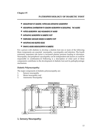
  • 1. 13 Chapter IV PATHOPHYSIOLOGY OF DIABETIC FOOT  DEVELOPMENT OF SENSORY, MOTOR AND AUTONOMIC NEUROPATHY  ASSOCIATIONS CONTRIBUTORY TO SENSORY NEUROPATHY IN DEVELOPING THE ULCERS  MOTOR NEUROPATHY AND MECHANISMS OF INJURY  AUTONOMIC NEUROPATHY IN DIABETIC FOOT  PERIPHERAL VASCULAR DISEASE IN DIABETIC FOOT  INFECTIONS AND RELATED ISSUES  PAINFUL NEURO-RADICULOPATHY IN DIABETES For a person with diabetes to develop a diabetic foot one or more of the following three components are essential neuropathy, vasculopathy and infection. The fourth extremely important risk factor is increased plantar pressure leading to ulceration. Neuropathy and peripheral vasculopathy can be independent risk factors or could be responsible in combination.12 Following is a description of what each of these components contributes to the development of diabetic foot and its pathophysiologic mechanism. Diabetic Polyneuropathy: The major components of diabetic polyneuropathy are: 1. Sensory neuropathy 2. Motor neuropathy and 3. Autonomic Neuropathy 1. Sensory Neuropathy:
  • 2. 14 Hyperglycemia is 'the' trigger that starts the cascade of neuropathy. It is well known that the subsequent progressive unrelenting deterioration of sensorimotor and autonomic function is a phenomenon related to the diabetic state per say and even the rigorous control of blood glucose after the neuropathy sets in does not often help. Given this, every effort at tight control which may include early aggressive life style modification, drugs, diet and insulinization from the stage of diagnosis may go a long way. The sensory neuropathy can cause loss of a variety of sensation like touch, pressure, temperature, vibration, position and pain. When the sensation of pain is lost it gives rise to an insensate foot. The loss of pain sensation makes the foot vulnerable to trivial trauma as it is not sensed. This causes a break in the skin and even if it is inconspicuous and miniscule, it can become a portal of entry for the bacteria to enter. As many as 81% amputations have been found to be due to initial minor trauma.14 Several mechanisms detailed below finally lead to the development of an ulcer. Trauma, whether trivial or more substantial, needs to be of the repetitive nature to become responsible for ulceration. Because the foot is insensate, the same type of trauma can occur without the patient realizing that it is recurring. That leads to a break in the skin and will lead to a noticeable ulcer that causes further facilitation to the entry of bacteria and for infection to set in. Distal symmetric sensory motor polyneuropathy is the most common form of neuropathy in diabetes resulting either in pain, parasthesias, muscle atrophy or loss of variety of sensations mentioned above. Radiculopathy due to thrombosis of vasa nervorum leads to unilateral leg pain and weakness. Entrapment syndromes are common in persons with diabetes like tarsal tunnel syndrome causing sensory impairment of plantar skin and the weakness of the intrinsic pedal musculature.12 An insensate foot also gets pressure necrosis. Because of loss of sensation a patient tends to keep his feet in the same position whether he stands for a long time or while he is lying in the hospital bed or at home. This leads to the necrosis of the foot skin under pressure at the pressure point. This happens particularly to heels in patients lying in. The necrosis causes the skin to break down and an ulcer forms. Glycosilation and the change in the shape of foot: The glycosilation in the ligaments changes its properties. This results in restricted movements of joints. This Limited Joint Mobility (LJM) affects the cycle of walking. At the take off stage in the cycle of walking the normal extension of 1st MTP joint is restricted. The subcutaneous pad of fat is thinner and it slips forward and does not cover the joint. Therefore the joint bears extra pressure. This repetitive trauma of walking starts the process of inflammation as during every cycle of walking there is anoxia that does not recover fully in diabetes due to metabolic and vascular factors. This causes warmth and erythema. Other associations of sensory neuropathy: Persons with diabetes loose proprioception, i.e., the sense of foot position with loss of pain. This does not allow the patient to know how he places his feet. That results in abnormal foot positions. An unevenly placed foot in varying or in the similar kind of
  • 3. 15 position, repetitively, develops unequal pressure areas on the plantar surface of the foot. The part of the foot that gets excessive pressure and unnoticed repetitive trauma can develop an ulcer. There are other causes of such unequal pressures also. If the loss of pain sensation, numbness and loss of proprioception are present, there will also be pressure unawareness. These patients do not tend to move their feet from any one position. This causes continuous pressure. Loss of temperature sensation: The heat and cold sensations are carried by C fibers. Small Fibers sub-serve warm, cold and pain sensation also. The rate of conduction for these sensations is slower, have a period of latency of about 500 milliseconds before the impulse gets initiated. Consequently there is a compounded delay in the sensation reaching the cortical areas, its registration of the sensation and response to it. The rate of rise or fall of temperature in degrees as well as time is also a determinant of cognition of these sensations. Heat and Cold Pain: In addition to the sensation of heat and cold there is the concept of heat pain and cold pain that should be understood. These thresholds are higher and lower than heat and cold sensation thresholds respectively. C Fibers are unmyelinated, without specialized nerve endings, lying naked in tissues and are distinct from A delta fibers that are responsible for deep aches and pain. Abnormalities of C fibers are supposedly responsible for early occurrence of symptoms. Other somatic sensations of pressure, touch, vibration, proprioception are likely as not, may not be affected at this stage.29 Some authors have considered C fibers to be the first to be affected in diabetic neuropathy.30 Symptomatic progression: Various symptoms occur initially when there is on going damage to the nerves. Later relief of symptoms to a pain free state signifies further damage and not amelioration. Amelioration of pain appears due to depletion of the neuropeptides, substance P that are needed for the pain sensations as well as the decrease in Nerve Growth Factors that maintain the C fibers. Adequacy of insulin, either endogenous or exogenously administered in diabetic state maintains the integrity of the nerve fibers. Vasomotion: Diabetes affects rhythmic vasomotion of small arterioles due to sympathetic damage early in disease. Loss of warm thermal threshold also occurs early with C fiber damage and correlates significantly to reduced vasomotion.31 Exact cause effect or association between vasomotion and C fiber damage as a time relationship is not established.31 Skin biopsies in persons with Diabetes show uniform depletion of substance P, CGRP and the cytoplasmic proteins, for small fiber specificity.32 Glabrous skin of the foot is far more affected by Diabetes than that of hand. Since these deficits may occur as a part of sensory neuropathy a patient is not likely to be aware if his feet come in contact with a hot object. This causes necrosis of the skin leading to ulcers. Some of the examples of this kind of thermal injuries cited by Indian workers are the contact of feet on the hot exhausts of motor-bikes or the cover of the engine in the driver's cabin in public transport system, where people tend to rest their feet after removing whatever foot wear they have. Another common cause
  • 4. 16 of thermal injury is that Indians to go for hot fomentations or immersing the feet in hot water. There are numerous examples of people with diabetes developing ulcers walking on hot tiles barefoot within temple complexes, as religious reasons forbid use of footwear. (Fig 8) The nature of repetitive trauma: Vigorous massage with counter irritants causes sliding of skin over fascia. This possibly happens because of loss of subcutaneous fat tissue due to lipolysis. This factor damages the underlying blood vessels. This in turn causes areas of patchy gangrene in the skin. (Fig 9) Foreign bodies in shoes are not sensed by the diabetic foot and can remain in the shoe causing repeated trauma giving rise to high pressure at certain points. It may cause small puncture wounds. (Fig 10) Walking is the most frequent example of repetitive stress and trauma. In normal foot, the pressure on the forefoot at its peak while walking is 600 kilo Pascals. To get an idea of the kilopascal, we should know that the systolic pressure of 120mm of mercury is an equivalent of 15 kilopascals. So it is easy to imagine how easily the forefoot gets ischemic during walking, repeatedly. Absence of recovery from anoxia and ischemia leads to inflammation that manifests as warmth and erythema. This has important clinical implication. If the patient monitors the temperature of the foot by back of the hand at 1st, 3rd and 5th metatarsal heads daily, especially at the end of the day, he will immediately notice the increased warmth. At this point of time if patient restricts the activity then the inflammation resolves. If patient continues the same level of activity, then the exudate collects and the blister forms which breaks down resulting in to a classical diabetic foot ulcer, commonly on 1st MTP joint. Therefore it is necessary to remember that majority of the foot ulcer in diabetes are from within out and not from outside in. Thus majority of these can be averted by simply educating the patient in basic foot care. Other pathogenetic mechanisms: A-V shunting: These cycles of anoxia are enhanced by arteriovenous shunting if it is present. The mechanism of A-V shunting is described elsewhere. The anoxia will lead to inflammation in the deeper tissues but there is no pain and patients keep walking. The inflammatory process then leads to tissue necrosis that leads to the formation of ulcer. Shear Forces: In addition to this, the feet are subjected to shear forces that are comparable to walking on the rollers. Shear forces are perpendicular to the vertical pressure exerted on the feet by patient's weight. There are anteroposterior and mediolateral shears that are roughly a sixth of the vertical pressure forces. The longer the stride, the higher are the shear forces. The faster one walks or longer, the greater will be the shear forces. These forces are also quite damaging to the feet. (Fig 11) The other causes of developing pressure points are found primarily in the motor component of neuropathy. We will elaborate on foot pressures and its correlations in biomechanics later.
  • 5. 17 Injuries of other kinds: The various types of the injuries which act like trigger factors in the presence of concomitant neuropathy and vasculopathy are home surgery, insect bites, chemical injuries, footwear injury, pressure injury and thermal injury. The insect bites occur in insensitive foot because the foot is exposed. The rodents are attracted by the smell of the callous of the exposed foot and nibble at it, as the foot is insensate. The simple precaution of routine use of socks can prevent this type of injury to an extent. Use of strong chemicals and counter irritants to relive pain could be damaging. 2. Motor Neuropathy: Motor neuropathy and some components of autonomic neuropathy cause change in the shape of the foot. Motor neuropathy leads to the weakness and / or atrophy of the interosseous muscles of the foot, resulting in the loss of ability of the small or the intrinsic muscles to hold the foot in normal shape and place it in the correct position. This leads to the deformity of foot. Once a foot is deformed then the pressure distribution is unequal and some points develop excessive pressure. Such a foot then becomes maladapted to the shoes one wears and the abnormal point of contact becomes a pressure area. The repetitive pressure leads to keratosis and callus formation. (Fig 12) The keratosis is said to develop more inappropriately in the presence of sensory neuropathy that is almost always present. Development of a callous is a good indicator of severity of neuropathy. The high pressure at the callus results in the damage to the tissues of the foot and can start the formation of ulcer below the callus that later on, breaks through. The pressure at callus point is nearly 20 times higher than the surrounding skin. Allowing the patient to walk with callus is akin to patient walking with pebble in his shoes. It is expressed by authorities like AJM Boulton that motor neuropathy and some deformity as a consequence thereof can precede the development of sensory neuropathy. It means that motor neuropathic abnormalities also would be high in prevalence like sensory neuropathy which is known to be frequent. The common deformities due to motor neuropathy: Crowding of toes: The crowded tows tend to get above or below the adjacent toes or come to lie in an oblique direction, while in the same plain. These tows may then hurt the adjacent toes with their nails. (Fig 13) Crowding of toes in the presence of sensory loss and poor foot hygiene can cause extensive web space fungal infection that leads to gangrene. Teaching the patient to separate the toes with cotton wool, foam pads or commercially available toe separators can avert this. Cock up toes: The toes, especially the great toe could cock up and is not level with the other toes of the foot. This makes them susceptible to trauma if shoes are habitually worn. They come in repetitive traumatic contact with the upper of the shoes and develop ulceration. There are other reasons for developing ulcers if there are cock up toes. In these cases the fibro-fatty pad of the metatarsal head moves forward and exposes metatarsal head to direct pressure and trauma. (Fig 14) Hallux valgus is a great toe not in alignment with the next and causes deformity. (Fig 15)
  • 6. 18 Hallux rigidus does not move with the movement of the foot does not bend or extend hence becomes the site for repetitive injury and ulcer. (Fig 16) Clawing of toes: Due to an imbalance in the muscle tone of the intrinsic muscles of the foot the toes bend down. (Fig. 25) This brings the tips of the toes in contact with the surface and bear pressure. The tips do not have the fatty insulation the toes in normal position have. There is little between the skin and the bone and the pressure thus tends to ulcerate the tips of the toes. The cock up toes, clawed toes, hallux valgus or hallux rigidus develop an ulcer by one more mechanism. The diabetic state is known to give rise to the glycation of long lasting collagen fibres of the ligaments that hold various joints. The subsequent inflammatory changes leading to advanced glycated end products make the ligaments inflexible resulting in various abnormal postures of feet and toes making them susceptible to ulceration. Persons with diabetes tend to choose tighter shoes because then only they can feel the presence of it on their foot. This, along with deformities causes pressure points, bunions and ulcerations. (Fig 17) The hammer toes (Fig 11) cause the tip of the Hallux to bear the pressure as the toe flexes. At this point the bone is directly in contact with the ground with no cushion of fat in between. This leads to ulceration. 3. The pathophysiologic contribution of autonomic neuropathy: Autonomic neuropathy results in two distinct mechanisms that contribute to diabetic foot ulceration, i.e. sudomotor dysfunction and arterio-venous shunting in the foot. Sudomotor dysfunction: It is the dysfunction of the sympathetic nerves supplying the sweat glands in the foot. This results in the reduction or absence of sweating which normally keeps the feet moist. Feet become dry and the skin cracks more easily.12 It is said that feet that sweat do not ulcerate. Autonomic Neuropathy: Decrease in the flair reaction to a noxious stimulus is also due to autonomic neuropathy resulting in reducing blood flow to the wound or infected area. 13 Arteriovenous shunting: It has two aspects - The mechanism by which it develops and the disastrous effects it causes in the development of deformities leading to the development of diabetic foot. Development of AV shunts: Arteriovenous shunts connecting small arterioles to small veins and small venules normally exist. They are preferentially either closed or are regulated under rigid sympathetic control so that excess blood does not flow through them but is preferentially directed to arterioles and from there to veins through capillary bed. (Fig18.) Arterioles are less richly innervated by sympathetic nerves than are the arteriovenous shunts. When sympathetic degeneration takes place the arteriovenous shunts open much more, the rigid control closing them down normally, is now gone.
  • 7. 19 But the arterioles do not dilate to the similar extent causing greater resistance to blood flow than the wider open AV shunt offers. That is how the blood gets shunted through AV shunts. The plantar circulation is organized with the small subcutaneous arteries penetrating the dermis and anastomizing in the cutaneous arteriolar plexus. The arterioles arise from these plexus and ascend to form sub-papillary plexuses. If AV shunting is present they are likely to be deprived of the normal blood supply causing ischemia to the skin and subcutaneous areas. The effects of AV shunting on diabetic foot: Since the blood is not perfused through the capillary bed via the arteriole but gets shunted off earlier to that, at the level of larger arteriole, the tissues are left hypoxic, the dermal capillary plexuses do not get perfused with blood resulting in lower transcutaneous oxygen tension and lower skin temperature. The veins become turgid, venous oxygen tension is high and in an operative setting the foot that is hypoxic, paradoxically bleeds more profusely. Hypoxia affects the bones of the feet. There is resorption of the bone that leads to osteopenia, i.e. decrease in calcium content of the bones. These changes lead to increase in frequency of fractures of the small bones. Since it is an insensate foot, the patient continues to walk despite the fractures, as there is no pain. These two factors can lead to considerable joint deformities, subluxation of talonavicular joint and lateral migration of toes. These changes lead to more deformities and greater susceptibility to ulceration as described above. This is the setting of Charcot's joint. (Fig 19, 20) The two classic deformities that finally lead to the Charcot's arthropathy are: The rocker bottom deformity in which there is sub-luxation and displacement of tarsal bones downwards. The medial convexity that results from the displacement of talonavicular joint or from tarso-metatarsal dislocations, results in toes overlapping each other. Such toes can cause injury to the adjacent toes by their nails and this can happen repeatedly leading to small puncture wound and ulceration. Alternatively, if the shoe size is small then a bunion may form on the medial side of 1st metatarsophalangeal joint that can then get injured and infection may start. (Fig 21) Nails and diabetic foot ulcers: Hypertrophied nails are possibly an effect of neuropathy. They can cause sub- ungual ulcerations, or could be in growing to cause ulceration there. The irregularly grown nails can and do injure adjacent toes to cause ulcers. (Fig 22) A word on painless / painful foot and leg: Persons with diabetes may have feet that are numb and insensate but get severe nocturnal pains in their lower limbs. Deep calf pains are characteristic at night and can be extremely severe. Prevalence of pain has been reported to be as high as 33 %
  • 8. 20 in diabetic feet.10 Sudden increase of pain in a painless chronic ulcer is indicative of worsening of infection or reduced vascularity. If there is global pain of sudden onset with cold feet and is of severe nature it could indicate an acute embolization or thrombosis in a large artery. All persons with diabetes who have painful legs and feet do not have diabetic polyneuropathy. In one series of 117 patients, 65.5 % had peripheral neuropathy of possibly diabetic origin of the distal symmetric type. 7% had femoral neuropathy 11% had peripheral vascular disease, 4% had tarsal tunnel syndrome, 4% had lumbar canal stenosis, 3% had reflex sympathetic dystrophies and 6% had other miscellaneous causes. Hence it is essential to keep other causes in mind.10 4. Peripheral (macro)vascular disease causing diabetic foot ulceration: The most significant lesion in peripheral vessel that contributes to the development of ulceration is the atherosclerotic disease of tibial and peroneal vessels that leads to decreased flow of blood through the foot, resulting in decreased delivery of oxygen, nutrients and antibiotics to the foot further hampering the chances of healing, although it does not affect the plantar arch. Some of the characteristics of the diabetic peripheral (macro)vascular disease as described in the international consensus of the diabetic foot are, 16 more common (than normal population), affects younger individuals. There is clear evidence from studies in India that the diabetes detection age is lowered by a decade now. There is no sex difference among persons with diabetes.15 Women with diabetes had more PAD than non-diabetic women in Framingham study with greater claudication as well as higher cardiac disease.17 There is faster progress, the affliction is multi- sequential, i.e. tandem lesions, is more distal, (more typically the infra-popliteal, tibial, peroneal segments)15 Vascular surgery is more common on persons with diabetes.18 Smoking complicates the PVD, doubles the risk in both sexes and is dose related. The impact was discernible in advanced age.19,20,12 Smoking is atherogenic, decreases the blood flow through vasospasm and increases blood viscosity and clotting factors. Low HDL-cholesterol in Indians is now well-documented as is hypertriglyceridemia. This pattern is considered atherogenic. Understanding macrovascular disease in diabetes: While diffuse atherosclerotic tandem lesions in diabetes can occur and do occur in the proximal femoropopliteal arteries the characteristic arteries involved in diabetes are below the trifurcation of the popliteal arteries tibial and peroneal.33 Reconstructive surgery is most frequently done for these vessels. The autonomic neuropathy leads to the decreased flair reaction. Microangiopathy is not believed to be the cause of foot ulceration.15 Monckeberg sclerosis affects the vascular media by calcification is considered to leave the lumen intact and therefore does not jeopardize the foot from the ischemic point of view, though in 15% of cases with diabetes where ankle pressure is measured medial sclerosis gives an abnormally high ankle pressure due to the non compressibility of the vessel.35 The smaller arteries: It is not uncommon to have ischemia in the arteries of toe as evident from toe pressure measurements while the ankle pressure may be normal.34
  • 9. 21 Cholesterol emboli can affect the smaller vessels and convert them in end arteries resulting in blue toe syndrome that will lead to gangrene. Hypertension is well known to be associated with atherosclerosis.16a Microangiopathy: It is not a significant factor in the development of foot ischemia. In many instances there will be enough patent vessels below the ankle to allow for reconstructive surgery on tibioperoneal vessels. Another protective factor is the really rich collateral circulation within the plantar and dorsal arches and between the two arches, as well as smaller unnamed communications all over the foot tissues usually allows major operations and make bypasses useful. 34 Infections and compromise of the foot vessels: Puncture or penetrating wounds of the plantar region or the web space infections may go up in the central non expansible plantar space. The inflammatory exudate that collects causes pressure on the small arteries in the tissues and will lead to thrombosis or obliteration. This will lead to gangrene. In diabetes the choice of treatment is important. ACE inhibitors are preferred. The role of a small dose of diuretics has been recognized in therapy. Despite the beneficial effects of beta-blockers to the cardiac outcome within the subgroup of diabetes it will be very contentious to use beta blockers in significant peripheral arterial disease. If it is used it will have to have adequate justification.21,12 Pathogenetic contribution of vasculopathy to the development of neuropathy: 50% of diabetic patients who have had ulceration / amputation have (in the western countries at least) co-existing neuropathy and peripheral vascular disease.1,2 Peripheral ischemia of lower limb can lead to ischemic nerve conduction failure.3 The lower endoneurial hypoxia is the possible mechanism in human beings.4 The presence of peripheral vascular disease co-relates with the peripheral neuropathy.5 Neuropathy is also caused by the lower transcutaneous oxygen tension but is probably due to shunting and not due to peripheral vascular disease.6 The abnormal vasa nervorum causing local ischemia have been proposed to be responsible for this since vasodilators and vasoconstrictor PGI2 antagonists have been shown to produce improvement.7,8,9 As discussed earlier - the recurrent ischemia of the forefoot on walking cannot be compensated fully and immediately in the presence of peripheral vascular disease and results in chronic hypoxia, inflammation etc. Similarly use of beta blockers in significant peripheral arterial disease will have to have adequate justification, due its propensity to vasoconstriction, despite its beneficial effects in cardiac disease, although selective beta blockers like metoprolol or bisoprolol do not seem to be contraindicated.21, 12 5. The infection and related issues: The source of infection is usually the contamination of the break in the skin, which may be imperceptible like cracks or fissures, puncture wounds or a major wound in a neuropathic foot due to trauma of any cause. Staphylococcus aureus and beta haemolytic streptococci rapidly colonise the break in the skin. The other sources would the web space fungal infections and paronychia, viz, tinea pedis,
  • 10. 22 Onychomycosis. The devastating developments subsequent to an infected ulcer that lead to the development of gangrene, necrotising fascitis and life threatening situations like multi-organ failure should be guarded against. The pathophysiology of these events can be constructed in the following sequence: In persons with diabetes, infection results in microthrombi formation in the smaller vessels unlike persons without diabetes where it results in vasodilatation. This impairs blood flow in diabetes, converting the small arteries of the toes into end arteries resulting in gangrene of the toes. Osteomyelitis can be difficult to diagnose and remains a focus of uneradicated infection and fails to indicate to the physician the need for longer antibiotic regimen. The diagnosis of Osteomyelitis was missed in as many as two thirds of bone culture proven cases.22 Excessive reliance on plain X rays by primary care physicians does not help.23 Simple probing the bone can make a diagnosis of Osteomyelitis, while scanning techniques are not always successful, some like Tc99 lack specificity, but MRI is proving helpful.24, 25, 26 Clinically the trophic ulcers in diabetic neuropathy occur in the area of metatarsophalangeal joints. The flexor tendons of the corresponding joint are involved. The infected tendon carries the infection proximally in the leg, may become devasularized and lead to loss of the sling support of the arch. This can enhances the chances of the collapse of the arch. This is more likely to happen in case of ulcers over the 1st MTP joints. Therefore eradication of the infection and salvage of the foot is at the cost of the collapse of the arch. It is known that surgical procedures on the foot in a person with diabetes can accelerate the process developing neuroarthropathy. Diabetes mellitus as an immunocompromized state Failure of persons with diabetes to control the spread Poor granuloma formation and inability to localise the infection is probably due to the immunocompromized state, along with impaired wound healing and persistence of abscesses.29 As a result of that an ulcer spreading deeper can contaminate a tendon causing tenosinovitis. Tendons are avascular and are enclosed in loose facial sheaths. They do not offer resistance to upward spread of the infection towards mid leg. The soft tissue, which could be hypoxic either because of AV shunting or peripheral vascular disease, does not offer resistance to infection. There is a concept of plantar spaces like the palmer spaces. These are spaces that contain the nerves and blood vessels and communicate with each other. Usually it is the medial or lateral plantar space that gets infected and forms an abscess corresponding to the original ulcer through which bacteria have entered. Infections along the Flexor Hallucis Longus can go up to the level of middle of the leg. Pathogenetically it is important to remember that the ulcers may look small or benign but may indicate deeper abscesses. They are like a tip of the iceberg. Necrotising fascitis, one of the dreaded complications is an aftermath of this upwardly spreading infection.
  • 11. 23 Hyperglycaemia is responsible for alternations of immune functions of the white cells.27 Some of the commonly mentioned abnormalities are as follows: Bacterial endotoxemia, in presence of hyperglycaemia causes slower response in the number of polymorphonuclear cells and also the speed of response. In one study where granulocyte colony stimulating factor was used to augment the granulocyte response in diabetic infected foot lesions suggested that along with increase in the number of polymorphs, the study indicated that G-CSF may be deficient or functionally sub-optimal in diabetic infection.11 Cruciani has indicated that use of G- CSF may not reduce the duration of healing but significantly reduces the need for both surgical intervention and amputation on a need to treat basis. There is no mention about costs involved.36 Diabetes or uncontrolled hyperglycaemia cause decreased diapedesis towards the site of infection, decreased chemotaxis, lower adherence that is normalised by correction of hyperglycaemia. The phagocytosis is supposedly poor, especially in diabetic ketoacidosis (an infected foot is likely to present with DKA).28 Killing after phagocytosis is also poor due to affectation possibly of superoxide bursts. Natural killer cells have a reduced capacity to kill in presence of hyperglycaemia that gets restored by normoglycaemia. CD4+ lymphocytes and antibody dependant cellular cytotoxicity is supposed to decrease in the presence of hyperglycaemia. The monocytes have decreased lectin like receptors with decreased affinity. An overview of the pathophysiology of diabetic foot: The diabetic state per se will result in peripheral neuropathy, peripheral vasculopathy also and autonomic neuropathy leading to deformities, abnormal pressure dynamics and ulcer. The ischemic-immunocompromized state in the face of anatomical peculiarities of foot will cause spread of infection. The development of gangrene: It is always the end result of the vascular closure to a part of the body. In spreading infection, either vasculitis, or microthrombi or pressure of an inflammatory exudate in plantar spaces could lead to vascular closure. References: 1. Edmonds ME, Blundell MP, Morris ME, et al. Improved survival of the diabetic foot: The role of the specialist foot clinic. QJ Med 1986; 232:763-771. 2. Thomson FJ, VevesA, Ashe H,et al. A team approach to diabetic foot care: The Manchester experience. Foot1991; 2:75-82. 3. Low PA Recent advances in the pathogenesis of diabetic neuropathy. Muscle nerve 1987; 10:121-128.
  • 12. 24 4. Newrick PG, Wilson AJ, Jakubowski J, et al. Sural nerve oxygen tension in persons with diabetes. BMJ1986; 293:1053-1054. 5. Ram Z, Sadeh M, Walden R, et al. Vascular insufficiency quantitatively aggravates diabetic neuropathy. Arch Neurol 1991; 48:1239-1242. 6. Young MJ, Veves A, Walker MG, et al. Correlations between nerve function and tissue oxygenation in diabetic patients: Further clues to diabetic neuropthy? Diabetologia1992; 35:1146-1150. 7. Auwerx J, Bouillion R, Collen D, et al. Tissue type plasminogen activator antigen and plasminogen activator inhibitor in diabetes mellitus. Arterisclerosis 1988; 8:68-72. 8. Beach KW, Strandness DE, Arteriosclerosis obliterans and associated risk factors in insulin dependant and non-insulin dependant diabetes. Diabetes 1980; 29:882-888. 9. Fontboonne AM, et al. Insulin and cardiovascular disease: a Paris prospective study. Diabetes Care 1991; 6:461-469. 10. Marvin E Levin. Overview of the diabetic foot: Pathogenesis, management and prevention of Lesions, In International J of Diabetes in Developing countries. 11. Andrew Gogh, Mary Clapperton, Nancy Rolando, Alethea V M Foster, John Philpott- Howard, Michael Edmunds, randomised Placebo Controlled Trial of Granulocyte Colony Stimulating Factor in Diabetic Foot Infection. The Lancet, Vol 350, September 30, 1997 pp 855 - 859. 12. Bowker, John H and Pfeifer, Michael A (2001) Levin and O'Neal's The Diabetic Foot. 6th Edition,. Mosby St.Louis, pp - 219. 13. Parkhouse N, LeQuesne PM: Impaired neurogenic vascular response inpatients with diabetes and neurogenic foot lesions. N Engl J Med 318:1306, 1988. 14. Pecoraro RE, Rieber GE, Burgess EM: Pathways to diabetic limb amputation: basis for prevention. Diabetes Care 16:1187-1189, 1993. 15. LoGerfo FW, Coffman JD: Vascular and microvascular disease of the foot in diabetes. N Engl J Med 311:1516, 1984. 16. International Consensus on the Diabetic foot by the International Working Group on the Diabetic Foot 1999. 16a. Wilson PWF, Kannel WB, Epidemiology of hyperglycemia and atherosclerosis. In Ruderman N, Williamson J, Brownlee M, (eds): Hyperglycemia, Diabetes and Vascular Disease, New York, Oxford University Press, 1992. 17. Malone JM, Snyder M, Anderson G et al: Prevention of amputation by diabetic education Am J Surg, 158:520, 1989. 18. Delbridge L, Appleberg M, Reeve TS: Factors associated with the development of foot lesions in the diabetic. Surgery 93:78, 1983. 19. Beach KW, Strandness DE Jr: Arteriosclerosis obliterans and associated risk factors in insulin dependent diabetes. Diabetes 29:882, 1980.
  • 13. 25 20. Bowker, John H and Pfeifer, Michael A (2001) Levin and O'Neal's The Diabetic Foot. 6th Edition. Mosby, St.Louis, pp 225 21. Newman LG, Walker J, Palestro CJ, Unsuspected Osteomyelitis in diabetic foot ulcers. JAMA, 266:1246-1251, 1991. 22. Edelman D, Matchar DB: Clinical and radiographic findings that lead to intervention in Diabetic patients with foot ulcers: A nationwide survey of primary care physicians. Diabetes Care 19:755-757, 1996. 23. Grayson ML, Gibbons GW, Balogh K, et al: probing to bone in infected pedal ulcers: A clinical sign of underlying Osteomyelitis in diabetic patients. JAMA 273:712-728, 1995. 24. Littenberg B, Mushalin AI: The diagnostic technology assessment consortium: Technetium bone scanning in the diagnosis of Osteomyelitis: A meta analysis of the test performance. J Gen Intern Med 7:158-163, 1992. 25. Durham JR: The role of magnetic resonance imaging in the management of foot abscess in the diabetic patients. In Yao JST, Pearce WH (eds): The ishcemic extremity: Advances in treatment. East Norwalk, CT: Appleton & Lange, 1995. 26. Mac Mahon MM, Bistrian BR: Host defenses and susceptibility to infection in patients with diabetes mellitus. Infect Dis Clin North Am 9:1-10, 1995. 27. Sentochnik DE, Eliopoulos GM: Infection and Diabetes. In Kahn CR, Weir GC, (eds): Joslin's Diabetes Mellitus, 13th ed. Philadelphia: Lea and Febiger, 1994, pp 867-888. 28. Lipsky BA, Pecoraro RE, Larson SA et al: Outpatient management of uncomplicated lower extremity infections in diabetic patients. Arch Intern Med 150:790-797, 1990. 29. Aron Vinik Exp Clin Endocrinol Diabetes, 109 (2001) Suppl 2). 30. Jamal et al, 1987, Dyck, 1988, Hanson et al, 1992, as quoted by Vinik vide above. 31. (Vinik ibid). 32. Levy et al, 1992,Wallargren et al, 1995, Lauria et al, 1998, and others. 33. Levin ME, Sicard GA: Evaluating and treating diabetic peripheral vascular disease: Part 1. Clin Diabetes 5:62, 1987. 34. Chapter 9 pp 223 as quoted by Bowker and Pfeifer in The Diabetic Foot, Sixth edition. 35. LoGerfo FW, Coffman JD: Vascular and microvascular disease of the foot in diabetes. N Engl J Med 311:1516, 1984. 36. Cruciany et al, Diabetes Care 28:454-460, 2005.