SlideShare une entreprise Scribd logo
1  sur  45
PERIODONTAL POCKET
PRESENTED BY –
SRISHTI SRIVASTAVA
BDS
• When the sulcular depth is more then
three millimeters, will allow food debris
and microbes to accumulate. This poses a
danger to the periodontal ligament (PDL)
fibers that attach the gingiva to the tooth.
If accumulated microbes remain
undisturbed in a sulcus for an extended
period of time, they will penetrate and
ultimately destroy the delicate soft tissue
and periodontal attachment fibers. If left
untreated, this process may lead to a
deepening of the sulcus.
Introduction
• Pocket can be defined as
pathologically deepened of the
gingival sulcus. (CARRANZA)
• Pocket occur with the destruction of
the supporting periodontal tissues.
Definition
• The periodental pockets are caused
by micro-organism and their
products,which produce pathologic
changes that lead to the deepening
of the gingival sulcus.
Etiology
Classification -
• GINGIVAL POCKET (PSEUDO
POCKET):
• This type of pocket is formed by gingival
enlargement without destruction of the
underlying periodontal tissues. The sulcus
is deepened because of the increased bulk
of the gingiva
• Periodontal pocket:
• This type of pocket occurs with
destruction of the supporting
periodontal tissues. Progressive
pocket deepening leads to destruction
of the supporting periodontal tissues
and loosening and exfoliation of the
teeth.
• Suprabony (supracrestal or
supraalveolar), in which the base of
the pocket is coronal to the underlying
alveolar bone.
• Intrabony (infrabony, subcrestal or
intraalveolar), in which the base of the
pocket is apical to the level of the
adjacent alveolar bone.
Different types of
periodontal pockets
A, Gingival pocket-There is no destruction of the
supporting periodontal tissues
B, Suprabony pocket-The base of the pocket is coronal to the
level of the underlying bone.
C, Intrabony pocket. The base of the pocket is apical to the level of the
adjacent bone. Bone loss is vertical.
• SIMPLE POCKET-involving one tooth
surface
• COMPOUND POCKET- involving two or
more tooth surface
• COMPLEX POCKET-base of the pocket is
not in direct communication with the
gingival margin, also c/a spiral pocket.
A-Simple pocket.
B-Compound pocket.
C-Complex pocket
Signs
• enlarged,bluish red marginal gingiva with a
rolled edge seprated from the tooth surface.
• Bluish red vertical zone extending from the
gingival margin to the alveolar mucosa.
• A break in the faciolingual continuity of the
interdental gingiva.
• Shiny,discolored and puffy gingiva
associated with exposed root surface.
clinical features
• Gingival bleeding , purulent ` from
the gingival margin.
• Mobility , extrusion and migration of
teeth.
• The development of diastema.
• Localised pain or a sensation of
pressure in the gingiva after
eating,which gradually dimnishes.
• A foul taste in localized areas
• Radiating pain “deep in the bone”.
• Feeling of itching in the gums
• The urge to dig a pointed instrument
into the gums and relief is obtained
from the resultant bleeding.
• Sensitivity to heat and cold.
symptoms
The gram-positive bacteria lays
down on the on the supragingival
tooth surface and extentd in to the
gingival sulcus.
As a result of inflammation,the
following changes are seen in the
junctional epithelium-
PATHOGENESIS
changes in junctional epithelium
prolifeartes along the root surface
(finger like projections)
coronal portion detaches apical portion of
from the root junctional epithelium migrates
due to bacterial enzymes and physical replaced by pocket epithelium
forces exerted by them
First event
• Colonization of Gram-positive
bacteria supragingivally and its
extension into the gingival sulcus
and conversion of Gram-positive
aerobes to Gram-negative
anaerobs.
aggressive growth and action of Gram-
negative bacteria
Emigration of neutrophils in large
numbers
Disruption of epithelial barrier
causing open communication
Second event
loss of chemotactic gradient
tissue destruction due to products
released by neutrophils as well as
bacteria
resorption of alveolar bone
periodontal pocket is established
• Once the pocket is formed , the
following microscopic features are
present-
Changes in the soft tissue wall:
 the blood vessels are dilated.
 The connective tissue is edematous and
densely infiltrated with plasma cells ,
lymphocytes.
HISTOPATHOLOGY
• Filaments , rods , cocoid organisms
with predominent gram (-) cell walls
have been found in intercellular spaces
of the epithelium.
• Bacteria may invade the intercellular
space under epithelial cells but they
are also found between deeper
epithelial cells and accumulating on
the basement lamina.
BACTERIAL INVASION
• The inflammatory response triggered
by bacterial plaque unleashes a
complex cascade of events , aimed at
destroying and removing bacteria ,
necrotic cells .
• The host cells such as neutrophiles ,
macrophages , fibroblasts , epithelial
cells and others , produce proteinases ,
cytokines and prostaglandins that can
damage or destroy the tissue .
MECHANISM OF TISSUE
DISTRUCTION-
• Under scanning electron microscope the
following areas have been noted:
1.areas of bacterial accumulation- which
appears as depression on the epithelial
surface with abundant debris and bacterial
clumps penetrating into the enlarged
intercellular spaces.
2.areas of emergence of leukocytes –
leukocytes appear in pocket wall through
holes located in the intracellular spaces .
3. Area of leukocytes – bacteria interaction
numerous leukocytes are present and
covered with bacteria in an apparent process
of phagocytosis .
MICROTOPOGRAPHY OF THE GINGIVAL
WALL OF THE POCKET
4. Area of intense epithelial
desquamation -consist of semiattached
and folded epithelial squames .
5.area of ulceration with exposed
connective tissue
6.area of hemorrhage with numerous
erythrocytes.
• IT is characterized by interplay of destructive
and constructive tissue changes .
- The destructive changes are characterized by
fluid and cellular inflammatory exudates and
by the associated changes initiated by the
plaque bacteria .
- The constructive changes consist of the
formation of blood vessels in an effort to
repair the tissue damage caused by
inflammation .
• The balance between the destructive and
constructive changes determines the clinical
feature such as colour , consistency , and
surface texture .
PERIODOTAL POCKET AS HEALING
LESION
• It consists of –
debris - containing micro-organisms
and their products (enzyme , endotoxin
, and other metabolic products).
dental plaque
Gingival fluid
food remnants
salivary mucin
desqumated epithelial cell and
leucocytes
POCKET CONTENTS
• If the purulent exudate is present , it
consist of-
living , degenerated and necrotic
leukocytes , living and dead bacteria ,
serum and a scanty amount of fibrin .
pus formation is common feature in
periodontal disease but it only
secondary sign .
• 1- STRUCTURAL CHANGES –
A- presence of pathologic granules
B- areas of increase mineralization
C- areas of demineralization / root caries
• 2- CHEMICAL CHANGES –
the mineral content of exposed cementum
increased .
Mineral increased in root surface – calcium ,
magnesium , phosphate , floride and others .
• 3- CYTOTOXIC CHANGES – bacterial
penetration into cementum can be found as
deep as cementodentinal junction .
in addition , bacterial products such as
endotoxins have also been detected .
CHANGES IN ROOT SURFACE WALL
• 1-cementum covered by calculus.
• 2-attached plaque.
• 3-the zone of unattached plaque.
• 4-the zone where the junctional
epithelium is attached to the tooth.
• 5-the zone of semi destroyed C.T.
fibers.
Surface morphology of the tooth wall of the
periodontal pockets
ZONES IN THE BASE OF PERIODONTAL POCKET
Periodontal probes are used to locate,
measure, and mark pockets, as well
as determine their course on
individual tooth surfaces.
DIAGNOSIS
• Periodontal probes may be divided
into:
First generation probes are
conventional,
and hand held probes, e.g.
conventional periodontal probes.
Second generation probes are
pressure –sensitive probes. It has been
shown that, with forces up to 30gms
the probe tip remains within
junctional epithelium and forces up to
50gms are necessary to diagnose
osseous defects. This probe did solve
many problems of the conventional
probes, but lacked tactile sensitivity.
 Third generation probes are computerized
probes. Gibbs et al designed Florida probe.
E.g.-Foster Miller Probe, Toronto
Automated Probes, which can detect the
cemento-enamel junction.
 Fourth generation probes are the three
dimensional probes in which sequential
probe positions are measured.
 Fifth generation probes are
ultrasonographic probes which provides
painless probing to the patient. The
guidance path is predetermined in these
probes.
There are three types of periodontal
probes. They are:
1. Calibrated periodontal probes
2. Naber’s furcation probe
3. Computer assisted probes
TYPES OF PERIODONTAL PROBES
• There are two different pocket depths:
- The biologic depth is the distance
between the gingival margin and the
base of the pocket (coronal end of
junctional epithelium). This can be
measured only by histological sections.
- The probing depth is the distance to
which the probe penetrates into the
pocket.
POCKET PROBING
• The depth of penetration of the
probe in the connective tissue
apical to the junctional epithelium
in a periodontal pocket is about
0.3mm.
• The probing forces of 0.75N have
been found to be well tolerated
and accurate.
• The probe should be inserted parallel to the
vertical axis of the tooth and “walked”
circumferentially around each tooth to detect
the areas of deepest penetration.
• To detect an interdental crater the probe
should be placed obliquely from both the
facial and the lingual surface to explore the
deepest point of the pocket located beneath
the contact point.
• To detect furcation involvement in multi-
rooted teeth, use of specially designed
Naber’s probe allows an easier and more
accurate exploration of the horizontal
component of furcation lesion.
PROBING TECHNIQUE
• Probing of pockets is done at various
times for diagnosis and for monitoring
the course of treatment and
maintenance.
• Initial probing: Done to determine
whether the tooth can be saved or
should be extracted.
• Second probing: Done to establish
accurately the level of attachment and
degree of involvement of roots and
furcations.
When to probe
TREATMENT
Periodontal Pockets
Periodontal Pockets

Contenu connexe

Tendances

Periodontal dressings
Periodontal dressingsPeriodontal dressings
Periodontal dressings
Parth Thakkar
 

Tendances (20)

Diseases of pulp
Diseases of pulpDiseases of pulp
Diseases of pulp
 
Gingival inflammation and features
Gingival inflammation and featuresGingival inflammation and features
Gingival inflammation and features
 
Gingival Recession
Gingival RecessionGingival Recession
Gingival Recession
 
Trauma from occlusion
Trauma from occlusionTrauma from occlusion
Trauma from occlusion
 
Gingival crevicular fluid
Gingival crevicular fluidGingival crevicular fluid
Gingival crevicular fluid
 
"GINGIVAL-ENLARGEMENT"
"GINGIVAL-ENLARGEMENT""GINGIVAL-ENLARGEMENT"
"GINGIVAL-ENLARGEMENT"
 
Periodontal Case History
Periodontal Case HistoryPeriodontal Case History
Periodontal Case History
 
Theories of calculus formation.pptx
Theories of calculus formation.pptxTheories of calculus formation.pptx
Theories of calculus formation.pptx
 
Biologic width
Biologic widthBiologic width
Biologic width
 
Resective osseous surgery
Resective osseous surgeryResective osseous surgery
Resective osseous surgery
 
Pulpal pahology
Pulpal pahologyPulpal pahology
Pulpal pahology
 
Cracked tooth syndrome
Cracked tooth syndromeCracked tooth syndrome
Cracked tooth syndrome
 
Periodontal pocket and CAL
Periodontal pocket and CALPeriodontal pocket and CAL
Periodontal pocket and CAL
 
Periodontal pocket
Periodontal pocketPeriodontal pocket
Periodontal pocket
 
local drug delivery in periodontics
local drug delivery in periodonticslocal drug delivery in periodontics
local drug delivery in periodontics
 
Periodontal dressings
Periodontal dressingsPeriodontal dressings
Periodontal dressings
 
2017 classification of periodontal and periimplant diseases
2017 classification of periodontal and periimplant diseases2017 classification of periodontal and periimplant diseases
2017 classification of periodontal and periimplant diseases
 
Root biomodification
Root biomodificationRoot biomodification
Root biomodification
 
Plaque control
Plaque controlPlaque control
Plaque control
 
Trauma from occlusion
Trauma from occlusionTrauma from occlusion
Trauma from occlusion
 

En vedette (9)

Perio Endo Inter-Relationship
Perio Endo Inter-RelationshipPerio Endo Inter-Relationship
Perio Endo Inter-Relationship
 
Periodontal pockets
Periodontal pocketsPeriodontal pockets
Periodontal pockets
 
Periodontal pockets
Periodontal pocketsPeriodontal pockets
Periodontal pockets
 
Endo perio
Endo perioEndo perio
Endo perio
 
Endo perio seminar
Endo perio seminarEndo perio seminar
Endo perio seminar
 
Endo-Perio relationship
Endo-Perio relationshipEndo-Perio relationship
Endo-Perio relationship
 
Endodontic Periodontal Relationship, ENDO PERIO LESION
Endodontic Periodontal Relationship, ENDO PERIO LESIONEndodontic Periodontal Relationship, ENDO PERIO LESION
Endodontic Periodontal Relationship, ENDO PERIO LESION
 
The Perio-Endo Relationship
The Perio-Endo RelationshipThe Perio-Endo Relationship
The Perio-Endo Relationship
 
Endo perio lesions
Endo perio lesionsEndo perio lesions
Endo perio lesions
 

Similaire à Periodontal Pockets

The periodontal pocket.pptx
The periodontal pocket.pptxThe periodontal pocket.pptx
The periodontal pocket.pptx
malti19
 

Similaire à Periodontal Pockets (20)

Periodontal Pocket
Periodontal PocketPeriodontal Pocket
Periodontal Pocket
 
PERIODONTAL_POCKET pathogenesis and management
PERIODONTAL_POCKET pathogenesis and managementPERIODONTAL_POCKET pathogenesis and management
PERIODONTAL_POCKET pathogenesis and management
 
The Periodontal Pocket
The Periodontal Pocket The Periodontal Pocket
The Periodontal Pocket
 
Periodontal pocket
Periodontal pocketPeriodontal pocket
Periodontal pocket
 
Periodontal pocket
Periodontal pocketPeriodontal pocket
Periodontal pocket
 
Peridontal pocket
Peridontal pocketPeridontal pocket
Peridontal pocket
 
(11) PERIODONTAL POCKET lecture.pptx
(11) PERIODONTAL POCKET lecture.pptx(11) PERIODONTAL POCKET lecture.pptx
(11) PERIODONTAL POCKET lecture.pptx
 
PERIODONTAL_POCKET-16-12-14.ppt
PERIODONTAL_POCKET-16-12-14.pptPERIODONTAL_POCKET-16-12-14.ppt
PERIODONTAL_POCKET-16-12-14.ppt
 
Periodontal pocket activity
Periodontal pocket activityPeriodontal pocket activity
Periodontal pocket activity
 
PERIODONTAL POCKET.pptx
PERIODONTAL POCKET.pptxPERIODONTAL POCKET.pptx
PERIODONTAL POCKET.pptx
 
The periodontal pocket - lec 2
The periodontal pocket - lec 2The periodontal pocket - lec 2
The periodontal pocket - lec 2
 
PERIODONTAL POCKET.pptx
PERIODONTAL POCKET.pptxPERIODONTAL POCKET.pptx
PERIODONTAL POCKET.pptx
 
Periodontal pockets.pptx
Periodontal  pockets.pptxPeriodontal  pockets.pptx
Periodontal pockets.pptx
 
Periodontal pocket
Periodontal pocketPeriodontal pocket
Periodontal pocket
 
The periodontal pocket.pptx
The periodontal pocket.pptxThe periodontal pocket.pptx
The periodontal pocket.pptx
 
perioDONTAL pocket
perioDONTAL pocketperioDONTAL pocket
perioDONTAL pocket
 
gingival disease...pptx
gingival disease...pptxgingival disease...pptx
gingival disease...pptx
 
Management of Periodontal Pockets
Management of Periodontal PocketsManagement of Periodontal Pockets
Management of Periodontal Pockets
 
Periodontal pocket
Periodontal pocketPeriodontal pocket
Periodontal pocket
 
Gingiva ppt
Gingiva pptGingiva ppt
Gingiva ppt
 

Dernier

🍑👄Ludhiana Escorts Service☎️98157-77685🍑👄 Call Girl service in Ludhiana☎️Ludh...
🍑👄Ludhiana Escorts Service☎️98157-77685🍑👄 Call Girl service in Ludhiana☎️Ludh...🍑👄Ludhiana Escorts Service☎️98157-77685🍑👄 Call Girl service in Ludhiana☎️Ludh...
🍑👄Ludhiana Escorts Service☎️98157-77685🍑👄 Call Girl service in Ludhiana☎️Ludh...
dilpreetentertainmen
 
❤️ Chandigarh Call Girls Service☎️9878799926☎️ Call Girl service in Chandigar...
❤️ Chandigarh Call Girls Service☎️9878799926☎️ Call Girl service in Chandigar...❤️ Chandigarh Call Girls Service☎️9878799926☎️ Call Girl service in Chandigar...
❤️ Chandigarh Call Girls Service☎️9878799926☎️ Call Girl service in Chandigar...
daljeetkaur2026
 
❤️ Zirakpur Call Girl Service ☎️9878799926☎️ Call Girl service in Zirakpur ☎...
❤️ Zirakpur Call Girl Service  ☎️9878799926☎️ Call Girl service in Zirakpur ☎...❤️ Zirakpur Call Girl Service  ☎️9878799926☎️ Call Girl service in Zirakpur ☎...
❤️ Zirakpur Call Girl Service ☎️9878799926☎️ Call Girl service in Zirakpur ☎...
daljeetkaur2026
 

Dernier (18)

The Events of Cardiac Cycle - Wigger's Diagram
The Events of Cardiac Cycle - Wigger's DiagramThe Events of Cardiac Cycle - Wigger's Diagram
The Events of Cardiac Cycle - Wigger's Diagram
 
🍑👄Ludhiana Escorts Service☎️98157-77685🍑👄 Call Girl service in Ludhiana☎️Ludh...
🍑👄Ludhiana Escorts Service☎️98157-77685🍑👄 Call Girl service in Ludhiana☎️Ludh...🍑👄Ludhiana Escorts Service☎️98157-77685🍑👄 Call Girl service in Ludhiana☎️Ludh...
🍑👄Ludhiana Escorts Service☎️98157-77685🍑👄 Call Girl service in Ludhiana☎️Ludh...
 
❤️ Chandigarh Call Girls Service☎️9878799926☎️ Call Girl service in Chandigar...
❤️ Chandigarh Call Girls Service☎️9878799926☎️ Call Girl service in Chandigar...❤️ Chandigarh Call Girls Service☎️9878799926☎️ Call Girl service in Chandigar...
❤️ Chandigarh Call Girls Service☎️9878799926☎️ Call Girl service in Chandigar...
 
❤️Amritsar Escort Service☎️98151-129OO☎️ Call Girl service in Amritsar☎️ Amri...
❤️Amritsar Escort Service☎️98151-129OO☎️ Call Girl service in Amritsar☎️ Amri...❤️Amritsar Escort Service☎️98151-129OO☎️ Call Girl service in Amritsar☎️ Amri...
❤️Amritsar Escort Service☎️98151-129OO☎️ Call Girl service in Amritsar☎️ Amri...
 
💸Cash Payment No Advance Call Girls Kolkata 🧿 9332606886 🧿 High Class Call Gi...
💸Cash Payment No Advance Call Girls Kolkata 🧿 9332606886 🧿 High Class Call Gi...💸Cash Payment No Advance Call Girls Kolkata 🧿 9332606886 🧿 High Class Call Gi...
💸Cash Payment No Advance Call Girls Kolkata 🧿 9332606886 🧿 High Class Call Gi...
 
💸Cash Payment No Advance Call Girls Kanpur 🧿 9332606886 🧿 High Class Call Gir...
💸Cash Payment No Advance Call Girls Kanpur 🧿 9332606886 🧿 High Class Call Gir...💸Cash Payment No Advance Call Girls Kanpur 🧿 9332606886 🧿 High Class Call Gir...
💸Cash Payment No Advance Call Girls Kanpur 🧿 9332606886 🧿 High Class Call Gir...
 
💞 Safe And Secure Call Girls gaya 🧿 9332606886 🧿 High Class Call Girl Service...
💞 Safe And Secure Call Girls gaya 🧿 9332606886 🧿 High Class Call Girl Service...💞 Safe And Secure Call Girls gaya 🧿 9332606886 🧿 High Class Call Girl Service...
💞 Safe And Secure Call Girls gaya 🧿 9332606886 🧿 High Class Call Girl Service...
 
2024 PCP #IMPerative Updates in Rheumatology
2024 PCP #IMPerative Updates in Rheumatology2024 PCP #IMPerative Updates in Rheumatology
2024 PCP #IMPerative Updates in Rheumatology
 
❤️ Zirakpur Call Girl Service ☎️9878799926☎️ Call Girl service in Zirakpur ☎...
❤️ Zirakpur Call Girl Service  ☎️9878799926☎️ Call Girl service in Zirakpur ☎...❤️ Zirakpur Call Girl Service  ☎️9878799926☎️ Call Girl service in Zirakpur ☎...
❤️ Zirakpur Call Girl Service ☎️9878799926☎️ Call Girl service in Zirakpur ☎...
 
💞 Safe And Secure Call Girls Mysore 🧿 9332606886 🧿 High Class Call Girl Servi...
💞 Safe And Secure Call Girls Mysore 🧿 9332606886 🧿 High Class Call Girl Servi...💞 Safe And Secure Call Girls Mysore 🧿 9332606886 🧿 High Class Call Girl Servi...
💞 Safe And Secure Call Girls Mysore 🧿 9332606886 🧿 High Class Call Girl Servi...
 
❤️Chandigarh Escort Service☎️9815457724☎️ Call Girl service in Chandigarh☎️ C...
❤️Chandigarh Escort Service☎️9815457724☎️ Call Girl service in Chandigarh☎️ C...❤️Chandigarh Escort Service☎️9815457724☎️ Call Girl service in Chandigarh☎️ C...
❤️Chandigarh Escort Service☎️9815457724☎️ Call Girl service in Chandigarh☎️ C...
 
💸Cash Payment No Advance Call Girls Surat 🧿 9332606886 🧿 High Class Call Girl...
💸Cash Payment No Advance Call Girls Surat 🧿 9332606886 🧿 High Class Call Girl...💸Cash Payment No Advance Call Girls Surat 🧿 9332606886 🧿 High Class Call Girl...
💸Cash Payment No Advance Call Girls Surat 🧿 9332606886 🧿 High Class Call Girl...
 
❤️ Call Girls service In Panchkula☎️9815457724☎️ Call Girl service in Panchku...
❤️ Call Girls service In Panchkula☎️9815457724☎️ Call Girl service in Panchku...❤️ Call Girls service In Panchkula☎️9815457724☎️ Call Girl service in Panchku...
❤️ Call Girls service In Panchkula☎️9815457724☎️ Call Girl service in Panchku...
 
💸Cash Payment No Advance Call Girls Pune 🧿 9332606886 🧿 High Class Call Girl ...
💸Cash Payment No Advance Call Girls Pune 🧿 9332606886 🧿 High Class Call Girl ...💸Cash Payment No Advance Call Girls Pune 🧿 9332606886 🧿 High Class Call Girl ...
💸Cash Payment No Advance Call Girls Pune 🧿 9332606886 🧿 High Class Call Girl ...
 
👉Bangalore Call Girl Service👉📞 6378878445 👉📞 Just📲 Call Manisha Call Girls Se...
👉Bangalore Call Girl Service👉📞 6378878445 👉📞 Just📲 Call Manisha Call Girls Se...👉Bangalore Call Girl Service👉📞 6378878445 👉📞 Just📲 Call Manisha Call Girls Se...
👉Bangalore Call Girl Service👉📞 6378878445 👉📞 Just📲 Call Manisha Call Girls Se...
 
💞 Safe And Secure Call Girls Coimbatore 🧿 9332606886 🧿 High Class Call Girl S...
💞 Safe And Secure Call Girls Coimbatore 🧿 9332606886 🧿 High Class Call Girl S...💞 Safe And Secure Call Girls Coimbatore 🧿 9332606886 🧿 High Class Call Girl S...
💞 Safe And Secure Call Girls Coimbatore 🧿 9332606886 🧿 High Class Call Girl S...
 
💸Cash Payment No Advance Call Girls Hyderabad 🧿 9332606886 🧿 High Class Call ...
💸Cash Payment No Advance Call Girls Hyderabad 🧿 9332606886 🧿 High Class Call ...💸Cash Payment No Advance Call Girls Hyderabad 🧿 9332606886 🧿 High Class Call ...
💸Cash Payment No Advance Call Girls Hyderabad 🧿 9332606886 🧿 High Class Call ...
 
💸Cash Payment No Advance Call Girls Nagpur 🧿 9332606886 🧿 High Class Call Gir...
💸Cash Payment No Advance Call Girls Nagpur 🧿 9332606886 🧿 High Class Call Gir...💸Cash Payment No Advance Call Girls Nagpur 🧿 9332606886 🧿 High Class Call Gir...
💸Cash Payment No Advance Call Girls Nagpur 🧿 9332606886 🧿 High Class Call Gir...
 

Periodontal Pockets

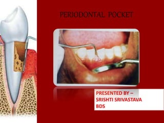
  • 1. PERIODONTAL POCKET PRESENTED BY – SRISHTI SRIVASTAVA BDS
  • 2. • When the sulcular depth is more then three millimeters, will allow food debris and microbes to accumulate. This poses a danger to the periodontal ligament (PDL) fibers that attach the gingiva to the tooth. If accumulated microbes remain undisturbed in a sulcus for an extended period of time, they will penetrate and ultimately destroy the delicate soft tissue and periodontal attachment fibers. If left untreated, this process may lead to a deepening of the sulcus. Introduction
  • 3. • Pocket can be defined as pathologically deepened of the gingival sulcus. (CARRANZA) • Pocket occur with the destruction of the supporting periodontal tissues. Definition
  • 4. • The periodental pockets are caused by micro-organism and their products,which produce pathologic changes that lead to the deepening of the gingival sulcus. Etiology
  • 6. • GINGIVAL POCKET (PSEUDO POCKET): • This type of pocket is formed by gingival enlargement without destruction of the underlying periodontal tissues. The sulcus is deepened because of the increased bulk of the gingiva
  • 7. • Periodontal pocket: • This type of pocket occurs with destruction of the supporting periodontal tissues. Progressive pocket deepening leads to destruction of the supporting periodontal tissues and loosening and exfoliation of the teeth.
  • 8. • Suprabony (supracrestal or supraalveolar), in which the base of the pocket is coronal to the underlying alveolar bone. • Intrabony (infrabony, subcrestal or intraalveolar), in which the base of the pocket is apical to the level of the adjacent alveolar bone.
  • 9. Different types of periodontal pockets A, Gingival pocket-There is no destruction of the supporting periodontal tissues B, Suprabony pocket-The base of the pocket is coronal to the level of the underlying bone. C, Intrabony pocket. The base of the pocket is apical to the level of the adjacent bone. Bone loss is vertical.
  • 10. • SIMPLE POCKET-involving one tooth surface • COMPOUND POCKET- involving two or more tooth surface • COMPLEX POCKET-base of the pocket is not in direct communication with the gingival margin, also c/a spiral pocket.
  • 12. Signs • enlarged,bluish red marginal gingiva with a rolled edge seprated from the tooth surface. • Bluish red vertical zone extending from the gingival margin to the alveolar mucosa. • A break in the faciolingual continuity of the interdental gingiva. • Shiny,discolored and puffy gingiva associated with exposed root surface. clinical features
  • 13. • Gingival bleeding , purulent ` from the gingival margin. • Mobility , extrusion and migration of teeth. • The development of diastema.
  • 14. • Localised pain or a sensation of pressure in the gingiva after eating,which gradually dimnishes. • A foul taste in localized areas • Radiating pain “deep in the bone”. • Feeling of itching in the gums • The urge to dig a pointed instrument into the gums and relief is obtained from the resultant bleeding. • Sensitivity to heat and cold. symptoms
  • 15. The gram-positive bacteria lays down on the on the supragingival tooth surface and extentd in to the gingival sulcus. As a result of inflammation,the following changes are seen in the junctional epithelium- PATHOGENESIS
  • 16. changes in junctional epithelium prolifeartes along the root surface (finger like projections) coronal portion detaches apical portion of from the root junctional epithelium migrates due to bacterial enzymes and physical replaced by pocket epithelium forces exerted by them First event
  • 17. • Colonization of Gram-positive bacteria supragingivally and its extension into the gingival sulcus and conversion of Gram-positive aerobes to Gram-negative anaerobs.
  • 18. aggressive growth and action of Gram- negative bacteria Emigration of neutrophils in large numbers Disruption of epithelial barrier causing open communication Second event
  • 19. loss of chemotactic gradient tissue destruction due to products released by neutrophils as well as bacteria resorption of alveolar bone periodontal pocket is established
  • 20.
  • 21.
  • 22. • Once the pocket is formed , the following microscopic features are present- Changes in the soft tissue wall:  the blood vessels are dilated.  The connective tissue is edematous and densely infiltrated with plasma cells , lymphocytes. HISTOPATHOLOGY
  • 23. • Filaments , rods , cocoid organisms with predominent gram (-) cell walls have been found in intercellular spaces of the epithelium. • Bacteria may invade the intercellular space under epithelial cells but they are also found between deeper epithelial cells and accumulating on the basement lamina. BACTERIAL INVASION
  • 24. • The inflammatory response triggered by bacterial plaque unleashes a complex cascade of events , aimed at destroying and removing bacteria , necrotic cells . • The host cells such as neutrophiles , macrophages , fibroblasts , epithelial cells and others , produce proteinases , cytokines and prostaglandins that can damage or destroy the tissue . MECHANISM OF TISSUE DISTRUCTION-
  • 25. • Under scanning electron microscope the following areas have been noted: 1.areas of bacterial accumulation- which appears as depression on the epithelial surface with abundant debris and bacterial clumps penetrating into the enlarged intercellular spaces. 2.areas of emergence of leukocytes – leukocytes appear in pocket wall through holes located in the intracellular spaces . 3. Area of leukocytes – bacteria interaction numerous leukocytes are present and covered with bacteria in an apparent process of phagocytosis . MICROTOPOGRAPHY OF THE GINGIVAL WALL OF THE POCKET
  • 26. 4. Area of intense epithelial desquamation -consist of semiattached and folded epithelial squames . 5.area of ulceration with exposed connective tissue 6.area of hemorrhage with numerous erythrocytes.
  • 27. • IT is characterized by interplay of destructive and constructive tissue changes . - The destructive changes are characterized by fluid and cellular inflammatory exudates and by the associated changes initiated by the plaque bacteria . - The constructive changes consist of the formation of blood vessels in an effort to repair the tissue damage caused by inflammation . • The balance between the destructive and constructive changes determines the clinical feature such as colour , consistency , and surface texture . PERIODOTAL POCKET AS HEALING LESION
  • 28. • It consists of – debris - containing micro-organisms and their products (enzyme , endotoxin , and other metabolic products). dental plaque Gingival fluid food remnants salivary mucin desqumated epithelial cell and leucocytes POCKET CONTENTS
  • 29. • If the purulent exudate is present , it consist of- living , degenerated and necrotic leukocytes , living and dead bacteria , serum and a scanty amount of fibrin . pus formation is common feature in periodontal disease but it only secondary sign .
  • 30. • 1- STRUCTURAL CHANGES – A- presence of pathologic granules B- areas of increase mineralization C- areas of demineralization / root caries • 2- CHEMICAL CHANGES – the mineral content of exposed cementum increased . Mineral increased in root surface – calcium , magnesium , phosphate , floride and others . • 3- CYTOTOXIC CHANGES – bacterial penetration into cementum can be found as deep as cementodentinal junction . in addition , bacterial products such as endotoxins have also been detected . CHANGES IN ROOT SURFACE WALL
  • 31. • 1-cementum covered by calculus. • 2-attached plaque. • 3-the zone of unattached plaque. • 4-the zone where the junctional epithelium is attached to the tooth. • 5-the zone of semi destroyed C.T. fibers. Surface morphology of the tooth wall of the periodontal pockets
  • 32. ZONES IN THE BASE OF PERIODONTAL POCKET
  • 33. Periodontal probes are used to locate, measure, and mark pockets, as well as determine their course on individual tooth surfaces. DIAGNOSIS
  • 34. • Periodontal probes may be divided into: First generation probes are conventional, and hand held probes, e.g. conventional periodontal probes. Second generation probes are pressure –sensitive probes. It has been shown that, with forces up to 30gms the probe tip remains within junctional epithelium and forces up to 50gms are necessary to diagnose osseous defects. This probe did solve many problems of the conventional probes, but lacked tactile sensitivity.
  • 35.  Third generation probes are computerized probes. Gibbs et al designed Florida probe. E.g.-Foster Miller Probe, Toronto Automated Probes, which can detect the cemento-enamel junction.  Fourth generation probes are the three dimensional probes in which sequential probe positions are measured.  Fifth generation probes are ultrasonographic probes which provides painless probing to the patient. The guidance path is predetermined in these probes.
  • 36. There are three types of periodontal probes. They are: 1. Calibrated periodontal probes 2. Naber’s furcation probe 3. Computer assisted probes TYPES OF PERIODONTAL PROBES
  • 37. • There are two different pocket depths: - The biologic depth is the distance between the gingival margin and the base of the pocket (coronal end of junctional epithelium). This can be measured only by histological sections. - The probing depth is the distance to which the probe penetrates into the pocket. POCKET PROBING
  • 38.
  • 39. • The depth of penetration of the probe in the connective tissue apical to the junctional epithelium in a periodontal pocket is about 0.3mm. • The probing forces of 0.75N have been found to be well tolerated and accurate.
  • 40. • The probe should be inserted parallel to the vertical axis of the tooth and “walked” circumferentially around each tooth to detect the areas of deepest penetration. • To detect an interdental crater the probe should be placed obliquely from both the facial and the lingual surface to explore the deepest point of the pocket located beneath the contact point. • To detect furcation involvement in multi- rooted teeth, use of specially designed Naber’s probe allows an easier and more accurate exploration of the horizontal component of furcation lesion. PROBING TECHNIQUE
  • 41.
  • 42. • Probing of pockets is done at various times for diagnosis and for monitoring the course of treatment and maintenance. • Initial probing: Done to determine whether the tooth can be saved or should be extracted. • Second probing: Done to establish accurately the level of attachment and degree of involvement of roots and furcations. When to probe