SlideShare une entreprise Scribd logo
1  sur  31
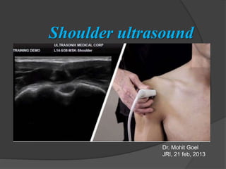
Shoulder ultrasound
Dr. Mohit Goel
JRI, 21 feb, 2013
Anatomy
Four muscles attach as tendons onto the proximal humerus and make up the rotator
cuff: supraspinatus, subscapularis, infraspinatus, and teres minor.
• The supraspinatus is located in the
suprascapular fossa of the scapula
and inserts onto the greater tuberosity
of the proximal humerus.
• The subscapularis is located anterior
to the scapula and inserts onto the
lesser tuberosity of the proximal humerus.
• The rotator interval -
In this space, the long head of the biceps
brachii tendon becomes intraarticular as it
courses toward the supraglenoid tubercle
of the scapula
• The infraspinatus is located posterior
to the scapula inferior to the scapular
spine and inserts onto the posterior
aspect of the greater tuberosity, and
• The teres minor is located just inferior
to the infraspinatus and also inserts
onto the greater tuberosity.
Positioning
It is strongly recommend to examine the patient while seated on a revolving stool.
This position allows the examiner to reach the anterior, lateral and posterior
aspects of the shoulder with the probe by simply asking the patient to rotate on
the chair.
The basic objectives for each step are to
(a) identify the tendon of interest,
(b) image the tendon in two planes (long and short axis),
(c) eliminate artifact by directing the ultrasound beam perpendicular to the
tendon, and
(d) diagnose disease.
Step 1: Biceps brachii tendon, long head.
The patient is asked to place his or her
hand palm up on the lap. The transducer is
placed in the axial plane on the body over
the anterior shoulder.
Within the bicipital groove lies the long
head of the biceps brachii tendon, seen in
short axis.
Because the biceps tendon is coursing
deep away from the skin surface, it is
common for the tendon to appear
artifactually hypoechoic from anisotropy.
This artifact is eliminated by making the transducer along the long axis of the
tendon so that the sound beam is angled superiorly. The normal tendon will then
appear hyperechoic and fibrillar.
The transducer is then turned 90° to visualize the biceps
tendon in long axis.
Transducer pressure distally is usually needed to aim the
ultrasound beam cephalad and perpendicular to the biceps
tendon, which will appear hyperechoic and fibrillar.
If the biceps brachii tendon is oblique to the sound beam, it
will appear hypoechoic from anisotropy.
Step 2: Subscapularis and biceps tendon subluxation
/dislocation.
With the patient’s hand remaining palm up on his or her
lap, the transducer is again placed over the anterior
shoulder in the axial plane to visualize the bicipital
groove.
The transducer is then centered over the lesser
tuberosity at the medial aspect of the
bicipital groove.
The patient is then asked to externally
rotate the shoulder. As the lesser
tuberosity rotates laterally, the
subscapularis located inferior to the
coracoid is pulled laterally.
The US transducer is then rotated 90° along the
long axis of the subscapularis and moved laterally
over the bicipital groove to ensure that the long
head of the biceps brachii tendon is normally
located in the bicipital groove.
Partial displacement of the biceps tendon from the
bicipital groove is termed subluxation, while
complete medial displacement
is termed dislocation.
Such abnormal position of the
biceps tendon may only
occur transiently during
external shoulder rotation.
Subscapularis tendon in short axis.
Step 3: Supraspinatus and rotator interval.
Corresponding US image over humeral head shows hyperechoic and fibrillar
supraspinatus tendon (SS). Note biceps brachii tendon (B) in the rotator interval
with superficial coracohumeral ligament (arrowhead) and medial superior
glenohumeral ligament (arrow). SC = subscapularis tendon, curved arrow =
hyaline articular cartilage, wavy arrow = subacromial-subdeltoid bursa, H =
humeral head. Right side of image is anterior.
Step 4:
Acromioclavicular joint, subacromial-subdeltoid bursa,
and dynamic evaluation for
subacromial impingement.
To locate the acromioclavicular joint, one may simply
palpate the clavicle and move laterally toward the
acromion, with the transducer in the coronal plane on
the body.
Corresponding US image shows
acromioclavicular joint (arrow) with
characteristic hyperechoic bone
contours of the distal clavicle (C) and
acromion (A).
Note echogenic fibrocartilage disc
(arrowhead). Left side of image is
lateral.
The acromioclavicular joint is evaluated for bone irregularity, narrowing, widening,
or offset.
If the acromioclavicular joint is widened or if there is clinical suspicion for
acromioclavicular joint disruption, dynamic evaluation should be used to assess
for changes in alignment.
While assessing the acromioclavicular joint in long axis relative to the clavicle, the
patient is asked to move his or her ipsilateral hand to the opposite shoulder.
With this maneuver, the acromioclavicular joint may abnormally widen or offset or
may cause a bone-on-bone contact between the acromion and clavicle, which
can be associated with symptoms.
With the bone landmarks of the greater tuberosity and the lateral acromion in view,
the patient is asked to actively elevate the arm to his or her side.
Dynamic evaluation is then used to assess for subacromial
impingement.
The transducer is moved laterally from the acromioclavicular joint and is
positioned over the lateral edge of the acromion.
Corresponding US image shows acromion (A) and greater tuberosity (GT) with
supraspinatus tendon (S) and collapsed subacromial-subdeltoid bursa (arrow).
US image shows acromion (A), greater tuberosity (GT), and normal collapsed
subacromial-subdeltoid bursa (arrow). Left side of images is lateral.
During active arm elevation, the supraspinatus tendon and overlying subacromial-
subdeltoid bursa should slide smoothly under the acromion and out of view.
Pooling of bursal fluid at the lateral acromion edge or snapping of bursal tissue
indicates subacromial impingement.
Other findings of impingement include interposition of the supraspinatus tendon
between the greater tuberosity and the acromion, as well as direct contact
between the greater tuberosity and the acromion.
Dynamic evaluation for subacromial impingement can also be completed with the
patients raising their arm anterolateral in front of their body, with their hand in
pronation.
Step 5: Infraspinatus, teres minor, and posterior
labrum
To evaluate the infraspinatus tendon, the hand
is returned to the patient’s lap, palm up.
In this neutral position, the transducer is placed
just below the scapular spine over the posterior
shoulder in a slightly oblique axial plane that
parallels the orientation of the scapular spine.
This position will produce a long-axis view of the infraspinatus tendon, which
is assessed at its insertion on the posterior aspect of the greater tuberosity.
Moving the transducer medial toward the scapula, other structures to be evaluated
include the posterior labrum (for labral tear), the spinoglenoid notch (for paralabral
cyst), and the posterior glenohumeral joint recess (for joint fluid or synovitis)
US image medial to b shows spinoglenoid notch (arrowheads) of scapula with adjacent
suprascapular vessels. Note infraspinatus musculotendinous junction (straight arrows)
and central tendon (curved arrows). H = humeral head, L = labrum. Left side of image
is medial.
The transducer is then rotated 90° to
assess the infraspinatus tendon in short
axis.
Corresponding US image shows infraspinatus (straight arrows) and central tendon
(curved arrow). S = scapular spine. Left side of image is superior.
In addition to evaluating the infraspinatus tendon for tear, it is important to
evaluate for fatty degeneration and atrophy of the infraspinatus muscle in the
setting of a rotator cuff tear as this finding indicates higher likelihood of failure
after rotator cuff repair.
The transducer is moved medially over the
musculotendinous junction of the
infraspinatus in short axis.
The diagnosis of fatty degeneration or atrophy is made by comparing the size
and echogenicity of the infraspinatus muscle at the muscle-tendon junction with
the adjacent teres minor muscle.
Normally, the infraspinatus muscle size should be approximately twice that of the
teres minor at the musculotendinous junction and the hypoechoic muscle
echogenicity should be similar.
Corresponding US image shows infraspinatus (straight arrows) with central tendon (curved arrow)
and teres minor (arrowheads) with more superficial tendon (squiggly arrow).
Left side of image
is cephalad.
THANK YOU

Contenu connexe

Tendances

Radiology of the Elbow Joint. Dr. Sumit Sharma
Radiology of the Elbow Joint. Dr. Sumit SharmaRadiology of the Elbow Joint. Dr. Sumit Sharma
Radiology of the Elbow Joint. Dr. Sumit SharmaSumit Sharma
 
Presentation1.pptx, ultrasound examination of the wrist joint.
Presentation1.pptx, ultrasound examination of the wrist joint.Presentation1.pptx, ultrasound examination of the wrist joint.
Presentation1.pptx, ultrasound examination of the wrist joint.Abdellah Nazeer
 
Doppler ultrasound of carotid arteries
Doppler ultrasound of carotid arteriesDoppler ultrasound of carotid arteries
Doppler ultrasound of carotid arteriesSamir Haffar
 
Presentation2.pptx wrist joint.
Presentation2.pptx wrist joint.Presentation2.pptx wrist joint.
Presentation2.pptx wrist joint.Abdellah Nazeer
 
Doppler ultrasound of lower limb arteries
Doppler ultrasound of lower limb arteriesDoppler ultrasound of lower limb arteries
Doppler ultrasound of lower limb arteriesSamir Haffar
 
Role of ultrasound in clinical evaluation of shoulder Dr. Muhammad Bin Zulfiqar
Role of ultrasound in clinical evaluation of shoulder Dr. Muhammad Bin ZulfiqarRole of ultrasound in clinical evaluation of shoulder Dr. Muhammad Bin Zulfiqar
Role of ultrasound in clinical evaluation of shoulder Dr. Muhammad Bin ZulfiqarDr. Muhammad Bin Zulfiqar
 
Presentation1.pptx, radiological anatomy of the shoulder joint.
Presentation1.pptx, radiological anatomy of the shoulder joint.Presentation1.pptx, radiological anatomy of the shoulder joint.
Presentation1.pptx, radiological anatomy of the shoulder joint.Abdellah Nazeer
 
Mri anatomy of knee Dr. Muhammad Bin Zulfiqar
Mri anatomy of knee Dr. Muhammad Bin ZulfiqarMri anatomy of knee Dr. Muhammad Bin Zulfiqar
Mri anatomy of knee Dr. Muhammad Bin ZulfiqarDr. Muhammad Bin Zulfiqar
 
Presentation1.pptx, ultrasound examination of the knee joint.
Presentation1.pptx, ultrasound examination of the knee joint.Presentation1.pptx, ultrasound examination of the knee joint.
Presentation1.pptx, ultrasound examination of the knee joint.Abdellah Nazeer
 
MRI KNEE JOINT ANATOMY
MRI KNEE JOINT ANATOMYMRI KNEE JOINT ANATOMY
MRI KNEE JOINT ANATOMYNikhil Bansal
 
Imaging in Skull base
Imaging in Skull baseImaging in Skull base
Imaging in Skull baseRakesh Ca
 
Presentation1, radiological imaging of popliteal fossa masses.
Presentation1, radiological imaging of popliteal fossa masses.Presentation1, radiological imaging of popliteal fossa masses.
Presentation1, radiological imaging of popliteal fossa masses.Abdellah Nazeer
 
Mri shoulder joint with common pathologies
Mri shoulder joint with common pathologiesMri shoulder joint with common pathologies
Mri shoulder joint with common pathologiesGobardhan Thapa
 

Tendances (20)

Radiology of the Elbow Joint. Dr. Sumit Sharma
Radiology of the Elbow Joint. Dr. Sumit SharmaRadiology of the Elbow Joint. Dr. Sumit Sharma
Radiology of the Elbow Joint. Dr. Sumit Sharma
 
Presentation1.pptx, ultrasound examination of the wrist joint.
Presentation1.pptx, ultrasound examination of the wrist joint.Presentation1.pptx, ultrasound examination of the wrist joint.
Presentation1.pptx, ultrasound examination of the wrist joint.
 
Mri knee trauma
Mri knee traumaMri knee trauma
Mri knee trauma
 
Doppler ultrasound of carotid arteries
Doppler ultrasound of carotid arteriesDoppler ultrasound of carotid arteries
Doppler ultrasound of carotid arteries
 
Presentation2.pptx wrist joint.
Presentation2.pptx wrist joint.Presentation2.pptx wrist joint.
Presentation2.pptx wrist joint.
 
Doppler ultrasound of lower limb arteries
Doppler ultrasound of lower limb arteriesDoppler ultrasound of lower limb arteries
Doppler ultrasound of lower limb arteries
 
Role of ultrasound in clinical evaluation of shoulder Dr. Muhammad Bin Zulfiqar
Role of ultrasound in clinical evaluation of shoulder Dr. Muhammad Bin ZulfiqarRole of ultrasound in clinical evaluation of shoulder Dr. Muhammad Bin Zulfiqar
Role of ultrasound in clinical evaluation of shoulder Dr. Muhammad Bin Zulfiqar
 
Presentation1.pptx, radiological anatomy of the shoulder joint.
Presentation1.pptx, radiological anatomy of the shoulder joint.Presentation1.pptx, radiological anatomy of the shoulder joint.
Presentation1.pptx, radiological anatomy of the shoulder joint.
 
Spine radiography
Spine radiographySpine radiography
Spine radiography
 
MRI of the shoulder
MRI of the shoulderMRI of the shoulder
MRI of the shoulder
 
Mri anatomy of knee Dr. Muhammad Bin Zulfiqar
Mri anatomy of knee Dr. Muhammad Bin ZulfiqarMri anatomy of knee Dr. Muhammad Bin Zulfiqar
Mri anatomy of knee Dr. Muhammad Bin Zulfiqar
 
Renal doppler
Renal dopplerRenal doppler
Renal doppler
 
Ultrasound - Knee
Ultrasound - KneeUltrasound - Knee
Ultrasound - Knee
 
Presentation1.pptx, ultrasound examination of the knee joint.
Presentation1.pptx, ultrasound examination of the knee joint.Presentation1.pptx, ultrasound examination of the knee joint.
Presentation1.pptx, ultrasound examination of the knee joint.
 
MRI KNEE JOINT ANATOMY
MRI KNEE JOINT ANATOMYMRI KNEE JOINT ANATOMY
MRI KNEE JOINT ANATOMY
 
Imaging in Skull base
Imaging in Skull baseImaging in Skull base
Imaging in Skull base
 
Presentation1, radiological imaging of popliteal fossa masses.
Presentation1, radiological imaging of popliteal fossa masses.Presentation1, radiological imaging of popliteal fossa masses.
Presentation1, radiological imaging of popliteal fossa masses.
 
MRI Knee trauma
MRI Knee traumaMRI Knee trauma
MRI Knee trauma
 
Mri shoulder joint with common pathologies
Mri shoulder joint with common pathologiesMri shoulder joint with common pathologies
Mri shoulder joint with common pathologies
 
Hip joint
Hip jointHip joint
Hip joint
 

En vedette

Imaging of shoulder - Dr. Vishal Sankpal
Imaging of shoulder - Dr. Vishal SankpalImaging of shoulder - Dr. Vishal Sankpal
Imaging of shoulder - Dr. Vishal SankpalVishal Sankpal
 
Presentation1.pptx. ultrasound examination of the ankle joint.
Presentation1.pptx. ultrasound examination of the ankle joint.Presentation1.pptx. ultrasound examination of the ankle joint.
Presentation1.pptx. ultrasound examination of the ankle joint.Abdellah Nazeer
 
Post-graduate Certifcate Musculoskeletal Ultrasound - The Shoulder
Post-graduate Certifcate Musculoskeletal Ultrasound - The ShoulderPost-graduate Certifcate Musculoskeletal Ultrasound - The Shoulder
Post-graduate Certifcate Musculoskeletal Ultrasound - The ShoulderDr. Peter Resteghini
 
Basics of msk ultrasound By Dr. Raham Bacha
Basics of msk ultrasound  By Dr. Raham BachaBasics of msk ultrasound  By Dr. Raham Bacha
Basics of msk ultrasound By Dr. Raham BachaMedical Ultrasound
 
Neonatal Cranial & Spinal Ultrasound
Neonatal Cranial & Spinal UltrasoundNeonatal Cranial & Spinal Ultrasound
Neonatal Cranial & Spinal UltrasoundJoan Zawin
 
Ultrasound of the shoulder 2012
Ultrasound of the shoulder 2012Ultrasound of the shoulder 2012
Ultrasound of the shoulder 2012Lennard Funk
 
20140913 basic musculoskeletal ultrasound abnormalities kailen tsai更正版
20140913 basic musculoskeletal ultrasound abnormalities kailen tsai更正版20140913 basic musculoskeletal ultrasound abnormalities kailen tsai更正版
20140913 basic musculoskeletal ultrasound abnormalities kailen tsai更正版Kailen Tsai
 
Updated lecture about Ultrasonography of the knee
Updated lecture about Ultrasonography of the kneeUpdated lecture about Ultrasonography of the knee
Updated lecture about Ultrasonography of the kneeAbdallah Allam
 
Ultrasonography of Hip joint
Ultrasonography of Hip jointUltrasonography of Hip joint
Ultrasonography of Hip jointAbdallah Allam
 
Basic sonoanatomy of ultrasound guided interventions of cervical and lumbar s...
Basic sonoanatomy of ultrasound guided interventions of cervical and lumbar s...Basic sonoanatomy of ultrasound guided interventions of cervical and lumbar s...
Basic sonoanatomy of ultrasound guided interventions of cervical and lumbar s...Abdallah Allam
 
MSK US of Wrist & Hand
MSK US of Wrist & HandMSK US of Wrist & Hand
MSK US of Wrist & HandAbdallah Allam
 
Ultrasound examination of the Ankle
Ultrasound examination of the AnkleUltrasound examination of the Ankle
Ultrasound examination of the AnkleAbdallah Allam
 
Shoulder impingement
Shoulder impingementShoulder impingement
Shoulder impingementMaher Assaf
 
Rotator cuff injuries
Rotator cuff injuriesRotator cuff injuries
Rotator cuff injuriesorthoprince
 

En vedette (20)

Ultrasound - Shoulder
Ultrasound - ShoulderUltrasound - Shoulder
Ultrasound - Shoulder
 
Ultrasound - Hip
Ultrasound - HipUltrasound - Hip
Ultrasound - Hip
 
Imaging of shoulder - Dr. Vishal Sankpal
Imaging of shoulder - Dr. Vishal SankpalImaging of shoulder - Dr. Vishal Sankpal
Imaging of shoulder - Dr. Vishal Sankpal
 
Presentation1.pptx. ultrasound examination of the ankle joint.
Presentation1.pptx. ultrasound examination of the ankle joint.Presentation1.pptx. ultrasound examination of the ankle joint.
Presentation1.pptx. ultrasound examination of the ankle joint.
 
Post-graduate Certifcate Musculoskeletal Ultrasound - The Shoulder
Post-graduate Certifcate Musculoskeletal Ultrasound - The ShoulderPost-graduate Certifcate Musculoskeletal Ultrasound - The Shoulder
Post-graduate Certifcate Musculoskeletal Ultrasound - The Shoulder
 
Basics of msk ultrasound By Dr. Raham Bacha
Basics of msk ultrasound  By Dr. Raham BachaBasics of msk ultrasound  By Dr. Raham Bacha
Basics of msk ultrasound By Dr. Raham Bacha
 
Neonatal Cranial & Spinal Ultrasound
Neonatal Cranial & Spinal UltrasoundNeonatal Cranial & Spinal Ultrasound
Neonatal Cranial & Spinal Ultrasound
 
Ultrasound of the shoulder 2012
Ultrasound of the shoulder 2012Ultrasound of the shoulder 2012
Ultrasound of the shoulder 2012
 
Ultrasoud hernia
Ultrasoud herniaUltrasoud hernia
Ultrasoud hernia
 
20140913 basic musculoskeletal ultrasound abnormalities kailen tsai更正版
20140913 basic musculoskeletal ultrasound abnormalities kailen tsai更正版20140913 basic musculoskeletal ultrasound abnormalities kailen tsai更正版
20140913 basic musculoskeletal ultrasound abnormalities kailen tsai更正版
 
Shoulder
ShoulderShoulder
Shoulder
 
Elbow
ElbowElbow
Elbow
 
Updated lecture about Ultrasonography of the knee
Updated lecture about Ultrasonography of the kneeUpdated lecture about Ultrasonography of the knee
Updated lecture about Ultrasonography of the knee
 
Ultrasonography of Hip joint
Ultrasonography of Hip jointUltrasonography of Hip joint
Ultrasonography of Hip joint
 
Basic sonoanatomy of ultrasound guided interventions of cervical and lumbar s...
Basic sonoanatomy of ultrasound guided interventions of cervical and lumbar s...Basic sonoanatomy of ultrasound guided interventions of cervical and lumbar s...
Basic sonoanatomy of ultrasound guided interventions of cervical and lumbar s...
 
MSK US of Wrist & Hand
MSK US of Wrist & HandMSK US of Wrist & Hand
MSK US of Wrist & Hand
 
Ultrasound examination of the Ankle
Ultrasound examination of the AnkleUltrasound examination of the Ankle
Ultrasound examination of the Ankle
 
MRI OF SHOULDER INJURY
MRI OF SHOULDER INJURYMRI OF SHOULDER INJURY
MRI OF SHOULDER INJURY
 
Shoulder impingement
Shoulder impingementShoulder impingement
Shoulder impingement
 
Rotator cuff injuries
Rotator cuff injuriesRotator cuff injuries
Rotator cuff injuries
 

Similaire à Shoulder ultrasound

Shoulder Examination.pdf
Shoulder Examination.pdfShoulder Examination.pdf
Shoulder Examination.pdfArshia Nozari
 
RADIOLOGICAL ANATOMY OF UPPER LIMB(SHOULDER@ELBOW)
RADIOLOGICAL ANATOMY OF UPPER LIMB(SHOULDER@ELBOW)RADIOLOGICAL ANATOMY OF UPPER LIMB(SHOULDER@ELBOW)
RADIOLOGICAL ANATOMY OF UPPER LIMB(SHOULDER@ELBOW)Bhaskar Sangamreddy
 
Shoulder girdle presentation
Shoulder girdle presentationShoulder girdle presentation
Shoulder girdle presentationEmmie Macabiog
 
Shoulder girdle presentation
Shoulder girdle presentationShoulder girdle presentation
Shoulder girdle presentationEmmie Macabiog
 
usg shoulder by dr. kanishka.pptx
usg shoulder by dr. kanishka.pptxusg shoulder by dr. kanishka.pptx
usg shoulder by dr. kanishka.pptxRajVaghasia
 
Shoulderclavicleribs.pptx
Shoulderclavicleribs.pptxShoulderclavicleribs.pptx
Shoulderclavicleribs.pptxNishanthRajan7
 
Clinical examination in orthopedics
Clinical examination in orthopedicsClinical examination in orthopedics
Clinical examination in orthopedicsSpringer
 
Shoulder dislocation: Types and Management Methods of Reduction
Shoulder dislocation: Types and Management Methods of ReductionShoulder dislocation: Types and Management Methods of Reduction
Shoulder dislocation: Types and Management Methods of ReductionUzair Siddiqui
 
Manualmusletesting 181 210
Manualmusletesting 181 210Manualmusletesting 181 210
Manualmusletesting 181 210Anaum1990
 
(4-M) 301 - A Shoulder to Clavicle.pptx
(4-M) 301 - A  Shoulder to Clavicle.pptx(4-M) 301 - A  Shoulder to Clavicle.pptx
(4-M) 301 - A Shoulder to Clavicle.pptxShiennaRoseAnnManalo1
 
(4-M) 301 - A Shoulder to Clavicle.pptx
(4-M) 301 - A  Shoulder to Clavicle.pptx(4-M) 301 - A  Shoulder to Clavicle.pptx
(4-M) 301 - A Shoulder to Clavicle.pptxShiennaRoseAnnManalo1
 
Radiographic positioning of humerus and shoulder
Radiographic positioning of humerus and shoulderRadiographic positioning of humerus and shoulder
Radiographic positioning of humerus and shouldershajitha khan
 
Rotatory cuff syndrome & Scapular Dyskinesia
Rotatory cuff syndrome & Scapular DyskinesiaRotatory cuff syndrome & Scapular Dyskinesia
Rotatory cuff syndrome & Scapular DyskinesiaDr. Manoj Parida
 
SHOULDER USG RAJ.pptx
SHOULDER USG RAJ.pptxSHOULDER USG RAJ.pptx
SHOULDER USG RAJ.pptxRajVaghasia
 
Deformities around elbow and management
Deformities around elbow and managementDeformities around elbow and management
Deformities around elbow and managementDR. D. P. SWAMI
 
Shoulder dislocation Co Norbert
Shoulder dislocation  Co Norbert Shoulder dislocation  Co Norbert
Shoulder dislocation Co Norbert Icyizanye Norbert
 
Presentation1.pptx, radiological anatomy of the upper limb joint.
Presentation1.pptx, radiological anatomy of the upper limb joint.Presentation1.pptx, radiological anatomy of the upper limb joint.
Presentation1.pptx, radiological anatomy of the upper limb joint.Abdellah Nazeer
 
Fractures of the distal humerus ppt
Fractures of the distal humerus pptFractures of the distal humerus ppt
Fractures of the distal humerus pptKunal Arora
 

Similaire à Shoulder ultrasound (20)

Shoulder Examination.pdf
Shoulder Examination.pdfShoulder Examination.pdf
Shoulder Examination.pdf
 
RADIOLOGICAL ANATOMY OF UPPER LIMB(SHOULDER@ELBOW)
RADIOLOGICAL ANATOMY OF UPPER LIMB(SHOULDER@ELBOW)RADIOLOGICAL ANATOMY OF UPPER LIMB(SHOULDER@ELBOW)
RADIOLOGICAL ANATOMY OF UPPER LIMB(SHOULDER@ELBOW)
 
Shoulder girdle presentation
Shoulder girdle presentationShoulder girdle presentation
Shoulder girdle presentation
 
Shoulder girdle presentation
Shoulder girdle presentationShoulder girdle presentation
Shoulder girdle presentation
 
usg shoulder by dr. kanishka.pptx
usg shoulder by dr. kanishka.pptxusg shoulder by dr. kanishka.pptx
usg shoulder by dr. kanishka.pptx
 
Shoulderclavicleribs.pptx
Shoulderclavicleribs.pptxShoulderclavicleribs.pptx
Shoulderclavicleribs.pptx
 
Clinical examination in orthopedics
Clinical examination in orthopedicsClinical examination in orthopedics
Clinical examination in orthopedics
 
Knee
KneeKnee
Knee
 
Shoulder dislocation: Types and Management Methods of Reduction
Shoulder dislocation: Types and Management Methods of ReductionShoulder dislocation: Types and Management Methods of Reduction
Shoulder dislocation: Types and Management Methods of Reduction
 
Manualmusletesting 181 210
Manualmusletesting 181 210Manualmusletesting 181 210
Manualmusletesting 181 210
 
Anterior knee pain
Anterior knee painAnterior knee pain
Anterior knee pain
 
(4-M) 301 - A Shoulder to Clavicle.pptx
(4-M) 301 - A  Shoulder to Clavicle.pptx(4-M) 301 - A  Shoulder to Clavicle.pptx
(4-M) 301 - A Shoulder to Clavicle.pptx
 
(4-M) 301 - A Shoulder to Clavicle.pptx
(4-M) 301 - A  Shoulder to Clavicle.pptx(4-M) 301 - A  Shoulder to Clavicle.pptx
(4-M) 301 - A Shoulder to Clavicle.pptx
 
Radiographic positioning of humerus and shoulder
Radiographic positioning of humerus and shoulderRadiographic positioning of humerus and shoulder
Radiographic positioning of humerus and shoulder
 
Rotatory cuff syndrome & Scapular Dyskinesia
Rotatory cuff syndrome & Scapular DyskinesiaRotatory cuff syndrome & Scapular Dyskinesia
Rotatory cuff syndrome & Scapular Dyskinesia
 
SHOULDER USG RAJ.pptx
SHOULDER USG RAJ.pptxSHOULDER USG RAJ.pptx
SHOULDER USG RAJ.pptx
 
Deformities around elbow and management
Deformities around elbow and managementDeformities around elbow and management
Deformities around elbow and management
 
Shoulder dislocation Co Norbert
Shoulder dislocation  Co Norbert Shoulder dislocation  Co Norbert
Shoulder dislocation Co Norbert
 
Presentation1.pptx, radiological anatomy of the upper limb joint.
Presentation1.pptx, radiological anatomy of the upper limb joint.Presentation1.pptx, radiological anatomy of the upper limb joint.
Presentation1.pptx, radiological anatomy of the upper limb joint.
 
Fractures of the distal humerus ppt
Fractures of the distal humerus pptFractures of the distal humerus ppt
Fractures of the distal humerus ppt
 

Plus de Anish Choudhary

Plus de Anish Choudhary (20)

Pulmonary embolism radiology
Pulmonary embolism radiologyPulmonary embolism radiology
Pulmonary embolism radiology
 
Anatomy of esophgus
Anatomy of esophgusAnatomy of esophgus
Anatomy of esophgus
 
Malignant liver masses
Malignant liver massesMalignant liver masses
Malignant liver masses
 
KEYS OF RADIOLOGY SPOTTERS GIT
KEYS OF RADIOLOGY SPOTTERS GITKEYS OF RADIOLOGY SPOTTERS GIT
KEYS OF RADIOLOGY SPOTTERS GIT
 
RADIOLOGY SPOTTERS GIT
RADIOLOGY SPOTTERS GITRADIOLOGY SPOTTERS GIT
RADIOLOGY SPOTTERS GIT
 
radiology Spotters mixed bag
radiology Spotters mixed bagradiology Spotters mixed bag
radiology Spotters mixed bag
 
Usg gb & biliary t
Usg gb & biliary tUsg gb & biliary t
Usg gb & biliary t
 
Tvs image gallery
Tvs image galleryTvs image gallery
Tvs image gallery
 
Treat. contrast reaction
Treat. contrast reactionTreat. contrast reaction
Treat. contrast reaction
 
Transitional vertebrae
Transitional vertebraeTransitional vertebrae
Transitional vertebrae
 
Spots with keys
Spots with keysSpots with keys
Spots with keys
 
Spots with keys (2)
Spots with keys (2)Spots with keys (2)
Spots with keys (2)
 
Sectional anatomy of abdomen
Sectional anatomy of abdomenSectional anatomy of abdomen
Sectional anatomy of abdomen
 
Retroperitoneal masses
Retroperitoneal massesRetroperitoneal masses
Retroperitoneal masses
 
Precocious puberty
Precocious pubertyPrecocious puberty
Precocious puberty
 
Pre fess pns ct
Pre fess pns ctPre fess pns ct
Pre fess pns ct
 
Pineal region masses
Pineal region massesPineal region masses
Pineal region masses
 
Peripheral arterial doppler
Peripheral  arterial dopplerPeripheral  arterial doppler
Peripheral arterial doppler
 
Pediatric stroke modified
Pediatric stroke modifiedPediatric stroke modified
Pediatric stroke modified
 
Pediatric chest
Pediatric chestPediatric chest
Pediatric chest
 

Dernier

Bangalore Call Girl Whatsapp Number 100% Complete Your Sexual Needs
Bangalore Call Girl Whatsapp Number 100% Complete Your Sexual NeedsBangalore Call Girl Whatsapp Number 100% Complete Your Sexual Needs
Bangalore Call Girl Whatsapp Number 100% Complete Your Sexual NeedsGfnyt
 
Call Girls Service Jaipur Grishma WhatsApp ❤8445551418 VIP Call Girls Jaipur
Call Girls Service Jaipur Grishma WhatsApp ❤8445551418 VIP Call Girls JaipurCall Girls Service Jaipur Grishma WhatsApp ❤8445551418 VIP Call Girls Jaipur
Call Girls Service Jaipur Grishma WhatsApp ❤8445551418 VIP Call Girls Jaipurparulsinha
 
High Profile Call Girls Coimbatore Saanvi☎️ 8250192130 Independent Escort Se...
High Profile Call Girls Coimbatore Saanvi☎️  8250192130 Independent Escort Se...High Profile Call Girls Coimbatore Saanvi☎️  8250192130 Independent Escort Se...
High Profile Call Girls Coimbatore Saanvi☎️ 8250192130 Independent Escort Se...narwatsonia7
 
Book Paid Powai Call Girls Mumbai 𖠋 9930245274 𖠋Low Budget Full Independent H...
Book Paid Powai Call Girls Mumbai 𖠋 9930245274 𖠋Low Budget Full Independent H...Book Paid Powai Call Girls Mumbai 𖠋 9930245274 𖠋Low Budget Full Independent H...
Book Paid Powai Call Girls Mumbai 𖠋 9930245274 𖠋Low Budget Full Independent H...Call Girls in Nagpur High Profile
 
Call Girls Horamavu WhatsApp Number 7001035870 Meeting With Bangalore Escorts
Call Girls Horamavu WhatsApp Number 7001035870 Meeting With Bangalore EscortsCall Girls Horamavu WhatsApp Number 7001035870 Meeting With Bangalore Escorts
Call Girls Horamavu WhatsApp Number 7001035870 Meeting With Bangalore Escortsvidya singh
 
Call Girls Siliguri Just Call 9907093804 Top Class Call Girl Service Available
Call Girls Siliguri Just Call 9907093804 Top Class Call Girl Service AvailableCall Girls Siliguri Just Call 9907093804 Top Class Call Girl Service Available
Call Girls Siliguri Just Call 9907093804 Top Class Call Girl Service AvailableDipal Arora
 
Russian Escorts Girls Nehru Place ZINATHI 🔝9711199012 ☪ 24/7 Call Girls Delhi
Russian Escorts Girls  Nehru Place ZINATHI 🔝9711199012 ☪ 24/7 Call Girls DelhiRussian Escorts Girls  Nehru Place ZINATHI 🔝9711199012 ☪ 24/7 Call Girls Delhi
Russian Escorts Girls Nehru Place ZINATHI 🔝9711199012 ☪ 24/7 Call Girls DelhiAlinaDevecerski
 
VIP Mumbai Call Girls Hiranandani Gardens Just Call 9920874524 with A/C Room ...
VIP Mumbai Call Girls Hiranandani Gardens Just Call 9920874524 with A/C Room ...VIP Mumbai Call Girls Hiranandani Gardens Just Call 9920874524 with A/C Room ...
VIP Mumbai Call Girls Hiranandani Gardens Just Call 9920874524 with A/C Room ...Garima Khatri
 
(👑VVIP ISHAAN ) Russian Call Girls Service Navi Mumbai🖕9920874524🖕Independent...
(👑VVIP ISHAAN ) Russian Call Girls Service Navi Mumbai🖕9920874524🖕Independent...(👑VVIP ISHAAN ) Russian Call Girls Service Navi Mumbai🖕9920874524🖕Independent...
(👑VVIP ISHAAN ) Russian Call Girls Service Navi Mumbai🖕9920874524🖕Independent...Taniya Sharma
 
Call Girls Faridabad Just Call 9907093804 Top Class Call Girl Service Available
Call Girls Faridabad Just Call 9907093804 Top Class Call Girl Service AvailableCall Girls Faridabad Just Call 9907093804 Top Class Call Girl Service Available
Call Girls Faridabad Just Call 9907093804 Top Class Call Girl Service AvailableDipal Arora
 
Bangalore Call Girls Nelamangala Number 7001035870 Meetin With Bangalore Esc...
Bangalore Call Girls Nelamangala Number 7001035870  Meetin With Bangalore Esc...Bangalore Call Girls Nelamangala Number 7001035870  Meetin With Bangalore Esc...
Bangalore Call Girls Nelamangala Number 7001035870 Meetin With Bangalore Esc...narwatsonia7
 
Night 7k to 12k Navi Mumbai Call Girl Photo 👉 BOOK NOW 9833363713 👈 ♀️ night ...
Night 7k to 12k Navi Mumbai Call Girl Photo 👉 BOOK NOW 9833363713 👈 ♀️ night ...Night 7k to 12k Navi Mumbai Call Girl Photo 👉 BOOK NOW 9833363713 👈 ♀️ night ...
Night 7k to 12k Navi Mumbai Call Girl Photo 👉 BOOK NOW 9833363713 👈 ♀️ night ...aartirawatdelhi
 
Call Girls Jabalpur Just Call 9907093804 Top Class Call Girl Service Available
Call Girls Jabalpur Just Call 9907093804 Top Class Call Girl Service AvailableCall Girls Jabalpur Just Call 9907093804 Top Class Call Girl Service Available
Call Girls Jabalpur Just Call 9907093804 Top Class Call Girl Service AvailableDipal Arora
 
Call Girls Kochi Just Call 9907093804 Top Class Call Girl Service Available
Call Girls Kochi Just Call 9907093804 Top Class Call Girl Service AvailableCall Girls Kochi Just Call 9907093804 Top Class Call Girl Service Available
Call Girls Kochi Just Call 9907093804 Top Class Call Girl Service AvailableDipal Arora
 
Low Rate Call Girls Kochi Anika 8250192130 Independent Escort Service Kochi
Low Rate Call Girls Kochi Anika 8250192130 Independent Escort Service KochiLow Rate Call Girls Kochi Anika 8250192130 Independent Escort Service Kochi
Low Rate Call Girls Kochi Anika 8250192130 Independent Escort Service KochiSuhani Kapoor
 
(Rocky) Jaipur Call Girl - 09521753030 Escorts Service 50% Off with Cash ON D...
(Rocky) Jaipur Call Girl - 09521753030 Escorts Service 50% Off with Cash ON D...(Rocky) Jaipur Call Girl - 09521753030 Escorts Service 50% Off with Cash ON D...
(Rocky) Jaipur Call Girl - 09521753030 Escorts Service 50% Off with Cash ON D...indiancallgirl4rent
 
Best Rate (Guwahati ) Call Girls Guwahati ⟟ 8617370543 ⟟ High Class Call Girl...
Best Rate (Guwahati ) Call Girls Guwahati ⟟ 8617370543 ⟟ High Class Call Girl...Best Rate (Guwahati ) Call Girls Guwahati ⟟ 8617370543 ⟟ High Class Call Girl...
Best Rate (Guwahati ) Call Girls Guwahati ⟟ 8617370543 ⟟ High Class Call Girl...Dipal Arora
 
Call Girls Aurangabad Just Call 9907093804 Top Class Call Girl Service Available
Call Girls Aurangabad Just Call 9907093804 Top Class Call Girl Service AvailableCall Girls Aurangabad Just Call 9907093804 Top Class Call Girl Service Available
Call Girls Aurangabad Just Call 9907093804 Top Class Call Girl Service AvailableDipal Arora
 
VIP Service Call Girls Sindhi Colony 📳 7877925207 For 18+ VIP Call Girl At Th...
VIP Service Call Girls Sindhi Colony 📳 7877925207 For 18+ VIP Call Girl At Th...VIP Service Call Girls Sindhi Colony 📳 7877925207 For 18+ VIP Call Girl At Th...
VIP Service Call Girls Sindhi Colony 📳 7877925207 For 18+ VIP Call Girl At Th...jageshsingh5554
 

Dernier (20)

Bangalore Call Girl Whatsapp Number 100% Complete Your Sexual Needs
Bangalore Call Girl Whatsapp Number 100% Complete Your Sexual NeedsBangalore Call Girl Whatsapp Number 100% Complete Your Sexual Needs
Bangalore Call Girl Whatsapp Number 100% Complete Your Sexual Needs
 
Call Girls Service Jaipur Grishma WhatsApp ❤8445551418 VIP Call Girls Jaipur
Call Girls Service Jaipur Grishma WhatsApp ❤8445551418 VIP Call Girls JaipurCall Girls Service Jaipur Grishma WhatsApp ❤8445551418 VIP Call Girls Jaipur
Call Girls Service Jaipur Grishma WhatsApp ❤8445551418 VIP Call Girls Jaipur
 
High Profile Call Girls Coimbatore Saanvi☎️ 8250192130 Independent Escort Se...
High Profile Call Girls Coimbatore Saanvi☎️  8250192130 Independent Escort Se...High Profile Call Girls Coimbatore Saanvi☎️  8250192130 Independent Escort Se...
High Profile Call Girls Coimbatore Saanvi☎️ 8250192130 Independent Escort Se...
 
Book Paid Powai Call Girls Mumbai 𖠋 9930245274 𖠋Low Budget Full Independent H...
Book Paid Powai Call Girls Mumbai 𖠋 9930245274 𖠋Low Budget Full Independent H...Book Paid Powai Call Girls Mumbai 𖠋 9930245274 𖠋Low Budget Full Independent H...
Book Paid Powai Call Girls Mumbai 𖠋 9930245274 𖠋Low Budget Full Independent H...
 
Call Girls Horamavu WhatsApp Number 7001035870 Meeting With Bangalore Escorts
Call Girls Horamavu WhatsApp Number 7001035870 Meeting With Bangalore EscortsCall Girls Horamavu WhatsApp Number 7001035870 Meeting With Bangalore Escorts
Call Girls Horamavu WhatsApp Number 7001035870 Meeting With Bangalore Escorts
 
Call Girls Siliguri Just Call 9907093804 Top Class Call Girl Service Available
Call Girls Siliguri Just Call 9907093804 Top Class Call Girl Service AvailableCall Girls Siliguri Just Call 9907093804 Top Class Call Girl Service Available
Call Girls Siliguri Just Call 9907093804 Top Class Call Girl Service Available
 
Russian Escorts Girls Nehru Place ZINATHI 🔝9711199012 ☪ 24/7 Call Girls Delhi
Russian Escorts Girls  Nehru Place ZINATHI 🔝9711199012 ☪ 24/7 Call Girls DelhiRussian Escorts Girls  Nehru Place ZINATHI 🔝9711199012 ☪ 24/7 Call Girls Delhi
Russian Escorts Girls Nehru Place ZINATHI 🔝9711199012 ☪ 24/7 Call Girls Delhi
 
VIP Mumbai Call Girls Hiranandani Gardens Just Call 9920874524 with A/C Room ...
VIP Mumbai Call Girls Hiranandani Gardens Just Call 9920874524 with A/C Room ...VIP Mumbai Call Girls Hiranandani Gardens Just Call 9920874524 with A/C Room ...
VIP Mumbai Call Girls Hiranandani Gardens Just Call 9920874524 with A/C Room ...
 
(👑VVIP ISHAAN ) Russian Call Girls Service Navi Mumbai🖕9920874524🖕Independent...
(👑VVIP ISHAAN ) Russian Call Girls Service Navi Mumbai🖕9920874524🖕Independent...(👑VVIP ISHAAN ) Russian Call Girls Service Navi Mumbai🖕9920874524🖕Independent...
(👑VVIP ISHAAN ) Russian Call Girls Service Navi Mumbai🖕9920874524🖕Independent...
 
Call Girls Faridabad Just Call 9907093804 Top Class Call Girl Service Available
Call Girls Faridabad Just Call 9907093804 Top Class Call Girl Service AvailableCall Girls Faridabad Just Call 9907093804 Top Class Call Girl Service Available
Call Girls Faridabad Just Call 9907093804 Top Class Call Girl Service Available
 
Bangalore Call Girls Nelamangala Number 7001035870 Meetin With Bangalore Esc...
Bangalore Call Girls Nelamangala Number 7001035870  Meetin With Bangalore Esc...Bangalore Call Girls Nelamangala Number 7001035870  Meetin With Bangalore Esc...
Bangalore Call Girls Nelamangala Number 7001035870 Meetin With Bangalore Esc...
 
Night 7k to 12k Navi Mumbai Call Girl Photo 👉 BOOK NOW 9833363713 👈 ♀️ night ...
Night 7k to 12k Navi Mumbai Call Girl Photo 👉 BOOK NOW 9833363713 👈 ♀️ night ...Night 7k to 12k Navi Mumbai Call Girl Photo 👉 BOOK NOW 9833363713 👈 ♀️ night ...
Night 7k to 12k Navi Mumbai Call Girl Photo 👉 BOOK NOW 9833363713 👈 ♀️ night ...
 
Call Girls Jabalpur Just Call 9907093804 Top Class Call Girl Service Available
Call Girls Jabalpur Just Call 9907093804 Top Class Call Girl Service AvailableCall Girls Jabalpur Just Call 9907093804 Top Class Call Girl Service Available
Call Girls Jabalpur Just Call 9907093804 Top Class Call Girl Service Available
 
Call Girls Kochi Just Call 9907093804 Top Class Call Girl Service Available
Call Girls Kochi Just Call 9907093804 Top Class Call Girl Service AvailableCall Girls Kochi Just Call 9907093804 Top Class Call Girl Service Available
Call Girls Kochi Just Call 9907093804 Top Class Call Girl Service Available
 
Low Rate Call Girls Kochi Anika 8250192130 Independent Escort Service Kochi
Low Rate Call Girls Kochi Anika 8250192130 Independent Escort Service KochiLow Rate Call Girls Kochi Anika 8250192130 Independent Escort Service Kochi
Low Rate Call Girls Kochi Anika 8250192130 Independent Escort Service Kochi
 
(Rocky) Jaipur Call Girl - 09521753030 Escorts Service 50% Off with Cash ON D...
(Rocky) Jaipur Call Girl - 09521753030 Escorts Service 50% Off with Cash ON D...(Rocky) Jaipur Call Girl - 09521753030 Escorts Service 50% Off with Cash ON D...
(Rocky) Jaipur Call Girl - 09521753030 Escorts Service 50% Off with Cash ON D...
 
Best Rate (Guwahati ) Call Girls Guwahati ⟟ 8617370543 ⟟ High Class Call Girl...
Best Rate (Guwahati ) Call Girls Guwahati ⟟ 8617370543 ⟟ High Class Call Girl...Best Rate (Guwahati ) Call Girls Guwahati ⟟ 8617370543 ⟟ High Class Call Girl...
Best Rate (Guwahati ) Call Girls Guwahati ⟟ 8617370543 ⟟ High Class Call Girl...
 
Call Girls Aurangabad Just Call 9907093804 Top Class Call Girl Service Available
Call Girls Aurangabad Just Call 9907093804 Top Class Call Girl Service AvailableCall Girls Aurangabad Just Call 9907093804 Top Class Call Girl Service Available
Call Girls Aurangabad Just Call 9907093804 Top Class Call Girl Service Available
 
VIP Service Call Girls Sindhi Colony 📳 7877925207 For 18+ VIP Call Girl At Th...
VIP Service Call Girls Sindhi Colony 📳 7877925207 For 18+ VIP Call Girl At Th...VIP Service Call Girls Sindhi Colony 📳 7877925207 For 18+ VIP Call Girl At Th...
VIP Service Call Girls Sindhi Colony 📳 7877925207 For 18+ VIP Call Girl At Th...
 
Russian Call Girls in Delhi Tanvi ➡️ 9711199012 💋📞 Independent Escort Service...
Russian Call Girls in Delhi Tanvi ➡️ 9711199012 💋📞 Independent Escort Service...Russian Call Girls in Delhi Tanvi ➡️ 9711199012 💋📞 Independent Escort Service...
Russian Call Girls in Delhi Tanvi ➡️ 9711199012 💋📞 Independent Escort Service...
 

Shoulder ultrasound

  • 1. Shoulder ultrasound Dr. Mohit Goel JRI, 21 feb, 2013
  • 2.
  • 3. Anatomy Four muscles attach as tendons onto the proximal humerus and make up the rotator cuff: supraspinatus, subscapularis, infraspinatus, and teres minor. • The supraspinatus is located in the suprascapular fossa of the scapula and inserts onto the greater tuberosity of the proximal humerus. • The subscapularis is located anterior to the scapula and inserts onto the lesser tuberosity of the proximal humerus. • The rotator interval - In this space, the long head of the biceps brachii tendon becomes intraarticular as it courses toward the supraglenoid tubercle of the scapula
  • 4. • The infraspinatus is located posterior to the scapula inferior to the scapular spine and inserts onto the posterior aspect of the greater tuberosity, and • The teres minor is located just inferior to the infraspinatus and also inserts onto the greater tuberosity.
  • 5. Positioning It is strongly recommend to examine the patient while seated on a revolving stool. This position allows the examiner to reach the anterior, lateral and posterior aspects of the shoulder with the probe by simply asking the patient to rotate on the chair. The basic objectives for each step are to (a) identify the tendon of interest, (b) image the tendon in two planes (long and short axis), (c) eliminate artifact by directing the ultrasound beam perpendicular to the tendon, and (d) diagnose disease.
  • 6. Step 1: Biceps brachii tendon, long head. The patient is asked to place his or her hand palm up on the lap. The transducer is placed in the axial plane on the body over the anterior shoulder. Within the bicipital groove lies the long head of the biceps brachii tendon, seen in short axis. Because the biceps tendon is coursing deep away from the skin surface, it is common for the tendon to appear artifactually hypoechoic from anisotropy.
  • 7. This artifact is eliminated by making the transducer along the long axis of the tendon so that the sound beam is angled superiorly. The normal tendon will then appear hyperechoic and fibrillar.
  • 8. The transducer is then turned 90° to visualize the biceps tendon in long axis. Transducer pressure distally is usually needed to aim the ultrasound beam cephalad and perpendicular to the biceps tendon, which will appear hyperechoic and fibrillar. If the biceps brachii tendon is oblique to the sound beam, it will appear hypoechoic from anisotropy.
  • 9.
  • 10.
  • 11. Step 2: Subscapularis and biceps tendon subluxation /dislocation. With the patient’s hand remaining palm up on his or her lap, the transducer is again placed over the anterior shoulder in the axial plane to visualize the bicipital groove. The transducer is then centered over the lesser tuberosity at the medial aspect of the bicipital groove. The patient is then asked to externally rotate the shoulder. As the lesser tuberosity rotates laterally, the subscapularis located inferior to the coracoid is pulled laterally.
  • 12. The US transducer is then rotated 90° along the long axis of the subscapularis and moved laterally over the bicipital groove to ensure that the long head of the biceps brachii tendon is normally located in the bicipital groove. Partial displacement of the biceps tendon from the bicipital groove is termed subluxation, while complete medial displacement is termed dislocation. Such abnormal position of the biceps tendon may only occur transiently during external shoulder rotation. Subscapularis tendon in short axis.
  • 13.
  • 14. Step 3: Supraspinatus and rotator interval.
  • 15. Corresponding US image over humeral head shows hyperechoic and fibrillar supraspinatus tendon (SS). Note biceps brachii tendon (B) in the rotator interval with superficial coracohumeral ligament (arrowhead) and medial superior glenohumeral ligament (arrow). SC = subscapularis tendon, curved arrow = hyaline articular cartilage, wavy arrow = subacromial-subdeltoid bursa, H = humeral head. Right side of image is anterior.
  • 16.
  • 17. Step 4: Acromioclavicular joint, subacromial-subdeltoid bursa, and dynamic evaluation for subacromial impingement. To locate the acromioclavicular joint, one may simply palpate the clavicle and move laterally toward the acromion, with the transducer in the coronal plane on the body. Corresponding US image shows acromioclavicular joint (arrow) with characteristic hyperechoic bone contours of the distal clavicle (C) and acromion (A). Note echogenic fibrocartilage disc (arrowhead). Left side of image is lateral.
  • 18. The acromioclavicular joint is evaluated for bone irregularity, narrowing, widening, or offset. If the acromioclavicular joint is widened or if there is clinical suspicion for acromioclavicular joint disruption, dynamic evaluation should be used to assess for changes in alignment. While assessing the acromioclavicular joint in long axis relative to the clavicle, the patient is asked to move his or her ipsilateral hand to the opposite shoulder. With this maneuver, the acromioclavicular joint may abnormally widen or offset or may cause a bone-on-bone contact between the acromion and clavicle, which can be associated with symptoms.
  • 19. With the bone landmarks of the greater tuberosity and the lateral acromion in view, the patient is asked to actively elevate the arm to his or her side.
  • 20. Dynamic evaluation is then used to assess for subacromial impingement. The transducer is moved laterally from the acromioclavicular joint and is positioned over the lateral edge of the acromion. Corresponding US image shows acromion (A) and greater tuberosity (GT) with supraspinatus tendon (S) and collapsed subacromial-subdeltoid bursa (arrow).
  • 21. US image shows acromion (A), greater tuberosity (GT), and normal collapsed subacromial-subdeltoid bursa (arrow). Left side of images is lateral. During active arm elevation, the supraspinatus tendon and overlying subacromial- subdeltoid bursa should slide smoothly under the acromion and out of view.
  • 22. Pooling of bursal fluid at the lateral acromion edge or snapping of bursal tissue indicates subacromial impingement. Other findings of impingement include interposition of the supraspinatus tendon between the greater tuberosity and the acromion, as well as direct contact between the greater tuberosity and the acromion. Dynamic evaluation for subacromial impingement can also be completed with the patients raising their arm anterolateral in front of their body, with their hand in pronation.
  • 23.
  • 24. Step 5: Infraspinatus, teres minor, and posterior labrum To evaluate the infraspinatus tendon, the hand is returned to the patient’s lap, palm up. In this neutral position, the transducer is placed just below the scapular spine over the posterior shoulder in a slightly oblique axial plane that parallels the orientation of the scapular spine.
  • 25. This position will produce a long-axis view of the infraspinatus tendon, which is assessed at its insertion on the posterior aspect of the greater tuberosity.
  • 26. Moving the transducer medial toward the scapula, other structures to be evaluated include the posterior labrum (for labral tear), the spinoglenoid notch (for paralabral cyst), and the posterior glenohumeral joint recess (for joint fluid or synovitis) US image medial to b shows spinoglenoid notch (arrowheads) of scapula with adjacent suprascapular vessels. Note infraspinatus musculotendinous junction (straight arrows) and central tendon (curved arrows). H = humeral head, L = labrum. Left side of image is medial.
  • 27. The transducer is then rotated 90° to assess the infraspinatus tendon in short axis. Corresponding US image shows infraspinatus (straight arrows) and central tendon (curved arrow). S = scapular spine. Left side of image is superior.
  • 28. In addition to evaluating the infraspinatus tendon for tear, it is important to evaluate for fatty degeneration and atrophy of the infraspinatus muscle in the setting of a rotator cuff tear as this finding indicates higher likelihood of failure after rotator cuff repair. The transducer is moved medially over the musculotendinous junction of the infraspinatus in short axis.
  • 29. The diagnosis of fatty degeneration or atrophy is made by comparing the size and echogenicity of the infraspinatus muscle at the muscle-tendon junction with the adjacent teres minor muscle. Normally, the infraspinatus muscle size should be approximately twice that of the teres minor at the musculotendinous junction and the hypoechoic muscle echogenicity should be similar. Corresponding US image shows infraspinatus (straight arrows) with central tendon (curved arrow) and teres minor (arrowheads) with more superficial tendon (squiggly arrow). Left side of image is cephalad.
  • 30.