SlideShare une entreprise Scribd logo
1  sur  6
Dermatological causes of white patches

 Lichen planus:
      Lichen = primitive plants composed of symbiotic algae and fungi
      Lichen planus = dry lichen-like appearance of affected skin
      It is the commonest dermatological disease that may result in oral white
      lesions
      It is a chronic mucocutaneous disease that involves skin and mucous
      membranes
      Clinical features:
          Skin lesions:
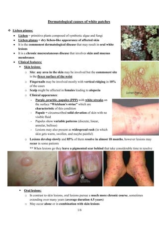
           o Site: any area in the skin may be involved but the commonest site
             is the flexer surface of the wrist
           o Fingernails may be involved mostly with vertical ridging in 10%
             of the cases
           o Scalp might be affected in females leading to alopecia
           o Clinical appearance:
                -   Purple, pruritic, papules (PPP) with white streaks on
                    the surface “Wickham’s striae” which are
                    characteristic of this condition
                -   Papule = circumscribed solid elevation of skin with no
                    visible fluid
                -   Papules show variable patterns (discrete, linear,
                    annular, bullous)
                -   Lesions may also present as widespread rash (in which
                    skin gets warm, swollen, and maybe painful)
           o Lesions develop slowly and 85% of them resolve in almost 18 months, however lesions may
             recur in some patients
               ** When lesions go they leave a pigmented scar behind that take considerable time to resolve


          Oral lesions:
           o In contrast to skin lesions, oral lesions pursue a much more chronic course, sometimes
             extending over many years (average duration 4.5 years)
           o May occur alone or in combination with skin lesions
                                                   1/6
o Site: lesions mostly affect buccal mucosa (90%), but may also affect the tongue, gingiva,
         palate, and lips. The floor of the mouth involvement is relatively uncommon
       o Lesions are often bilateral and show wide spectrum of presentations
       o Clinical appearance:
            -   Non-erosive lesions:
                 Reticular, annular, papular, plaque-like
                  ** Reticular lesions have white streaks arranged in lace-like pattern
                  ** Plaque-like lesions are white patches resembling leukoplakia clinically
                  ** Papular lesions are small white papules that may coalesce
                  ** Bullous lesions are subepithelial bullae
                 They are usually asymptomatic
                 They may show hyperkeratosis with NO atrophy & NO ulceration


Reticular                     Annular                      Papular                      Plaque-like

            -   Erosive/atrophic lesions:
                    ** Erosive lesions are extensive areas of shallow ulceration
                    ** Atrophic lesions are diffuse red lesions resembling Erythroplakia
                 Erosion and atrophy are usually present together and lesions have a red glazed
                  appearance with areas of superficial ulceration which may take several weeks to heal
                 Occasionally, ulcers are preceded by bullae (bullous type) that then rupture
                 Lesions are often associated with typical areas of non-erosive lichen planus
                 They are usually symptomatic
                 Pain and discomfort may be severe (especially when eating spicy/acidic foods)


            -   Gingival lesions:
                 Lichen planus involving the gingiva often presents as a desquamative gingivitis


                                              2/6
 Desquamative gingivitis is a clinical descriptive
                term that doesn’t infer any specific underlying
                pathology (so that it has many causes!)
               Gingival lesions may occur alone (in 10% of
                cases) or with other oral lesions
Prevalence:
o   Lichen planus is a relatively common disease (affects 0.5-2% of general population)
o   There's a worldwide distribution
o   Peak incidence is found between ages 30-50
o   Lesions are more common in females (60% of cases)
o   Oral lesions are detected in ~50% of patients with initial skin lesions
o   Skin lesions are detected in ~10-50% of patients with initial oral lesions!!
    This may be due to:
    - Asymptomatic nature of oral lesions in many cases
    - Inconstant relationship of oral and skin lesions (oral lesions may occur BEFORE, AFTER
        or AT THE SAME TIME as skin lesions)
Histopathological features:
o Orthokeratosis or Parakeratosis
o Epithelial atrophy or acanthosis
o Acanthosis results in irregular elongation and widening
  of the rete ridges in a “saw-tooth pattern”
o Dense, well-defined band of subepithelial mononuclear
  infiltrate (mainly T-lymphocytes)
o Liquefactive degeneration of the basal cell layer with
  edema and lymphocytic infiltration
o The degenerating cells appear as hyaline shrunken bodies,
  called Civatte bodies
o Basal cell degeneration may result in subepithelial bullae
  formation and ulceration because of the lack of cohesion
  between epithelium and lamina propria following the
  degeneration


                                            3/6
o Oral lesions may show little superficial resemblance to skin lesions clinically even though the
  basic histological changes are similar, because of the modifying environment of the oral cavity
  by:
    -   Continuous presence of saliva
    -   Secondary infection by oral organisms
    -   Repeated trauma
o Almost all cases run a benign course
o Malignant transformation has been described in a very small proportion (0.5%-2.5% over 5 years)
o Some studies suggest that atrophic/erosive forms are more likely for such transformation
  because of the decreased barrier presented to potential carcinogens
o Other studies found malignant transformation more likely with plaque lesions
Etiology & pathogenesis:
o It is NOT fully understood
o In most cases the precipitating factors are unknown and the disease is Idiopathic
o It is widely accepted that cell-mediated immune responses to an external antigen, or internal
  antigenic changes in epithelial cells, are involved since response resembles type IV
  hypersensitivity reaction which is T-cell mediated
o Cytotoxic lymphocytes damage the basal epithelium
    ** Possible immunological mechanism in lichen planus:
    External antigen challenge and/or modified antigenic structure of epithelial cells  induces
    cytokines release from langerhans cells & keratinocytes  chemotaxis of lymphocytes which
    accumulate in the basement membrane zone & basal epithelium  antigen presentation to CD4
    helper cells  activation of CD8 Cytotoxic cells  basal cell degeneration
o Lichen planus has been associated with some systemic diseases, in many of these, a cause-and-
  effect relationship has not been established (e.g. there is strong association of the disease with
  chronic liver disease especially hepatitis C virus)
o Oral & skin lesions resembling lichen planus are also seen as a part of graft-versus-host
  reaction (immune reactions in patients receiving transplants), in such cases, the transplanted T
  cells react to antigens on host epithelial cells
o Lichenoid reaction:
  - Lichenoid = lichen-like
  - In some patients, lesions similar to lichen planus may appear
      triggered by hypersensitivity reaction (IV) to certain drugs
      (NSAIDs) or dental materials (amalgam)
  - These lesions are clinically and histologically similar to
      Lichen planus, but tend to be reversible so that they resolve
      upon withdrawal of the offending agent
        ** Lichenoid reactions aren't idiopathic as lichen planus
        ** Lichenoid reactions are usually unilateral while lichen planus is usually bilateral


                                             4/6
 Lupus Erythematosus (LE):
      It is a chronic mucocutaneous disease that involves skin and mucous membranes and believed to be
      due to an autoimmune process
      It has two main forms:
       1. Chronic discoid lupus erythematosus  localized
       2. Systemic lupus erythematosus  disseminated
      Lesions are more common in females
      Clinical features:
       1. Chronic discoid lupus erythematosus (DLE):
           o It is the localized form of the disease in which skin lesions occur without any systemic
             involvement
           o Site: lesions are usually restricted to the skin, and confined to the face
           o Clinical presentation:
               -   Skin lesions:
                    Lesions appear as scaly or crusted red patches that heal with scar
                    Sometimes facial lesions have a symmetrical distribution over the nose and cheek,
                     the so-called (butterfly rash)
                       ** This butterfly rash can be seen in both forms
                    Follicular plugging “hair follicles being plugged and prevented from going out and
                     growing leading to hair loss)


               -   Oral lesions:
                    Lesions are found in up to 50% of cases
                    Buccal mucosa is most frequently affected
                    There's considerable variation in the usual
                     presentation of oral lesions but the most common
                     is a discoid area of Erythema or ulceration
                     surrounded by white Keratotic border
                     sometimes with radiating striae (resembling
                     lichen planus)
           o Histopathology:
               -   Orthokeratosis or Parakeratosis

                                                  5/6
-   Epithelial atrophy or acanthosis
       -   Keratin plugging
       -   The subepithelial lymphocytes are aggregated
           in follicles and don’t show the clear band-like
           arrangement as in lichen planus
       -   Liquefactive degeneration of basal cells
       -   Circulating autoantibodies are found in one-
           third of patients


2. Systemic Lupus Erythematous (SLE):
   o It is the disseminated form of the disease in which
     skin lesions occur with systemic involvement
   o Site: skin lesions typically affect the face
     “cheeks” and the hands
   o Photosensitivity may be implicated in lesions
     eruption
   o May be fatal
   o Clinical presentation:
       -   Skin lesions:
            Skin rashes (maculopapular)
            Sometimes facial lesions have a symmetrical distribution over the nose and cheek,
             the so-called (butterfly rash)
       -   Oral lesions:
            They are variable
            Superficial erosions and erythematous patches on the buccal mucosa
            White Keratotic areas are not so frequently seen as in DLE
   o Histopathology:
       -   Non specific diffuse inflammatory infiltrate
       -   A variety of circulating autoantibodies are almost always present (e.g. antinuclear
           antibodies (ANAs))


                                          6/6

Contenu connexe

Tendances (20)

Oral lichen planus
Oral lichen planusOral lichen planus
Oral lichen planus
 
Oral lichen planus
Oral lichen planusOral lichen planus
Oral lichen planus
 
Lichen planus
Lichen planusLichen planus
Lichen planus
 
White lesions (2)
White lesions (2)White lesions (2)
White lesions (2)
 
Lichen planus and lichenoid disorders
Lichen planus and lichenoid disordersLichen planus and lichenoid disorders
Lichen planus and lichenoid disorders
 
Leukoplakia
LeukoplakiaLeukoplakia
Leukoplakia
 
Oral lichen planus
Oral lichen planusOral lichen planus
Oral lichen planus
 
Lichen planus
Lichen planusLichen planus
Lichen planus
 
Lichen planus
Lichen planusLichen planus
Lichen planus
 
Leukoplakia
LeukoplakiaLeukoplakia
Leukoplakia
 
Oral lichen planus in children
Oral lichen planus in childrenOral lichen planus in children
Oral lichen planus in children
 
White sponge nevus
White sponge nevusWhite sponge nevus
White sponge nevus
 
Lichen planus
Lichen planusLichen planus
Lichen planus
 
Lichen planus
Lichen planusLichen planus
Lichen planus
 
Dermatology 5th year, 4ht lecture (Dr. Ali El-Ethawi)
Dermatology 5th year, 4ht lecture (Dr. Ali El-Ethawi)Dermatology 5th year, 4ht lecture (Dr. Ali El-Ethawi)
Dermatology 5th year, 4ht lecture (Dr. Ali El-Ethawi)
 
White lesions (2)
White lesions (2)White lesions (2)
White lesions (2)
 
management of Oral leukoplakia by cryotherapy
 management of Oral leukoplakia by cryotherapy management of Oral leukoplakia by cryotherapy
management of Oral leukoplakia by cryotherapy
 
Oral hairy leukoplakia 2
Oral hairy leukoplakia 2Oral hairy leukoplakia 2
Oral hairy leukoplakia 2
 
Disorders of eyelids
Disorders of eyelidsDisorders of eyelids
Disorders of eyelids
 
6th lecture papulosquamous ds-lp
6th lecture papulosquamous ds-lp 6th lecture papulosquamous ds-lp
6th lecture papulosquamous ds-lp
 

Similaire à 5 dermatological causes of white patches

oral lichen planus presentation
oral lichen planus  presentationoral lichen planus  presentation
oral lichen planus presentationKandasamy Murugan
 
Oral lichen planus
Oral lichen planusOral lichen planus
Oral lichen planusIman Zubair
 
Non infectivive stomatitis [autosaved]
Non infectivive stomatitis [autosaved]Non infectivive stomatitis [autosaved]
Non infectivive stomatitis [autosaved]Edward Kaliisa
 
6th lecture lichen-planus
6th lecture lichen-planus6th lecture lichen-planus
6th lecture lichen-planusLama K Banna
 
Diseases of skin 5/endodontic courses
Diseases of skin 5/endodontic coursesDiseases of skin 5/endodontic courses
Diseases of skin 5/endodontic coursesIndian dental academy
 
RED AND WHITE LESIONS OF THE ORAL MUCOSA.pptx
RED AND WHITE LESIONS OF THE ORAL MUCOSA.pptxRED AND WHITE LESIONS OF THE ORAL MUCOSA.pptx
RED AND WHITE LESIONS OF THE ORAL MUCOSA.pptxAbdellatif Elhadad
 
Desquamative gingivitis
Desquamative gingivitis Desquamative gingivitis
Desquamative gingivitis UTPALHALDER4
 
Oral cavity ppt- college seminar
Oral cavity ppt- college seminarOral cavity ppt- college seminar
Oral cavity ppt- college seminarHussien Ali
 
4 keratoses and related disorders of oral mucosa ii
4  keratoses and related disorders of oral mucosa ii4  keratoses and related disorders of oral mucosa ii
4 keratoses and related disorders of oral mucosa iiRmz Study
 
INTEGUMENTARY SYSTEM.pptx
INTEGUMENTARY SYSTEM.pptxINTEGUMENTARY SYSTEM.pptx
INTEGUMENTARY SYSTEM.pptxViola Esther
 
3 keratoses and related disorders of oral mucosa i
3  keratoses and related disorders of oral mucosa i3  keratoses and related disorders of oral mucosa i
3 keratoses and related disorders of oral mucosa iRmz Study
 
skin diseases shaffer................ppt
skin diseases shaffer................pptskin diseases shaffer................ppt
skin diseases shaffer................pptHimanshu Goyal
 
Red and White Lesions of the.pptx
Red and White Lesions of the.pptxRed and White Lesions of the.pptx
Red and White Lesions of the.pptx52581
 
Keratosis and-related-disorder-of-the-oral-mucosa
Keratosis and-related-disorder-of-the-oral-mucosaKeratosis and-related-disorder-of-the-oral-mucosa
Keratosis and-related-disorder-of-the-oral-mucosaHamzeh AlBattikhi
 

Similaire à 5 dermatological causes of white patches (20)

Lichen planus
Lichen planusLichen planus
Lichen planus
 
oral lichen planus presentation
oral lichen planus  presentationoral lichen planus  presentation
oral lichen planus presentation
 
Oral lichen planus
Oral lichen planusOral lichen planus
Oral lichen planus
 
sle dle.pptx
sle dle.pptxsle dle.pptx
sle dle.pptx
 
Non infectivive stomatitis [autosaved]
Non infectivive stomatitis [autosaved]Non infectivive stomatitis [autosaved]
Non infectivive stomatitis [autosaved]
 
6th lecture lichen-planus
6th lecture lichen-planus6th lecture lichen-planus
6th lecture lichen-planus
 
Diseases of skin 5/endodontic courses
Diseases of skin 5/endodontic coursesDiseases of skin 5/endodontic courses
Diseases of skin 5/endodontic courses
 
RED AND WHITE LESIONS OF THE ORAL MUCOSA.pptx
RED AND WHITE LESIONS OF THE ORAL MUCOSA.pptxRED AND WHITE LESIONS OF THE ORAL MUCOSA.pptx
RED AND WHITE LESIONS OF THE ORAL MUCOSA.pptx
 
Oral Lichen Planus.pptx
Oral Lichen Planus.pptxOral Lichen Planus.pptx
Oral Lichen Planus.pptx
 
Oral Lichen Planus
Oral Lichen PlanusOral Lichen Planus
Oral Lichen Planus
 
2 dr mario sideri vv
2  dr mario sideri  vv2  dr mario sideri  vv
2 dr mario sideri vv
 
Desquamative gingivitis
Desquamative gingivitis Desquamative gingivitis
Desquamative gingivitis
 
Oral cavity ppt- college seminar
Oral cavity ppt- college seminarOral cavity ppt- college seminar
Oral cavity ppt- college seminar
 
4 keratoses and related disorders of oral mucosa ii
4  keratoses and related disorders of oral mucosa ii4  keratoses and related disorders of oral mucosa ii
4 keratoses and related disorders of oral mucosa ii
 
INTEGUMENTARY SYSTEM.pptx
INTEGUMENTARY SYSTEM.pptxINTEGUMENTARY SYSTEM.pptx
INTEGUMENTARY SYSTEM.pptx
 
3 keratoses and related disorders of oral mucosa i
3  keratoses and related disorders of oral mucosa i3  keratoses and related disorders of oral mucosa i
3 keratoses and related disorders of oral mucosa i
 
skin diseases shaffer................ppt
skin diseases shaffer................pptskin diseases shaffer................ppt
skin diseases shaffer................ppt
 
Red and White Lesions of the.pptx
Red and White Lesions of the.pptxRed and White Lesions of the.pptx
Red and White Lesions of the.pptx
 
white lesions.pptx
white lesions.pptxwhite lesions.pptx
white lesions.pptx
 
Keratosis and-related-disorder-of-the-oral-mucosa
Keratosis and-related-disorder-of-the-oral-mucosaKeratosis and-related-disorder-of-the-oral-mucosa
Keratosis and-related-disorder-of-the-oral-mucosa
 

5 dermatological causes of white patches

  • 1. Dermatological causes of white patches  Lichen planus: Lichen = primitive plants composed of symbiotic algae and fungi Lichen planus = dry lichen-like appearance of affected skin It is the commonest dermatological disease that may result in oral white lesions It is a chronic mucocutaneous disease that involves skin and mucous membranes Clinical features:  Skin lesions: o Site: any area in the skin may be involved but the commonest site is the flexer surface of the wrist o Fingernails may be involved mostly with vertical ridging in 10% of the cases o Scalp might be affected in females leading to alopecia o Clinical appearance: - Purple, pruritic, papules (PPP) with white streaks on the surface “Wickham’s striae” which are characteristic of this condition - Papule = circumscribed solid elevation of skin with no visible fluid - Papules show variable patterns (discrete, linear, annular, bullous) - Lesions may also present as widespread rash (in which skin gets warm, swollen, and maybe painful) o Lesions develop slowly and 85% of them resolve in almost 18 months, however lesions may recur in some patients ** When lesions go they leave a pigmented scar behind that take considerable time to resolve  Oral lesions: o In contrast to skin lesions, oral lesions pursue a much more chronic course, sometimes extending over many years (average duration 4.5 years) o May occur alone or in combination with skin lesions 1/6
  • 2. o Site: lesions mostly affect buccal mucosa (90%), but may also affect the tongue, gingiva, palate, and lips. The floor of the mouth involvement is relatively uncommon o Lesions are often bilateral and show wide spectrum of presentations o Clinical appearance: - Non-erosive lesions:  Reticular, annular, papular, plaque-like ** Reticular lesions have white streaks arranged in lace-like pattern ** Plaque-like lesions are white patches resembling leukoplakia clinically ** Papular lesions are small white papules that may coalesce ** Bullous lesions are subepithelial bullae  They are usually asymptomatic  They may show hyperkeratosis with NO atrophy & NO ulceration Reticular Annular Papular Plaque-like - Erosive/atrophic lesions: ** Erosive lesions are extensive areas of shallow ulceration ** Atrophic lesions are diffuse red lesions resembling Erythroplakia  Erosion and atrophy are usually present together and lesions have a red glazed appearance with areas of superficial ulceration which may take several weeks to heal  Occasionally, ulcers are preceded by bullae (bullous type) that then rupture  Lesions are often associated with typical areas of non-erosive lichen planus  They are usually symptomatic  Pain and discomfort may be severe (especially when eating spicy/acidic foods) - Gingival lesions:  Lichen planus involving the gingiva often presents as a desquamative gingivitis 2/6
  • 3.  Desquamative gingivitis is a clinical descriptive term that doesn’t infer any specific underlying pathology (so that it has many causes!)  Gingival lesions may occur alone (in 10% of cases) or with other oral lesions Prevalence: o Lichen planus is a relatively common disease (affects 0.5-2% of general population) o There's a worldwide distribution o Peak incidence is found between ages 30-50 o Lesions are more common in females (60% of cases) o Oral lesions are detected in ~50% of patients with initial skin lesions o Skin lesions are detected in ~10-50% of patients with initial oral lesions!! This may be due to: - Asymptomatic nature of oral lesions in many cases - Inconstant relationship of oral and skin lesions (oral lesions may occur BEFORE, AFTER or AT THE SAME TIME as skin lesions) Histopathological features: o Orthokeratosis or Parakeratosis o Epithelial atrophy or acanthosis o Acanthosis results in irregular elongation and widening of the rete ridges in a “saw-tooth pattern” o Dense, well-defined band of subepithelial mononuclear infiltrate (mainly T-lymphocytes) o Liquefactive degeneration of the basal cell layer with edema and lymphocytic infiltration o The degenerating cells appear as hyaline shrunken bodies, called Civatte bodies o Basal cell degeneration may result in subepithelial bullae formation and ulceration because of the lack of cohesion between epithelium and lamina propria following the degeneration 3/6
  • 4. o Oral lesions may show little superficial resemblance to skin lesions clinically even though the basic histological changes are similar, because of the modifying environment of the oral cavity by: - Continuous presence of saliva - Secondary infection by oral organisms - Repeated trauma o Almost all cases run a benign course o Malignant transformation has been described in a very small proportion (0.5%-2.5% over 5 years) o Some studies suggest that atrophic/erosive forms are more likely for such transformation because of the decreased barrier presented to potential carcinogens o Other studies found malignant transformation more likely with plaque lesions Etiology & pathogenesis: o It is NOT fully understood o In most cases the precipitating factors are unknown and the disease is Idiopathic o It is widely accepted that cell-mediated immune responses to an external antigen, or internal antigenic changes in epithelial cells, are involved since response resembles type IV hypersensitivity reaction which is T-cell mediated o Cytotoxic lymphocytes damage the basal epithelium ** Possible immunological mechanism in lichen planus: External antigen challenge and/or modified antigenic structure of epithelial cells  induces cytokines release from langerhans cells & keratinocytes  chemotaxis of lymphocytes which accumulate in the basement membrane zone & basal epithelium  antigen presentation to CD4 helper cells  activation of CD8 Cytotoxic cells  basal cell degeneration o Lichen planus has been associated with some systemic diseases, in many of these, a cause-and- effect relationship has not been established (e.g. there is strong association of the disease with chronic liver disease especially hepatitis C virus) o Oral & skin lesions resembling lichen planus are also seen as a part of graft-versus-host reaction (immune reactions in patients receiving transplants), in such cases, the transplanted T cells react to antigens on host epithelial cells o Lichenoid reaction: - Lichenoid = lichen-like - In some patients, lesions similar to lichen planus may appear triggered by hypersensitivity reaction (IV) to certain drugs (NSAIDs) or dental materials (amalgam) - These lesions are clinically and histologically similar to Lichen planus, but tend to be reversible so that they resolve upon withdrawal of the offending agent ** Lichenoid reactions aren't idiopathic as lichen planus ** Lichenoid reactions are usually unilateral while lichen planus is usually bilateral 4/6
  • 5.  Lupus Erythematosus (LE): It is a chronic mucocutaneous disease that involves skin and mucous membranes and believed to be due to an autoimmune process It has two main forms: 1. Chronic discoid lupus erythematosus  localized 2. Systemic lupus erythematosus  disseminated Lesions are more common in females Clinical features: 1. Chronic discoid lupus erythematosus (DLE): o It is the localized form of the disease in which skin lesions occur without any systemic involvement o Site: lesions are usually restricted to the skin, and confined to the face o Clinical presentation: - Skin lesions:  Lesions appear as scaly or crusted red patches that heal with scar  Sometimes facial lesions have a symmetrical distribution over the nose and cheek, the so-called (butterfly rash) ** This butterfly rash can be seen in both forms  Follicular plugging “hair follicles being plugged and prevented from going out and growing leading to hair loss) - Oral lesions:  Lesions are found in up to 50% of cases  Buccal mucosa is most frequently affected  There's considerable variation in the usual presentation of oral lesions but the most common is a discoid area of Erythema or ulceration surrounded by white Keratotic border sometimes with radiating striae (resembling lichen planus) o Histopathology: - Orthokeratosis or Parakeratosis 5/6
  • 6. - Epithelial atrophy or acanthosis - Keratin plugging - The subepithelial lymphocytes are aggregated in follicles and don’t show the clear band-like arrangement as in lichen planus - Liquefactive degeneration of basal cells - Circulating autoantibodies are found in one- third of patients 2. Systemic Lupus Erythematous (SLE): o It is the disseminated form of the disease in which skin lesions occur with systemic involvement o Site: skin lesions typically affect the face “cheeks” and the hands o Photosensitivity may be implicated in lesions eruption o May be fatal o Clinical presentation: - Skin lesions:  Skin rashes (maculopapular)  Sometimes facial lesions have a symmetrical distribution over the nose and cheek, the so-called (butterfly rash) - Oral lesions:  They are variable  Superficial erosions and erythematous patches on the buccal mucosa  White Keratotic areas are not so frequently seen as in DLE o Histopathology: - Non specific diffuse inflammatory infiltrate - A variety of circulating autoantibodies are almost always present (e.g. antinuclear antibodies (ANAs)) 6/6