Usg applications in anaesthesia dr gs
•Télécharger en tant que PPTX, PDF•
9 j'aime•374 vues

basics of ultrasound, history, physics, applications of ultrasound in anesthesia practice

Recommandé
Recommandé
Contenu connexe
Tendances
Tendances (20)
Similaire à Usg applications in anaesthesia dr gs
Similaire à Usg applications in anaesthesia dr gs (20)
Plus de Gowri Shankar
Plus de Gowri Shankar (9)
Dernier
Dernier (20)
Usg applications in anaesthesia dr gs
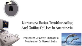
- 1. Ultrasound Basics, Troubleshooting And Outline Of Uses In Anaesthesia Presenter Dr Gowri Shankar B Moderator Dr Naresh babu
- 2. Outline History ofultrasound Physics of echo Ultrasound waveand itsinteractions Ultrasound machine anditsparts ArtifactsAndTheir ClinicalImportance DopplerUltrasound Current And Potential Applications Of Ultrasound Bioeffects Conclusion
- 3. Introduction • Anesthesiologists require quick and accurate diagnostic tools for the effective management of emergencies • Ultrasound (US) is a safe, easily accessible point-of- care imaging modality • US being increasingly adopted in modern anesthesiology practice • Anesthesiologists are aware of the expanding applications of this technology and the status of its use.
- 4. So What is ultrasound? Ultrasound or ultrasonography is a medical imaging technique that uses high frequency sound waves to obtain cross sectional images of the body. Also known as ‘pulse echo’ technique
- 5. History • In 1794 Spallanzani performed studies on bats that concluded that they could navigate using sound rather than sight. • In 1915 Inspired by the sinking of the Titanic, Physicist Paul Langevin invented a hydrophone – what the World Congress Ultrasound in Medical Education refers to as the “first transducer” • 1920s-1940s Sonography was used to treat members of European soccer teams as a form of physical therapy, to appease arthritic pain and eczema • In 1942 Neurologist Karl Dussik is credited with being the first to use sonography for medical diagnoses
- 9. Physics of echo Sound • Sound is a mechanical energy that travels through matter, as a result of vibration of the particles of the medium through which the sound wave is moving. • Transport the energy through medium > pressure wave • Change in pressure > sinusoidal waveform • Medical ultrasound Megahertz range(2-15 MHz)
- 10. • Velocity • Speed at which a sound wave travels through a medium • Determined by density and stiffness of media • Slowest in air/gas • Fastest in solids • Average speed of ultrasound in body is 1540m/sec Physics of echo
- 12. Frequency • Number of cycles per second, Units are Hertz • Ultrasound imaging frequency range 2-15Mhz • Low the frequency, higher the penetration and lower the resolution Higher the frequency, lower the penetration and higher the resolution Physics of echo
- 13. Amplitude • The strength/intensityof a sound waveatanygiven time • Represented as height of thewave • Decreases with increasingdepth • Amplitude determines brightness ofimage • The highertheamplitude, the brighter the imageand vice-versa Physics of echo
- 14. Electrical energy converted to sound waves Thesound waves are reflected by tissues Reflected sound waves are convertedto electrical signals and later toImage Physics of echo
- 15. • Image Formation Electrical signal produces ‘dots’ on thescreen Brightness of the dot is proportional to the strength of the returning echoes Location of the dot is determined by travel time Physics of echo
- 16. PrincipleOFULTRASOUND Ultrasoundworksontheprincipleof piezoelectriceffect. When acrystal is subjected to mechanical pressure, electric voltage is generatedand viceversa.
- 18. Interactions of ultrasound • Transmission • Reflection • Refraction • Scattering • Attenuation
- 19. Interactions of ultrasound Transmission Not all the sound wave is reflected, some continues deeper into thebody These waves will reflect from deeper tissue structures
- 20. Interactions of ultrasound Reflection Occurs at a boundary between 2 adjacent tissues or media The amount of reflection depends on differences in acoustic impedance (z) between media The ultrasound image is formed from reflected echoes Acoustic impedance=density of medium x velocity
- 21. Reflection • Iftwo materialshavesameimpedance,no echoproduced. • Ifthedifferenceinacousticimpedanceis: • Small–weakechoisproducedandmostof the soundwaveswill continue in second medium • Large-strongechoisproduced • Verylarge- all soundwaveswill betotally reflectedback.Example:ce 99%ofbeam isreflectedback. Interactions of ultrasound
- 22. Interactions of ultrasound Refraction This occurs when an ultrasound beam passes at an angle other than 90 degrees, from one tissue into another with change in velocity It increase with the increasing angle of incidence It passes deeper into the body where it gives rise to artifacts
- 24. Interactions of ultrasound Scattering Redirection of sound in several directions Caused by interaction with small reflector or rough surface Only portion of sound wave returns to transducer
- 25. Interactions of ultrasound Attenuation • The decrease in the intensity of the ultrasound wave as they pass through tissues • Results from absorption , reflection , scattering and beam divergence
- 27. Ultrasound machine and itsparts A basic ultrasound machine 1.Display 2.CPU 3.Transducer probes 4.Keyboard/control panel 5.Disc storage device 6.Printer
- 28. Ultrasound machine and itsparts
- 29. Ultrasound machine and itsparts Machine orientation: • -presets • -features • -save and print Machine features: • -Quality • -Mode • -Tools
- 30. Mode 2D/B mode Doppler -Color doppler -continuous wave doppler -pulse wave doppler M-mode 3D 4D Ultrasound machine and itsparts
- 31. Freeze Trackball Caliper Toggle key Text key Ultrasound machine and itsparts
- 36. Current and potential applications of ultrasound Regional Anesthesia Neuraxial And Chronic Pain Procedures Vascular Access Airway Assessment Lung Ultrasound Gastric Ultrasound Focused Transthoracic Echo (TTE) Ultrasound for the Anesthesiologists 36
- 37. Regional Anaesthesia Direct Observation Of The Nerves Surrounding Structures Direct Observation Of Local Anesthetic Spread Faster Onset 21 3 4
- 39. Neuraxial And Chronic Pain Procedures Neuraxial Blocks Nerve Root Blocks (E.G., Cervical And Lumbar); Stellate Ganglion Block US Guidance For Peripheral Nerve Stimulator Implantation Interventional Procedures For Patients With Chronic Pelvic Pain (E.G., Pudendal Neuralgia, Piriformis Syndrome , And “Border Nerve” Syndrome).
- 40. Vascular Access • Identification of the vein • Detection of variable anatomy, intravascular thrombosis • Avoidance of inadvertent arterial puncture • Safer and less time consuming than the traditional landmark technique • Patients with underlying coagulopathy or platelet dysfunction
- 41. A new 4- dimensional imaging (real-time 3-dimensional imaging) approach, using a matrix arrays transducer, for central venous cannulation, prevents “overshooting” the needle and provides better visualization of anatomy. • High-frequency (50mhz) micro- ultrasound (HFMU) may allow better visualization for the sub-10mm space. This could be a valuable tool for difficult vascular access in pediatric patients
- 42. Airway Assessment Ultrasound for the Anesthesiologists 42 visualize with US tongue oropharynx hypopharynx epiglottis Larynx & vocal cords cricoid cartilage cricothyroid membrane trachea Cervical esophagus NOT Visualize with US posterior pharynx Posterior commissure, posterior wall of the trachea
- 43. Airway Assessment Prediction of difficult airway Confirmation of proper endotracheal tube placement and ventilation Prediction of obstructive sleep apnea Airway Related nerve blocks Prediction of size of endotracheal, endobronchial, and tracheostomy tubes Assessing and guidance for proper percutaneous dilatational tracheostomy (PDT); Evaluation of airway pathologies :Mandate urgent securing of airway (e.G., Epiglottitis);
- 45. The probe is placed under the submandibular area in a longitudinal orientation Ultrasound for the Anesthesiologists 45 Post injection sonography. (1)Superior border of the thyroid cartilage. (2)Greater horn of the hyoid bone. (3)Thyrohyoid muscle. (4)Thyrohyoid membrane. (5)Thyroid cartilage lamina The probe is placed under the submandibular area in a longitudinal orientation Post injection sonography. (1)Superior border of the thyroid cartilage. (2)Greater horn of the hyoid bone. (3)Thyrohyoid muscle. (4)Thyrohyoid membrane. (5)Thyroid cartilage lamina
- 48. Lung Ultrasound In a number of emergency situations: Hypoxia Pneumothorax pulmonary edema pulmonary embolism ARDS ultrasound can be an important tool for diagnosis Ultrasound for the Anesthesiologists 48
- 49. LUNG ULTRASOUND • choice for detecting pleural line abnormalities A high frequency (7.5 to 10MHz) • diagnose pleural effusions and lung parenchymal abnormalities lower frequency (3.5MHz) convex and microconvex • virtual interplay of two elements: air and fluid. B- and M-mode Ultrasound for the Anesthesiologists 49
- 50. Normal Lung “Lung sliding” signs are sliding of visceral and parietal layers of pleura with respiration Seashore sign is a complex picture of parallel lines signifying the static thoracic wall and sandy “granulous” pattern, which reflect the normal pulmonary parenchyma. A-lines are a basic artifact of normally aerated lung Ultrasound for the Anesthesiologists 50
- 51. Normal Lung
- 52. Gastric ultrasound • Assessment of gastric content and diagnosis of full stomach • Confirmation of gastric tube placement Ultrasound for the Anesthesiologists 52
- 54. Gastric Ultrasound Ultrasound for the Anesthesiologists 54
- 55. Focus assessed transthoracic echo • Focus assessed transthoracic echo (FATE) was introduced by jensen et al for cardiopulmonary monitoring in ICU • Basically involves 4 standardized acoustic views for cardiopulmonary screening and monitoring • Recent studies show a great impact of FATE in preoperative assessment • Significantly alters perioperative management
- 56. Technological Advances • Matrix array is a new transducer with improved resolution.It has a lens that is placed in front of the piezoelectric element to allow a mechanical focusing in the y- and z-planes • Four-dimensional ultrasound provides real-time 3d images (the 4th “d” is time) and currently is used for fetal imaging, Ultrasound for the Anesthesiologists 56
- 57. Technological Advances • Mobile ultrasound guided peripheral nerve block has been developed • Sonixgps needle guidance system is a gps technology with a new needle tracking system, using sensors in both the needle and transducer to obtain a real-time image of needle shaft and tip position related to the us beam that is based on the needle trajectory Ultrasound for the Anesthesiologists 57
- 58. Conclusion • Ultrasound is a unique tool which optimization of perioperative management. • We believe that ultrasound can be the "third eye" of the anesthesiologist that helps in the performance of previously blind procedures and allows discovery of many hidden spaces to uncover their mysteries • Anesthesiologists, in the near future, may need to carry a portable ultrasound around their neck instead of a stethoscope. Ultrasound for the Anesthesiologists 58
- 60. Thermal bioeffect • Mechanism - with sound wave attenuation, energy is lost as heat • Longer use of ultrasound on the tissue longer the ultrasound has attenuated in the tissue • More the tissue attenuates ultrasound more the bioeffects • Higher the frequency/power/PRF/pulse duration , more the attenuation and thus the heating • NCRP guidelines – in ultrasound examinations, no temperature rise > 1°C
- 61. Mechanical bioeffects • Current USG systems can produce cavitation • Determines potential for bubble formation in vivo • Cavitation - Stable Inertial / Transient
- 62. Stable cavitation • A bubble forms as the peak low pressure region of an ultrasound pulse passes through a nucleation site • This bubble grows and shrinks with the high pressure and low pressure region of the ultrasound pulses • This growing and shrinking of the bubble causes fluid to flow around the bubble - micro-streaming • Causes mechanical damage, sometimes cell lysis
- 63. Inertial/transient cavitation • Here, the bubbles formed in a similar way but under high pressure or intensities, they expand very quickly following which they collapse violently • This collapse produces high temperature and pressure, which damages cells in the region of the collapse
- 65. Mechanical index It is an approximation of mechanical effects It is a calculated number based on ultrasound parameters and facts about the machine that the manufacturer built into the programming Mechanical index less than 1.9, no mechanical effects is seen
- 67. References Rumack , Diagnostic Ultrasound , FifthEdition WHO Manual of Diagnostic Ultrasound Internet
Notes de l'éditeur
- (e.g., subglottic hemangiomas and stenosis)
- Confirmation of a gastric tube placement is also possible using ultrasound which might replace the conventional radiography method
- National Council on Radiation Protection and Measurements