
Recommandé
Contenu connexe
Tendances
Tendances (20)
En vedette
En vedette (14)
Similaire à Complications of third stage of labou rppt
Similaire à Complications of third stage of labou rppt (20)
Plus de Drisya Nidhin
Dernier
Dernier (20)
Complications of third stage of labou rppt
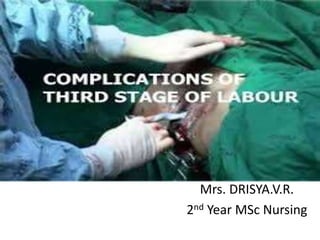
- 1. Mrs. DRISYA.V.R. 2nd Year MSc Nursing
- 2. • Postpartum hemorrhage • Placenta accreta, increta, and percreta • Inversion of the uterus
- 3. POSTPARTUM HEMORRHAGE Any amount of bleeding from and into the genital tract following the birth of the baby up to the end of the pueperium which adversely affects the general condition of the patient evidenced by rise in pulse rate and falling BP is called post partum haemorrhage”.
- 4. • Incidence 4-6% of all deliveries.
- 5. Types • Primary Third stage hemorrhage True PPH • Secondary PPH/ delayed/late
- 6. Primary post partum haemorrhage Causes • 4 T’s • Tone • Tissue • Trauma • Thrombin( blood coagulopathy)
- 7. • High parity • Over-distension of the uterus • Malnutrition and anemia • Antepartum hemorrhage • General anesthesia • Poorly perfused myometrium • Prolonged labour • Following augmented labour Uterine atony
- 8. • Uterine atony in previous labour • Placenta • Chorioamnionitis • Malformation of uterus • Uterine fibroid • Very rapid labour • Mismanaged third stage of labour
- 9. • Constriction ring: • Avulsed cotyledon, succenturiate lobe • Placenta previa • Placental abruption • A full bladder
- 10. Traumatic Trauma to the genital tract usually occurs following operative delivery; even after spontaneous delivery
- 11. • Retained tissues • Drugs : tocolytic drugs, MgSO4, nifedipine • Combination of atonic and traumatic causes • Blood coagulation disorders
- 12. Clinical Features • Visible bleeding • Maternal collapse • Pallor • Rising pulse rate • Falling BP • Altered level of consciousness • May restless/drowsy • Enlarged uterus, boggy on palpation
- 13. Diagnosis • Direct observation in open hemorrhage. • In concealed case, diagnosis is based on clinical effects. • In traumatic hemorrhage- uterus is contracted. • In atonic hemorrhage-uterus is flabby and becomes hard on massaging.
- 14. Investigations • Thorough examination of the lower genital tract. This may require theatre/anaesthesia. • CBC, clotting screen, cross match, Coagulation studies • Hourly urine output • Continuous pulse/blood pressure or central venous pressure monitoring • ECG, pulse oximetry
- 15. Prevention Antenatal • Improvement in health status, keep Hb level >10gm/dl. • Screen high risk clients. • Blood grouping • Unfractionated Heparin (UH) or Low Molecular Weight Heparin (LMWH).
- 16. Intranatal • In the event of a woman coming to delivery while receiving therapeutic heparin, the infusion should be stopped. Heparin activity will fall to safe levels within an hour. Protamine sulphate will reverse activity more rapidly, if required. • Slow delivery of baby. • Expert obstetric anesthetist. • Active management of 3rd stage of labour.
- 17. Immediate care in PPH • COMMUNICATE. • RESUSCITATE. • MONITOR / INVESTIGATE. • STOP THE BLEEDING.
- 18. • Communicate to clinical team—Call 6. • Call experienced midwife • Call obstetric registrar & alert consultant • Call anaesthetic registrar, alert consultant • Alert haematologist • Alert Blood Transfusion Service • Call porters for delivery of specimens / blood
- 19. Resuscitate • IV access with 14 G cannula X 2 • Head down tilt • Oxygen by mask, 8 litres / min • Transfuse • Crystalloid • Colloid
- 20. Management of 3rd stage hemorrhage The principles in the management are: • To empty the uterus of its content and to make it contract. • To replace the blood. If in shock, then manage shock. • To ensure effective hemostasis in traumatic bleeding.
- 21. Steps of management: Placental site bleeding • Palpate the fundus and massage the uterus to make it hard. • To start crystalloid solution (NS or RL) with oxytocin (1L with 20 units) at 60 drops per minute and to arrange for blood transfusion • Oxytocin 10U IM or Ergometrine 0.25mg or methergine 0.2mg is given intravenously. • Catheterise the bladder • Sedation with morphine 15mg intramuscularly. • To give antibiotics
- 22. Manual Removal of Placenta Step 1: The operation is done under GA. In extreme urgency where anaesthesia is not available, the operation may have to be done under deep sedation with IV diazepam 10mg. The patient is placed in lithotomy position. With all aseptic measures the bladder is catheterized.
- 23. Step 2: One hand is introduced into the uterus after smearing with the antiseptic solution in cone shaped manner following the cord, which is made taut by the other hand. While introducing the hand, the labia are separated by the fingers of the other hand. The fingers of the uterine hand should locate the margin of the placenta.
- 24. Step 3: Counter pressure on the uterine fundus is applied by the other hand placed over the abdomen. The abdominal hand should steady the fundus and guide the movements of the fingers inside the uterine cavity till the placenta is completely separated.
- 25. Step 4: As soon as the placental margin is reached, the fingers are insinuated between the placenta and the uterine wall with the back of the hand in contact with the uterine wall. The placenta is gradually separated with a sideways slicing movement of the fingers, until whole of the placenta is separated.
- 26. Step 5: When the placenta is completely separated, it is extracted by traction of the cord by the other hand. The uterine hand is still inside the uterus for exploration of the cavity to be sure that nothing is left behind.
- 27. Step 6: Intravenous methergine 0.2mg is given and the uterine hand is gradually removed while massaging the uterus by the external hand to make it hard. After the completion of the manual removal, inspection of the cervico- vaginal canal is to be made to exclude any injury.
- 28. Step 7: The placenta and membranes are to be inspected for completeness and be sure that the uterus remains hard and contracted.
- 29. Difficulties • Hour – glass contraction • Morbid adherent placenta
- 30. Complications • Haemorrhage due to incomplete removal • Shock • Injury to the uterus • Infection • Inversion • Subinvolution • Thrombophlebitis • Embolism.
- 31. Scheme of management of third stage haemorrhage. ● Control of the fundus, massage and make it hard ● Inj.Methergin 0.2mg I.V ●To start NS drip and arrange for blood transfusion ● Catheterise the bladder Placenta separated Not separated Express the placenta Manual removal under GA out by controlled cord traction
- 32. Management of true post partum haemorrhage Principles • To diagnose the cause of bleeding. • To take prompt and effective measures to control bleeding. • To correct hypovolemia.
- 33. Management • Call for help. • Put in two large bore, 14 gauge, cannulas. • Keep patient flat and warm. • Send blood for grouping and cross matching and ask for 2 units of blood. • Oxygen by mask, 10-15 litres / min • Start 20 units of oxytocin in 1 L of NS at the rate of 60 drops/mt. • Monitor vital signs • Monitor type and amount of fluids the patient has received, urine output, drugs- type, dose and time, CVP.
- 34. Actual Management: Atonic uterus Step 1: • Massage the uterus to make it hard and express the blood clot. • Inj. Methergin 0.2mg IV. • Start Inj.oxytocin drip (10 units in 500ml of NS) at the rate of 40- 60 drops per min. • Catheterise the bladder • Examine the expelled placenta and membranes for completeness. If the uterus fails to contract , proceed to the next step.
- 35. Step 2: • Explore the uterus under GA. Simultaneous inspection of the cervix, vagina specially the para- urethral region is to be done to exclude co- existent bleeding sites from the injured area. • Blood transfusion • Continue oxytocin drip.
- 36. In refractory cases: • Inj. 15 methyl PGF2α 250micro gram IM in the deltoid muscle every 15 minutes ( upto maximum of 2 mg) or Misoprostol (PGE1) 1000 microgram per rectum. • When uterine atony is due to tocolytic drugs, calcium gluconate (1 gm IV slowly)
- 37. • Step 3: Uterine massage and bimanual compression. • Step 4: Uterine tamponade (i) Tight intra uterine packing done uniformly under GA. (ii) Balloon tamponade
- 38. Step 5: Surgical methods • Ligation of uterine arteries • Ligation of the ovarian and uterine artery anastomosis • Ligation of the anterior division of internal iliac artery (unilateral or bilateral). • B-Lynch compression suture and multiple square sutures: • Angiographic arterial embolisation under fluoroscopy
- 39. • Step 6: Hysterectomy
- 40. Protocol • Stage 0: normal - treated with fundal massage and oxytocin. • Stage 1: more than normal bleeding - establish large-bore intravenous access, assemble personnel, increase oxytocin, consider use of methergine, perform fundal massage, prepare 2 units of packed red cells.
- 41. • Stage 2: bleeding continues - check coagulation status, assemble response team, move to operating room, place intrauterine balloon, administer additional uterotonics(misoprostol, carboprost tromethamine), consider: uterine artery embolization, dilatation and curettage, and laparotomy with uterine compression stitches or hysterectomy.
- 42. • Stage 3: bleeding continues - activate massive transfusion protocol, mobilize additional personnel, recheck laboratory tests, perform laparotomy, consider hysterectomy • Following PPH keep the patient in labour room and observe for 24- 48 hrs. • Traumatic PPH • The trauma to the perineum, vagina and the cervix is to be searched under good light by speculum examination and haemostasis is achieved by appropriate catgut sutures. The repair is done under GA, if necessary.
- 43. Secondary PPH Causes: • Retained bits of placenta or membranes. • Infection and separation of slough over a deep cervico- vaginal laceration. • Endometritis and subinvolution of the placental site • Withdrawal bleeding following oestrogen therapy for suppression of lactation. • carcinoma of cervix, infected fibroid or fibroid polyp and puerperal inversion of uterus.
- 44. Management • Principles— (1) To assess the amount of blood loss and to replace the lost blood. (2) To find out the cause and to take appropriate steps to rectify it.
- 45. • Call doctor • Reassure woman and support person • Rub up contraction by massaging uterus if it is still palpable • Express any clots • Encourage to empty bladder • Give an uterotonic • Keep all pads and linen to assess blood loss • If bleeding persists transfer women
- 46. • Supportive therapy: • Blood transfusion, if necessary; • Inj Ergometrine 0.5mg IM, if the bleeding is uterine in origin • Antibiotics as routine.
- 47. Complications • Shock • Collapse • Disseminated intravascular coagulation
- 48. RETAINED PLACENTA The placenta is said to be retained when it is not expelled out even 30 minutes after the birth of the baby.(WHO 15mnts)
- 49. Causes • There are three phases involved in the normal expulsion of placenta. • Separation through the spongy layer of the decidua • Descent into the lower segment and vagina • Finally the expulsion to outside
- 50. Management • Separated • Un-separated • Complicated
- 51. Complications • Haemorrhage • Shock • Puerperal sepsis • Risk of recurrence in next pregnancy.
- 52. PLACENTA ACCRETA, INCRETA, AND PERCRETA
- 53. • The term placenta accreta is used to describe any implantation in which there is abnormally firm adherence to the uterine wall. • With placenta increta, villi actually invade into the myometrium and anchored into the muscle bundles. • with placenta percreta, villi penetrate through the myometrium upto the serosal layer.
- 54. Management • In the focal placenta accreta • In the total placenta accreta • In rare cases
- 55. Complications • Haemorrhage • Shock • Infection • Inversion of uterus
- 56. INVERSION OF THE UTERUS • It is extremely rare but a life threatening complication in third stage in which the uterus is turned inside out partially or completely.
- 57. Varieties • First degree • Second degree • Third degree
- 58. Etiology • Spontaneous • Iatrogenic: This is due to the mismanagement of third stage of labour. Pulling the cord when the uterus is atonic specially when combined with fundal pressure. Fundal pressure while the uterus is relaxed Faulty technique in manual removal
- 59. Diagnosis Symptoms: • Acute lower abdominal pain with bearing down sensation Signs: • Varying degree of shock is a constant feature • Abdominal examination • Bimanual examination • In complete variety pear shaped mass protrudes outside the vulva with broad end pointing downwards and looking reddish purple in colour
- 60. Prevention • Do not employ any method to expel placenta out when the uterus is relaxed. • Pulling the cord simultaneously with fundal pressure should be avoided. • Manual removal in a safe manner
- 61. Management Before shock develops: • To replace the part first which is inverted last with the placenta attached to the uterus by steady firm pressure exerted by the fingers. • To apply counter support by the other hand placed on the abdomen. • After replacement the hand should remain inside the until the uterus become contracted by parentral oxytocin or PGF2α • The placenta is to be removed manually after the uterus became contracted to reduce the bulk which facilitates replacement or if partially separated to minimize blood loss. • Usual treatment of shock including blood transfusion should be arranged.
- 62. After shock develops: • urgent normal saline drip and blood transfusion • To push the uterus inside the vagina if possible and pack the vagina with antiseptic roller gauze. • Foot end of the bed is raised. • Replacement of uterus either manually or hydrostatic method (O Sullivan’s) under GA.
- 63. Complications • Shock • Peritoneal irritation • Haemorrhage • Pulmonary embolism • If left uncared it leads to: o Infection o Uterine sloughing o A chronic one