A Rare Case of a Late Onset Central Tendon Diaphragmatic Hernia

A congenital diaphragmatic hernia (CDH) is a rare condition which usually presents in the first few days of life. In older children and adults however, the diagnosis may be more difficult. Late-presenting CDH is less common and the majority present with non-specific gastrointestinal or respiratory symptoms later in childhood. For these reasons, late-presenting CDH is quite frequently misdiagnosed. Most CDH are of the Bochdalek or posterolateral type with much smaller numbers of anterior or Morgagni defects. We present a case report of this rare type of CDH in an older child and our experience managing the condition. The purpose of this report is to familiarize clinicians and surgeons with the clinical presentations that can occur in children with CDH, in order to facilitate early management and avoid possible life-threatening conditions.

Recommandé
Recommandé
Contenu connexe
Similaire à A Rare Case of a Late Onset Central Tendon Diaphragmatic Hernia
Similaire à A Rare Case of a Late Onset Central Tendon Diaphragmatic Hernia (15)
Plus de Associate Professor in VSB Coimbatore
Plus de Associate Professor in VSB Coimbatore (20)
Dernier
Dernier (20)
A Rare Case of a Late Onset Central Tendon Diaphragmatic Hernia
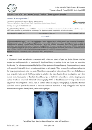
- 1. Asian Journal of Basic Science & Research Volume 4, Issue 2, Pages 106-109, April-June 2022 ISSN: 2582-5267 www.ajbsr.net 106 A Rare Case of a Late Onset Central Tendon Diaphragmatic Hernia Arifin BA1 & Mohanaprakash KRA2 1 International Islamic University Malaysia, Kuantan, Pahang, Malaysia. 2 Department of Paediatric Surgery, Hospital Tengku Ampuan Afzan, Kuantan, Pahang, Malaysia. DOI: http://doi.org/10.38177/AJBSR.2022.4208 Copyright: © 2022 Arifin BA & Mohanaprakash KRA. This is an open access article distributed under the terms of the Creative Commons Attribution License, which permits unrestricted use, distribution, and reproduction in any medium, provided the original author and source are credited. Article Received: 15 February 2022 Article Accepted: 20 April 2022 Article Published: 25 May 2022 1. Case A 10-year-old female was admitted to our centre with a recurrent history of pain and feeling fullness over her epigastrium, multiple episodes of vomiting with significant history of retching for the past 1 year and worsening last 1 week. The pain was constant and dull aching. Child denies any history of trauma. On examination, she was a well-nourished child, afebrile, not in respiratory distress or tachycardic. There were no abnormalities noted during her lungs examination; air entry was equal. The abdomen was scaphoid but non-tender. However, she had a mass over epigastric region about 15x15 cm, unable to get above the mass. Routine blood investigations are within normal limits. Radiography of the chest showed bowel gas in the left lower hemithorax with the diaphragmatic outline of left side is not well delineated. Ultrasonography (USG) of the abdomen showed large cystic mass in epigastrium measuring about 17x16x8.7 cm. A contrast enhanced computed tomography (CECT) of the abdomen done then showed part of the stomach is massively distended, herniation of body and pylorus into the left hemithorax through the defect (2.4 cm) of left hemidiaphragm. Fig.1. Chest X-ray showing loop of bowel gas in the left hemithorax ABSTRACT A congenital diaphragmatic hernia (CDH) is a rare condition which usually presents in the first few days of life. In older children and adults however, the diagnosis may be more difficult. Late-presenting CDH is less common and the majority present with non-specific gastrointestinal or respiratory symptoms later in childhood. For these reasons, late-presenting CDH is quite frequently misdiagnosed. Most CDH are of the Bochdalek or posterolateral type with much smaller numbers of anterior or Morgagni defects. We present a case report of this rare type of CDH in an older child and our experience managing the condition. The purpose of this report is to familiarize clinicians and surgeons with the clinical presentations that can occur in children with CDH, in order to facilitate early management and avoid possible life-threatening conditions. Keywords: Congenital diaphragmatic hernia, Central tendon defect, Morgagni defects.
- 2. Asian Journal of Basic Science & Research Volume 4, Issue 2, Pages 106-109, April-June 2022 ISSN: 2582-5267 www.ajbsr.net 107 Fig.2. Coronal view of CECT abdomen showing the herniation of the body and pylorus into the left hemithorax Fig.3. Axial view of the CECT abdomen showing stomach dilatation with wall thickening with herniation through a defect Fig.4. Intraoperative finding of a central tendon defect bounded by the medial side of left lobe of liver and the spleen We explored the patient with left transverse incision. There was a central tendon defect with thin sac medially with the defect measuring about 8x6 cm. The stomach herniated with dense adhesions especially to left lobe liver and spleen. After reduction of the content, defect was closed with prolene 2–0. Gastropexy was also done at 2 points to the anterior abdominal wall.
- 3. Asian Journal of Basic Science & Research Volume 4, Issue 2, Pages 106-109, April-June 2022 ISSN: 2582-5267 www.ajbsr.net 108 Postoperatively, the patient did extremely well. The patient was discharged 3 days after the operation. The patient was well at a 1-month follow-up visit. 2. Discussions The congenital defect that determines the congenital diaphragmatic hernias is due to the failed development of the septum transversum, the failed development of the pleuroperitoneal folds or improper or absent migration of the diaphragmatic musculature, or weaknesses in points of embryologic fusion. The septum transversum (central tendon) diaphragmatic hernia occurs in only 1% to 6% of CDHs [1]. The first case of isolated central anterior diaphragmatic hernia from a defect of the septum transversum dates back to 1966 [2]. Since then, other cases, all in infancy and childhood, have been reported [3]. Most of the cases of the congenital diaphragmatic hernia are diagnosed within the first few hours of life, with 5– 25% of diaphragmatic hernias appearing beyond the neonatal period, with age at discovery from 1 month to late adulthood [4]. In 349 late-presenting CDH children recently reviewed, the male-to-female ratio was close to 2:1 [5]. Late-onset CDH is an uncommon subset of CDH and distinct from neonatal CDH with respect to presenting symptoms, diagnosis, management, and prognosis. The presentation of late onset diaphragmatic defect is often intriguing as the child may present with various respiratory or gastrointestinal symptoms or may be absolutely asymptomatic. On chest X-ray, often bowel loops and fundic bubble are seen in the thoracic cavity with shift of the heart and mediastinum. Ultrasonography is useful in the diagnosis of congenital diaphragmatic hernia where uninterrupted contours of the diaphragm are not seen and peristalsis of the bowel can be observed in the thorax. Contrast CT scans of the thoracic and abdominal cavity are specific in making the diagnosis. Like in our case, a systemic approach to the child’s presentation has led to promptly diagnosing her rare condition and treating her effectively. 3. Conclusion Late-onset CDH is a tricky diagnosis with misleading symptoms and signs with a wide range of differential diagnoses. Therefore, we conclude that the central tendon diaphragmatic hernia, although extremely uncommon, should be considered in differential diagnosis. Thus, it is important, and may be lifesaving, to include this possibility in any patient; even in an older child with an unusual chest x-ray film, particularly if the diaphragm is indefinable and a 'cystic lesion' or an air-fluid level is seen in the left hemithorax. Declarations Source of Funding This research did not receive any grant from funding agencies in the public, commercial, or not-for-profit sectors. Competing Interests Statement The authors declare no competing financial, professional and personal interests. Consent for publication Authors declare that they consented for the publication of this research work.
- 4. Asian Journal of Basic Science & Research Volume 4, Issue 2, Pages 106-109, April-June 2022 ISSN: 2582-5267 www.ajbsr.net 109 Patient consent The authors certify that they have obtained all the necessary permissions to publish this case report. Acknowledgement The authors are highly thankful to the rest of the staff of Department of Paediatric Surgery, Hospital Tengku Ampuan Afzan, Kuantan for their tremendous support managing the case and contributing to the writing of this case. References [1] Clavert JM, De Geeter B, Bientz J, Sauvage P, Buck P. (1983). Abdominopericardial hernia as a result of an anomaly of the septum transversum. Apropos of a case in a newborn infant. Chir Pediatr., 24: 137-9. [2] Pomputius WF, Fisher ER. (1966). Intrapericardial fat pads and anomalous development of the septum transversum as a cause of pseudocardiomegaly. Am J Clin Pathol., 46: 92-98. [3] Wesselhoeft, DeLuca. (1984). Neonatal septum transversum diaphragmatic defects. Am J Surg., 147: 481-485. [4] Mishalany, Gordo (1986). Congenital diaphragmatic hernia in monozygotic twins. J Pediatr Surg, 21: 372-374. [5] Bagłaj M. (2004). Late-presenting congenital diaphragmatic hernia in children: a clinical spectrum. Pediatr Surg Int., 20(9): 658-669.