‘Not Your Usual Upper Gastrointestinal Bleeding’

Renal cell carcinoma (RCC) accounts above 3 percent of all cancers and often diagnosed incidentally. Highest incidence reported in Western countries. Classical tried of RCC now rarely seen, In about 25 percent of cases, patients often present with an advanced disease. Upper gastrointestinal (GI) bleeding due to stomach metastasis of RCC is rare and to the best of our knowledge, only a few cases are reported in the literature. Gastric metastasis of RCC is often associated with poor outcome. Managing such cases can be very tricky; hence it is imperative for surgeons to be very familiar and to have a high grade of suspicion in order to prevent delay and upgrade of tumour staging. We report the case of a patient presented to our centre with upper GI bleeding as the primary presenting complaint in an advanced RCC and our experience managing the condition.

Recommandé
Recommandé
Contenu connexe
Similaire à ‘Not Your Usual Upper Gastrointestinal Bleeding’
Similaire à ‘Not Your Usual Upper Gastrointestinal Bleeding’ (16)
Plus de Associate Professor in VSB Coimbatore
Plus de Associate Professor in VSB Coimbatore (20)
Dernier
Dernier (20)
‘Not Your Usual Upper Gastrointestinal Bleeding’
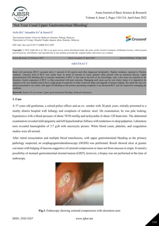
- 1. Asian Journal of Basic Science & Research Volume 4, Issue 2, Pages 110-114, April-June 2022 ISSN: 2582-5267 www.ajbsr.net 110 ‘Not Your Usual Upper Gastrointestinal Bleeding’ Arifin BA1 , Salauddin SA2 & Hamid G2 1 International Islamic University Malaysia, Kuantan, Pahang, Malaysia. 2 Department of Urology, Hospital Tengku Ampuan Afzan, Kuantan, Malaysia. DOI: http://doi.org/10.38177/AJBSR.2022.4209 Copyright: © 2022 Arifin BA et al. This is an open access article distributed under the terms of the Creative Commons Attribution License, which permits unrestricted use, distribution, and reproduction in any medium, provided the original author and source are credited. Article Received: 15 February 2022 Article Accepted: 22 April 2022 Article Published: 26 May 2022 1. Case A 57 years old gentleman, a retired police officer and an ex- smoker with 20 pack years, initially presented to a nearby district hospital with lethargy and complains of malenic stool. On examination, he was pale looking, hypotensive with a blood pressure of about 70/50 mmHg and tachycardia of about 120 beats/min. The abdominal examination revealed mild epigastric and left hypochondriac fullness with tenderness to deep palpation. Laboratory tests revealed haemoglobin of 3.7 g/dl with microcytic picture. White blood count, platelets, and coagulation studies were all normal. After initial resuscitation and multiple blood transfusions, with upper gastrointestinal bleeding as the primary pathology suspected, an esophagogastroduodenoscopy (OGDS) was performed. Result showed ulcer at greater curvature with bulging of mucosa suggestive of external compression or mass not from mucosa in origin. It raised a possibility of stomach gastrointestinal stromal tumour (GIST), however, a biopsy was not performed at the time of endoscopy. Fig.1. Endoscopy showing external compression with ulceration seen ABSTRACT Renal cell carcinoma (RCC) accounts above 3 percent of all cancers and often diagnosed incidentally. Highest incidence reported in Western countries. Classical tried of RCC now rarely seen, In about 25 percent of cases, patients often present with an advanced disease. Upper gastrointestinal (GI) bleeding due to stomach metastasis of RCC is rare and to the best of our knowledge, only a few cases are reported in the literature. Gastric metastasis of RCC is often associated with poor outcome. Managing such cases can be very tricky; hence it is imperative for surgeons to be very familiar and to have a high grade of suspicion in order to prevent delay and upgrade of tumour staging. We report the case of a patient presented to our centre with upper GI bleeding as the primary presenting complaint in an advanced RCC and our experience managing the condition. Keywords: Renal cell carcinoma, Upper gastrointestinal bleeding, Stomach metastasis.
- 2. Asian Journal of Basic Science & Research Volume 4, Issue 2, Pages 110-114, April-June 2022 ISSN: 2582-5267 www.ajbsr.net 111 He was then subjected to an urgent computerized tomography (CT) scan of the abdomen and pelvis revealing a large left upper abdominal mass with central necrotic component which is in close proximity with greater curvature of stomach with poor plane of demarcation between the mass and spleen, also involving the left kidney. Differentials at that time included GIST of stomach, left renal cell carcinoma or malignant splenic tumour suggesting a histopathological examination for further confirmation. He then underwent a CT guided biopsy which came back as a papillary renal cell carcinoma type 2. A CT of the thorax for completion of staging was performed showing multiple tiny right lung nodules likely to represent metastases. He was then referred to our urology centre for further management. Fig.2. CT showing a mass originating from the superior pole of left kidney Fig.3. CT showing no clear demarcation between the mass and the stomach He was hemodynamically stable in our ward, however, had persistent anaemia requiring multiple pack cells transfusions. Decision was made for an emergency nephrectomy with a suspicion of bleeding from the renal tumour. Intraoperatively, the highly vascular tumour was unresectable as the it was hard, fixed and seen invading into the greater curvature of the stomach and there was no clear demarcation seen between the tumour with the stomach and spleen.
- 3. Asian Journal of Basic Science & Research Volume 4, Issue 2, Pages 110-114, April-June 2022 ISSN: 2582-5267 www.ajbsr.net 112 Fig.4. Intra-operative image showing the hard mass fixed and plastered to the posterior aspect of stomach Immediately after surgery, patient was noted to have another bout of fresh bleeding from his nasogastric tube and was then subjected to another OGDS which showed a large blood clot in the stomach. He was then referred to an interventional radiology centre for angioembolisation where angioembolisation was performed to left superior segmental renal artery, splenic artery and branches, left gastric artery and gastroepiploic artery. He was also seen by the oncology team and was planned for a course of oral Sunitinib. Unfortunately for the patient, he succumbed due to his disease progression even prior to commencing of his targeted therapy. 2. Discussions Renal cell carcinoma (RCC) represents 2-3% of all adult malignant neoplasm and presents with different symptoms [1]. RCC most commonly develops in the sixth and seventh decade of life. The male to female incidence ratio is about 2:1. RCC may manifest with a variety of symptoms due to local growth, metastasis, haemorrhage and sometimes paraneoplastic syndrome. Classical tried of flank pain, palpable abdominal mass and gross haematuria is now rarely seen. Spread in RCC is lymphatic, haematogenous, trans-coelomic, or by direct invasion [1]. Most common sites of metastasis from RCC includes lung (75%), soft tissue (36%), bone (20%), liver (18%), cutaneous sites (8%) and central nervous system (8%) [2]. Primary RCC presenting as either an upper or lower gastrointestinal bleeding as a result of direct extension is rare. There are only few cases being reported in the literature [3]. Gastrointestinal bleeding is more commonly presenting feature of known patient of metastatic RCC or as a recurrence many years after nephrectomy for RCC [4]. Here we report an uncommon case of RCC where the primary manifestation of disease was with symptoms of upper gastrointestinal bleeding. Diagnosis was confirmed by upper gastrointestinal endoscopy, histopathological examination from the renal biopsy and imaging performed. The current diagnostic paradigm for evaluating renal masses is primarily dependent on cross-sectional imaging modalities: ultrasonography (US), computed tomography (CT) and magnetic resonance (MR) imaging. When a mass demonstrates characteristic features of malignancy, surgical resection, if warranted, can be performed without a preoperative biopsy because the prior probability of disease is sufficiently high; a negative biopsy result would
- 4. Asian Journal of Basic Science & Research Volume 4, Issue 2, Pages 110-114, April-June 2022 ISSN: 2582-5267 www.ajbsr.net 113 not likely alter management [5]. Unlike in our case, performing a biopsy was vital as there was a suspicion of the tumour arising from the GIST of stomach. Otherwise, renal mass biopsy often only reserved for a limited number of indications including diagnosis of metastatic disease, infection, lymphoma and to diagnose masses in patients who are poor surgical candidates [6]. In patients presenting with massive gastrointestinal bleeding owing to either a gastric or duodenal metastases from RCC, emergency arteriography and embolization of the gastroduodenal artery is an alternative life-saving treatment. Lynch-Nyhan et al. [7] reported successful embolization via the gastroduodenal artery in two cases of massive gastrointestinal bleeding due to duodenal metastases of RCC. Palliative nephrectomy has shown to be effective in alleviating local symptoms and can be undertaken in selected cases, but should always be weighed against the burden of surgical morbidity and mortality [8]. Hence the rationale behind attempting a palliative nephrectomy in this case although in the presence of lung metastasis. The management of metastatic RCC (mRCC) however, has evolved considerably over the past 15 years with the introduction of tyrosine kinase inhibitors (TKIs), and more recently, immunotherapy with immune checkpoint inhibitors, a combination of immunotherapy or combined with TKIs, has increased the lifespan of patients with metastatic clear cell renal cell cancer (mccRCC) [9]. The CARMENA trial showed that sunitinib alone was not inferior to nephrectomy followed by sunitinib in patients with metastatic renal-cell carcinoma who were in the MSKCC intermediate-risk or poor-risk groups [10]. This is important as initial nephrectomy can delay the start of systemic targeted therapies where the patient may die even prior to receiving this therapy as in our case. Apart from that, avoiding nephrectomy also avoids surgical complications involving blood transfusions, further operations, or intensive care, which may also delay systemic therapy [11]. Hence, the decision of whether to perform a nephrectomy or not has to be made earlier when managing a case of a metastatic renal-cell carcinoma. Outcome in metastatic RCC is generally poor, with one-year survival rate less than 50% and five-year survival rate of 5–30% [1]. Although evidence for the role of metastasectomy in RCC having multiple metastasis is lacking, if patients are fit for surgery, there should be no hesitation about performing surgical exploration and metastasectomy [12]. 3. Conclusion From the above case report, we demonstrated that patients presenting with gastrointestinal symptoms like upper gastrointestinal bleeding may have a primary pathology unrelated to that of the presenting organ hence is often under recognised early. Therefore, early suspicion and prompt diagnosis of the possibility of upper gastrointestinal bleeding due to RCC should be kept in mind, in order to facilitate effective management strategy and avoid possible life-threatening complications. Acknowledgement The authors are highly thankful to the rest of the staff of Department of Urology, Hospital Tengku Ampuan Afzan, Kuantan for their tremendous support managing the case and contributing to the writing of this case.
- 5. Asian Journal of Basic Science & Research Volume 4, Issue 2, Pages 110-114, April-June 2022 ISSN: 2582-5267 www.ajbsr.net 114 Declarations Source of Funding This research did not receive any grant from funding agencies in the public, commercial, or not-for-profit sectors. Competing Interests Statement The authors declare no competing financial, professional and personal interests. Consent for publication Authors declare that they consented for the publication of this research work. Patient consent The authors certify that they have obtained all the necessary permissions to publish this case report. References [1] Campbell SC, Andrew CN. (2007). Renal tumours. Campbell-Walsh Urology. 9th ed. Sounders Elsevier Pub., Pages. 1582-605. [2] Maldazys JD, DeKernion JB. (1986). Prognostic factors in metastatic renal cell carcinoma. J Urol., 136: 376-9. [3] Soin S, Verkhovsky E, (2019). Locally advanced renal cell carcinoma: cause of upper gastrointestinal bleeding. BMJ Case Reports, 12: e229992. [4] Harish K, Raju SL, Nagaraj HK. (2006). Recurrent renal cell carcinoma presenting as gastrointestinal bleed: A case report. Minerva Urol Nefrol., 58: 87-90. [5] An evaluation of the effectiveness of CT vs. other imaging modalities in the diagnosis of atypical renal masses. Curry NS, Reinig J, Schabel SI, Ross P, Vujic I, Gobien RP Invest Radiol. 1984 Sep-Oct; 19(5): 447-54. [6] Renal masses in the adult patient: the role of percutaneous biopsy. Silverman SG, Gan YU, Mortele KJ, Tuncali K, Cibas ES Radiology. 2006 Jul; 240(1): 6-22. [7] Lynch-Nyhan A, Fishman EK, Kadir S. (1987). Diagnosis and management of massive gastrointestinal bleeding owing to duodenal metastasis from renal cell carcinoma. J Urol., 138: 611-613. [8] Tiwari P, Tiwari A, Vijay M, Kumar S, Kundu AK. (2010). Upper gastro-intestinal bleeding - Rare presentation of renal cell carcinoma. Urol Ann., 2(3): 127-129. [9] Patel HV, Shinder, Srinivasan, Singer. (2020). Challenges and opportunities in the management of metastatic renal cell carcinoma: combination therapy and the role of cytoreductive surgery. Curr Opin Oncol., 32: 240-9. [10] Méjean, A., Ravaud, A., Thezenas, S., Colas, S., Beauval, J.-B., Bensalah, K., Escudier, B. (2018). Sunitinib Alone or after Nephrectomy in Metastatic Renal-Cell Carcinoma. New England Jour of Medicine, 379(5): 417-427. [11] Silberstein JL, Adamy A, Maschino AC, et al. (2012). Systematic classification and prediction of complications after nephrectomy in patients with metastatic renal cell carcinoma (RCC). BJU Int., 110: 1276-1282. [12] Chang WT, Chai CY, Lee KT. (2004). Unusual upper gastrointestinal bleeding due to late metastasis from renal cell carcinoma: a case report. Kaohsiung J Med Sci., 20(3): 137-141.