More Related Content
Similar to Mirror neurons in_autism (20)
More from merzak emerzak (16)
Mirror neurons in_autism
- 1. B R I E F C O M M U N I C AT I O N S
Understanding emotions in previously described a neural network in which the insula acts as
an interface between the frontal component of the MNS and the
others: mirror neuron dysfunction limbic system, thus enabling the translation of an observed or imitated
facial emotional expression into its internally felt emotional signi-
in children with autism ficance6. Three recent studies using different electrophysiological
© 2005 Nature Publishing Group http://www.nature.com/natureneuroscience
techniques have reported preliminary evidence for abnormal MNS
spectrum disorders functioning during action imitation8 and observation9,10 in adults with
ASD. However, a more definitive test of an MNS theory of autism
Mirella Dapretto1,2, Mari S Davies3, Jennifer H Pfeifer3,
would involve examining MNS activity in the context of a socio-
Ashley A Scott1, Marian Sigman2,3, Susan Y Bookheimer1,2 &
emotional task and in a sample of children.
Marco Iacoboni1,2
Here we used an event-related fMRI design to investigate neural
activity during the imitation and observation of facial emotional
To examine mirror neuron abnormalities in autism, high- expressions, in ten high-functioning children with ASD (9 males;
functioning children with autism and matched controls 12.05 ± 2.50 years of age) and ten typically developing children
underwent fMRI while imitating and observing emotional (9 males; 12.38 ± 2.22 years of age) matched by age and IQ (Supple-
expressions. Although both groups performed the tasks equally mentary Table 1 online). Subjects and their parents provided written
well, children with autism showed no mirror neuron activity in consent according to guidelines specified by the Institutional Review
the inferior frontal gyrus (pars opercularis). Notably, activity in Board at the University of California, Los Angeles. Stimuli consisted of
this area was inversely related to symptom severity in the social 80 faces expressing five different emotions: anger, fear, happiness,
domain, suggesting that a dysfunctional ‘mirror neuron system’ neutrality or sadness. Each face was presented for 2 s according to an
may underlie the social deficits observed in autism. optimized random sequence which included null events (that is, blank
screens with fixation crosses at eye level) and temporal jittering to
It has recently been proposed that dysfunction of the mirror neuron increase statistical efficiency. In two separate scans (with the order
system (MNS) early in development could give rise to the cascade of
impairments that are characteristic of autism spectrum disorders
(ASD)1, including deficits in imitation, theory of mind and social a
communication. First discovered in the ventral premotor cortex (area
F5) of the macaque, mirror neurons fire both while a monkey performs
goal-directed actions and while it observes the same actions performed
by others. This observation-execution matching system is thought to
provide a neural mechanism by which others’ actions and intentions
can be automatically understood2. The existence of an analogous MNS
in humans has been demonstrated by a number of independent b
investigations2: MNS activity in the human homolog of area F5—the
pars opercularis in the inferior frontal gyrus—has been consistently
reported during imitation3, action observation4 and intention under-
standing5. Relevant to an MNS theory of autism is further evidence
suggesting that the MNS, in concert with activity in limbic centers,
may mediate our understanding of the emotional states of others6,7.
Using functional magnetic resonance imaging (fMRI), we have
c
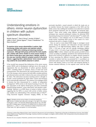
Figure 1 Reliable activity during imitation of emotional expressions.
(a,b) Activity in bilateral pars opercularis (stronger in the right) of the inferior
frontal gyrus is seen in the typically developing group (a) but not in the ASD
group (b). A between-group comparison (c) revealed that this difference was
significant (t 4 1.83, P o 0.05, corrected for multiple comparisons at the
cluster level). RH, right hemisphere; LH, left hemisphere. RH LH
1Ahmanson-Lovelace Brain Mapping Center, Semel Institute for Neuroscience and Human Behavior, 2Department of Psychiatry and Biobehavioral Sciences, David Geffen
School of Medicine and 3Department of Psychology, University of California Los Angeles, Los Angeles, California 90095, USA. Correspondence should be addressed to M.D.
(mirella@loni.ucla.edu).
Received 20 June; accepted 2 November; published online 4 December 2005; doi:10.1038/nn1611
NATURE NEUROSCIENCE ADVANCE ONLINE PUBLICATION 1
- 2. B R I E F C O M M U N I C AT I O N S
robust activation in visual cortices (including the fusiform gyrus),
t
4 premotor and motor regions of the face and the amygdala (Fig. 1b and
Supplementary Table 3 online). This indicated that these children
3 indeed attended to the stimuli and imitated the facial expressions.
Unlike the typically developing children, however, the ASD group
2 showed no activity in the mirror area in the pars opercularis (even
when results were examined at the most liberal thresholds). Direct
1
comparisons between the typically developing children and those with
ASD confirmed that activity in the anterior component of the MNS was
RH 0
reliably greater in typically developing children (Fig. 1c). Consistent
Figure 2 Mirror neuron system activity during observation of emotional with the neural model previously proposed6, whereby the frontal
expressions. The right pars opercularis showed significantly greater activity component of the MNS modulates limbic system activity via the insula,
in typically developing children than in children with ASD (t 4 1.83,
typically developing children also showed reliably greater activity in
P o 0.05, small volume corrected).
© 2005 Nature Publishing Group http://www.nature.com/natureneuroscience
insular and periamygdaloid regions as well as in the ventral striatum
and thalamus (Supplementary Table 3). In contrast, children with
counterbalanced within each group), subjects either imitated or simply ASD showed greater activity in left anterior parietal and right visual
observed the faces presented via high-resolution, magnet-compatible association areas (Supplementary Fig. 1 online).
goggles. For each subject, we acquired two sets of 96 whole-volume Individuals with autism typically show deficits in understanding the
images using a 3.0-Tesla head-only scanner (Siemens). The images were emotional states of others; thus dysfunction in the MNS should be
realigned, spatially normalized and smoothed using Automated Image manifest not only when these individuals explicitly imitate emotional
Registration. Random-effects analyses were implemented in SPM99. expressions but also when they merely observe emotions displayed by
All children practiced the tasks outside the scanner, thus demonstrating others. As predicted, activity in the right pars opercularis during the
that they were willing and able to comply with the task requirements. observation of facial expressions was reliably stronger in the typically
Subsequently, half the children in each group also performed both tasks developing group than in the ASD group (Fig. 2). Notably, this
during a videotaped session with an eye tracker. Analyses of these difference could not be attributed to a failure of the children with
behavioral data showed no group differences in the amount of time ASD to attend to the face stimuli, as both groups showed reliable acti-
spent fixating on the face and eye region, nor in how well the children vation in regions implicated in face processing, including the fusiform
imitated facial expressions (Supplementary Table 2 and Supplemen- gyrus and the amygdala (Supplementary Fig. 2 and Supplementary
tary Methods online). Table 4 online). Moreover, we observed no group differences in these
During the imitation of emotional expressions (versus null events), areas even when the data were explored at the most liberal thresholds.
the typically developing children activated a neural network very To further test the hypothesis that a dysfunctional MNS may
similar to that previously observed in adults6 (Fig. 1a and Supplemen- underlie the social deficits characteristic of ASD, we examined the
tary Table 2): there was extensive bilateral activation of striate and extra relationship between activity in regions with mirror neuron properties
striate cortices, primary motor and premotor regions, limbic structures and symptom severity, as indexed by children’s scores on the Autism
(amygdala, insula and ventral striatum) and the cerebellum. Notably, Diagnostic Observation Schedule–Generic (ADOS-G)11 and the Aut-
this group also showed strong bilateral activity within the pars ism Diagnostic Observation Interview–Revised (ADI-R)12. Controlling
opercularis of the inferior frontal gyrus (Brodmann’s area 44)—the for IQ, we found reliable negative correlations between activity in the
site with previously identified mirror properties—as well as in the pars opercularis and the children’s scores on the social subscales of the
neighboring pars triangularis (Brodmann’s area 45), with strongest ADOS-G and ADI-R (Fig. 3): the greater the activity in this critical
peaks in the right hemisphere. In the ASD group, we also observed component of the MNS during imitation, the higher a child’s level of
a t c Pars opercularis activity as a function of symptom severity
6 1.5
ADI: r (8) = –0.85, P < 0.002
4 1.0
Raw parameter estimates
0.5
2
0.0
0
RH RH
b t –0.5
12
–1.0
ADOS: r (8) = –0.70, P < 0.02
8
–1.5
4
–2.0
0 5 10 15 20 25 30
RH LH 0 ADOS and ADI scores (social subscales)
Figure 3 Mirror neuron system activity and symptom severity. (a–c) Negative correlations were found in the ASD group between activity in the pars opercularis
of the inferior frontal gyrus and scores on the social subscale of both ADOS-G (a,c) and ADI-R (b,c). t 4 1.83, P o 0.05, corrected for multiple comparisons
at the cluster level.
2 ADVANCE ONLINE PUBLICATION NATURE NEUROSCIENCE
- 3. B R I E F C O M M U N I C AT I O N S
functioning in the social domain. Activity in other components of the mechanism—interfacing with the limbic system via the insula—
normative network underlying emotion understanding via action rep- whereby the meaning of the imitated (or observed) emotion is directly
resentation (that is, the insula and limbic structures) was also negatively felt and hence understood. In contrast, this mirroring mechanism is
correlated with symptom severity (Supplementary Table 5 online). seemingly not engaged in children with ASD, who must then adopt an
To our knowledge, this is the first developmental fMRI study to show alternative strategy of increased visual and motor attention whereby the
reliable differences between typically developing children and children internally felt emotional significance of the imitated facial expression is
with ASD in the network comprising the frontal component of the probably not experienced. In line with previous findings in normal
MNS (namely, pars opercularis), the insula and the amygdala—that is, adults6,7, the fact that typically developing children showed increased
in the system thought to enable emotion understanding via action MNS activity even when simply observing an emotional expression
representation6. Although we were unable to monitor gaze fixation further indicates that this mirroring mechanism may underlie the
during scanning, a variable shown to affect brain activity in ASD13, it is remarkable ability to read others’ emotional states from a mere glance
extremely unlikely that our findings reflect between-group differences at their faces. The lack of MNS activity during both the imitation and
in the amount of time spent looking at the eye region. First, during the observation of emotional expressions in our sample of children
© 2005 Nature Publishing Group http://www.nature.com/natureneuroscience
neither the imitation or observation of facial expressions did we find with ASD provides strong support for the hypothesis that early
group differences in the fusiform gyrus, a region that is differentially dysfunction in the mirror neuron system may be at the core of the
activated in individuals with ASD as a function of gaze fixation13. This social deficits observed in autism.
suggests that—in keeping with other reports14,15—the inclusion of
Note: Supplementary information is available on the Nature Neuroscience website.
fixation crosses in the null events was successful at directing the subjects’
attention to this key region of the face, a conclusion supported by the ACKNOWLEDGMENTS
fact that no group differences were observed in eye fixation time Support for this work was provided by a grant from the National Institute
between the ASD and typically developing children who participated of Child Health and Human Development (P01 HD035470). The authors
thank the Brain Mapping Medical Research Organization, the Brain
in the eye-tracking session outside the scanner. Second, activity in the
Mapping Support Foundation, the Pierson-Lovelace Foundation, the
fusiform gyrus did not correlate with activity in MNS areas in either Ahmanson Foundation, the Tamkin Foundation, the Jennifer Jones-Simon
condition. Third, in the children with ASD for whom eye-tracking data Foundation, the Capital Group Companies Charitable Foundation, the
were available, there was no indication of a positive relationship Robson Family, the William M. and Linda R. Dietel Philanthropic Fund
between MNS activity in the pars opercularis and time spent fixating at the Northern Piedmont Community Foundation, the Northstar Fund
and the National Center for Research Resources (grants RR12169, RR13642
the eyes (see Supplementary Methods). and RR08655).
Similarly, despite the fact that we were unable to monitor task
performance during scanning, and although imitation deficits in COMPETING INTERESTS STATEMENT
autism are well documented1, we believe it implausible that our The authors declare that they have no competing financial interests.
findings merely reflect the children with ASD not performing the Published online at http://www.nature.com/natureneuroscience/
imitation task, or not performing it well. First, all the children with Reprints and permissions information is available online at http://npg.nature.com/
ASD were willing and able to perform the task just before scanning. reprintsandpermissions/
Second, children with ASD who performed the task outside the scanner
did so just as well as the typically developing children did, as rated by
1. Williams, J.H., Whiten, A., Suddendorf, T. & Perrett, D.I. Neurosci. Biobehav. Rev. 25,
independent observers blind to diagnosis but familiar with autism 287–295 (2001).
symptomatology (see Supplementary Methods). Third, in the ASD 2. Rizzolatti, G. & Craighero, L. Annu. Rev. Neurosci. 27, 169–192 (2004).
group, we observed robust activity in primary motor and premotor 3. Iacoboni, M. et al. Science 286, 2526–2528 (1999).
4. Johnson-Frey, S.H. et al. Neuron 39, 1053–1058 (2003).
areas of the face during the imitation task, with no evidence of between- 5. Iacoboni, M. in Perspectives on Imitation: From Neuroscience to Social Science
group differences in these regions even at the most liberal statistical (eds. Hurley, S. & Chater, N.) 77–99 (MIT Press, Cambridge, Massachusetts,
2005).
thresholds. Lastly, the children with ASD actually showed greater
6. Carr, L. et al. Proc. Natl. Acad. Sci. USA 100, 5497–5502 (2003).
activity than did the typically developing children in right visual and 7. Leslie, K.R., Johnson-Frey, S.H. & Grafton, S.T. Neuroimage 21, 601–607 (2004).
left anterior parietal areas, regions shown to be modulated by visual 8. Nishitani, N., Avikainen, S. & Hari, R. Ann. Neurol. 55, 558–562 (2004).
9. Oberman, L.M. et al. Brain Res. Cogn. Brain Res. 24, 190–198 (2005).
and motor attention, respectively. 10. Theoret, H. et al. Curr. Biol. 15, R84–R85 (2005).
The evidence thus suggests that although both groups performed the 11. Lord, C. et al. J. Autism Dev. Disord. 30, 205–223 (2000).
imitation task as requested, the neural strategies adopted by typically 12. Lord, C., Rutter, M. & Le Couteur, A. J. Autism Dev. Disord. 24, 659–685 (1994).
13. Dalton, K.M. et al. Nat. Neurosci. 8, 519–526 (2005).
developing children and those with ASD are quite different. Typically 14. Hadjikhani, N. et al. Neuroimage 22, 1141–1150 (2004).
developing children can rely upon a right hemisphere–mirroring neural 15. Pierce, K., Haist, F., Sedaghat, F. & Courchesne, E. Brain 127, 2703–2716 (2004).
NATURE NEUROSCIENCE ADVANCE ONLINE PUBLICATION 3