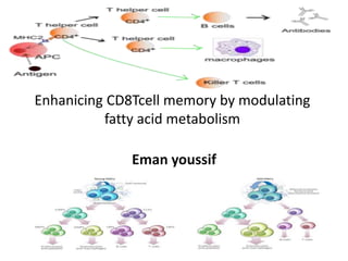
Enhanicing cd8 tcell memory by modulating fatty acid metabolism
•Télécharger en tant que PPTX, PDF•
1 j'aime•606 vues


Recommandé
Recommandé
Contenu connexe
Tendances
Tendances (20)
Similaire à Enhanicing cd8 tcell memory by modulating fatty acid metabolism
Similaire à Enhanicing cd8 tcell memory by modulating fatty acid metabolism (20)
Plus de eman youssif
Dernier
Dernier (20)
Enhanicing cd8 tcell memory by modulating fatty acid metabolism
- 1. Enhanicing CD8Tcell memory by modulating fatty acid metabolism Eman youssif
- 2. Notes: Entrez Gene summary for TRAF6 Gene: The protein encoded by this gene is a member of the TNF receptor associated factor (TRAF) protein family. TRAF proteins are associated with, and mediate signal transduction from, members of the TNF receptor superfamily. This protein mediates signaling from members of the TNF receptor superfamily as well as the Toll/IL-1 family. Signals from receptors such as CD40, TNFSF11/RANCE and IL-1 have been shown to be mediated by this protein. This protein also interacts with various protein kinases including IRAK1/IRAK, SRC and PKCzeta, which provides a link between distinct signaling pathways. This protein functions as a signal transducer in the NF-kappaB pathway that activates IkappaB kinase (IKK) in response to proinflammatory cytokines. The interaction of this protein with UBE2N/UBC13, and UBE2V1/UEV1A, which are ubiquitin conjugating enzymes catalyzing the formation of polyubiquitin chains, has been found to be required for IKK activation by this protein. This protein also interacts with the transforming growth factor (TGF) beta receptor complex and is required for Smad-independent activation of the JNK and p38 kinases. This protein has an amino terminal RING domain which is followed by four zinc-finger motifs, a central coiled-coil region and a highly conserved carboxyl terminal domain, known as the TRAF-C domain. Two alternatively spliced transcript variants, encoding an identical protein, have been reported.
- 3. T cell memory: Keeping energy levels up The generation of a long-lived memory CD8+ T-cell population depends on a switch in energy metabolism from glucose to fatty acid metabolism, suggesting that metabolism-altering drugs might be useful for boosting CD8+ T-cell responses.
- 4. A recent study published in Nature suggests that the generation of a long-lived memory CD8+ T cell population depends on a switch in energy metabolism from glucose to fatty acid metabolism. Modulation of this metabolic transition shows promise as an approach to boost vaccine efficacy
- 5. To explore the mechanisms regulating memory T cell development, Pearce et al. studied mice with a T cell-specific deletion of TNFR-associated factor 6 (TRAF6), which is known to negatively regulate antigen-specific T cell activation. These mice mounted normal antigen-specific effector CD8+ T cell responses following bacterial infection, but few memory CD8+ T cells could be detected 60 days after infection, and the mice failed to respond robustly to re-infection. The defect in memory T cell generation was shown to be CD8+ T cell intrinsic and not a result of insufficient CD4+ T cell help.
- 6. Using a systems biology approach to compare gene expression in wild-type and TRAF6-deficient CD8+ T cells, the authors found that TRAF6-deficient T cells had a defect in the expression of genes involved in several metabolic pathways, including fatty acid metabolism. In vitro experiments revealed that, unlike wild-type T cells, TRAF6-deficient CD8+ T cells had a reduced capacity to oxidize fatty acids after the withdrawal of interleukin-2 (IL-2). Withdrawal of growth factors such as IL-2 is known to induce metabolic stress in haematopoietic cells, and this causes cells to switch from glycolytic metabolism to catabolic metabolism (such as a fatty acid oxidation and autophagy) to generate energy that is necessary for survival. So the authors proposed that the inability of TRAF6-deficient T cells to switch to fatty acid metabolism impairs their chances of survival when metabolic stress is induced following the peak of the immune response, perhaps owing to limiting growth factors such as IL-2.
- 7. Consistent with this hypothesis, TRAF6-deficient CD8+ T cells generated lower levels of active AMP-activated kinase (AMPK), which is a key regulator of fatty acid oxidation, after IL-2 withdrawal compared with wild-type CD8+ T cells. Moreover, exposure to the widely prescribed anti-diabetic drug metformin, which promotes AMPK activation, enhanced fatty acid metabolism in TRAF6-deficient CD8+ T cells. Importantly, daily administration of metformin or rapamycin, another drug that promotes fatty acid metabolism, to mice 1 week following TRAF6-deficient T cell transfer and bacterial infection mitigated the defects in memory T cell development and promoted the survival of both the endogenous and transferred CD8+ T cells. Finally, the pharmacological potential of modulating fatty acid metabolism using metformin was further shown by the finding that metformin treatment of mice increased the efficacy of an experimental cancer vaccine; the increased survival of metformin-treated mice following injection with an otherwise lethal tumour was associated with increased memory CD8+ T cell numbers. This study highlights a previously unappreciated link between metabolic transition and cell fate determination and suggests that metabolism-altering drugs may be useful for boosting memory T cell responses.
- 12. Abstract of the paper CD8 T cells, which play a crucial role in immunity to infection and cancer, are maintained in constant numbers, but upon antigen stimulation undergo a developmental program characterized by distinct phases encompassing the expansion and then contraction of antigen-specific effector (TE) populations, followed by the persistence of long-lived memory (TM) cells1, 2. Although this predictable pattern of CD8 T cell responses is well established, the underlying cellular mechanisms regulating the transition to TM remain undefined1,
- 13. Here we show that TRAF6, an adapter protein in the TNF-receptor (TNFR) and IL-1R/TLR superfamily, regulates CD8 TM development following infection by modulating fatty acid metabolism. We show that mice with a T cell-specific deletion of TRAF6 mount robust CD8 TE responses, but have a profound defect in their ability to generate TM that is characterized by the disappearance of antigen-specific cells in the weeks following primary immunization. Microarray analyses revealed that TRAF6-deficient CD8 T cells exhibit altered expression of genes that regulate fatty acid metabolism.
- 14. Consistent with this, activated CD8 T cells lacking TRAF6 display defective AMPK- activation and mitochondrial fatty acid oxidation (FAO) in response to growth factor withdrawal. Administration of the anti-diabetic drug metformin restored FAO and CD8 TM generation in the absence of TRAF6. Remarkably, this treatment also increased CD8 TM in wild type mice, and consequently was able to significantly improve the efficacy of an experimental anti-cancer vaccine.
- 15. Introduction: A prevailing paradigm in immunology is that antigenic signal strength drives progressive T cell differentiation 3. To investigate this model regarding TM, we studied CD8 T cell responses to bacterial infection in mice with a T cell-specific deletion of TRAF6 (TRAF6-ΔT), a negative regulator of antigen-specific T cell activation
- 16. Despite having fewer total CD8 T cells (SI Fig. 1), TRAF6-ΔT mice mounted normal Ova-specific TE responses to attenuated L. monocytogenes expressing Ova (LmOva) (Fig. 1a and SI Fig. 2). To examine CD8 TM in TRAF6-ΔT mice, we immunized with LmOva and measured Ova-specific cells 60 days post- infection. Although Ova-specific TE responses were intact, CD8 TM generation in TRAF6-ΔT mice was severely compromised (Fig. 1b and SI Fig. 3), even with 10-fold higher or lower immunizing doses (not shown). In mice lacking cbl-b 5, a different negative regulator of antigen-specific T cell activation, TM developed normally (SI Fig. 4), indicating that failure of TM generation in TRAF6-ΔT mice cannot be entirely explained by loss of a negative regulator.
- 17. A hallmark of TM is the ability to mount accelerated recall responses to challenge infection. To confirm the impaired generation of CD8 TM in TRAF6- ΔT mice, we challenged previously immunized CTRL and TRAF6-ΔT mice with LmOva and measured Ova-specific cells 7 days later. TRAF6-ΔT mice failed to respond robustly to re-infection, suggesting that TM may not have been generated and that the smaller Ova-specific population present in TRAF6-ΔT mice was a new primary response (Fig. 1c). To determine if TRAF6-ΔT mice generate TM, we transferred equal numbers of Ova-specific cells from previously immunized CTRL and TRAF6-ΔT mice 28 days post-infection, at which time a small population of Ova-specific cells appeared to be present (SI Fig. 3),
- 18. compared functionality on a per cell basis in response to challenge. We reasoned that naïve donor cells transferred into immune-competent animals would not respond to infection because they would be vigorously outcompeted by endogenous naïve cells, and only TM donor cells would mount accelerated recall responses that could outcompete endogenous naïve cells. At day 7 post-challenge, donor-derived Ova-specific secondary TE cells from CTRL mice represented 11% of the engrafted population while TRAF6-ΔT donor cells were undetectable (SI Fig. 5), demonstrating a severe impairment in CD8 TM development in TRAF6-ΔT mice.
- 19. TRAF6-ΔT mice accumulate CD4 T cells and develop multi-organ inflammatory disease 4. Since CD4 T cells can influence CD8 T cells responses 6–8 we wanted to rule out effects from the TRAF6-deficient CD4 T cell compartment. To determine if defective CD8 TM generation in TRAF6-ΔT mice was intrinsic to CD8 T cells, we crossed TRAF6-ΔT mice with MHCI-restricted Ova-specific TCR-transgenic OT-I mice. Since OT-I mice lack CD4 T cells, generating OTI- TRAF6-ΔT mice allowed us to assay a pure population of uniformly naïve TRAF6-deficient CD8 T cells (SI Figs. 6–8). We transferred OTI-TRAF6-WT and OTI-TRAF6-ΔT cells into congenic recipients, which possess a normal CD4 T cell compartment, and then immunized with LmOva
- 20. By transferring only small numbers of transgenic cells we could measure endogenous Ova-specific cells at the same time as the engrafted donor response, allowing us to assume that the majority of donor cells were activated 9. In addition, following T cell responses by serially bleeding allowed us to compare the kinetics of responding CD8 T cells within individual animals. Although both OTI- TRAF6-WT and OTI-TRAF6-ΔT donor cells mounted strong TE responses (days 4– 6), OTI-TRAF6-ΔT cells were not maintained during contraction (days 10–21) and were undetectable by 3 weeks post-infection
- 21. Donor TE cells from both genotypes exhibited similar expression of classical activation markers following primary immunization (SI Figs. 10,11) however, consistent with published data regarding killer cell lectin-like receptor 1 (KLRG1) as a marker for short- lived TE cells that do not form TM, a greater percentage of the OTI-TRAF6-ΔT TE population expressed higher levels of KLRG1 (SI Fig. 12) 10. Following challenge infection, only donor OTI-TRAF6-WT TM cells responded robustly (Fig. 2b,c and SI Fig. 9). These results demonstrate that although TRAF6 is dispensable for CD8 TE responses, TRAF6 signaling within CD8 T cells is crucial to TM generation.
- 22. Figures of the page:
- 23. Comment on figure 1 TRAF6-ΔT mice mount normal TE responses, but have impaired TM development following immunization with L. monocytogenes Control (CTRL) and TRAF6-ΔT mice were LmOva-immunized and spleen (a and c) or spleen (SPL), lymph nodes (LN), and bone marrow (BM) cells (b) were Ova peptide-restimulated and analyzed for intracellular IFN-γ 7 days (a) (n=3–5 per group) or 60 days (n=3 per group) (b) post-infection. (c) 60 days post-immunization mice were challenged with LmOva and Ova-specific cells were analyzed 7 days post-challenge (n=3 per group). Dot plots and bar graphs show percentages of CD8 T cells producing IFN-γ (means ± standard deviation) *p=0.02 (b), 0.0005 (c).
- 24. figure
- 25. comment TRAF6 intrinsically regulates CD8 TM generation OT-I cells (<5000) from OTI-TRAF6-WT and OTI-TRAF6-ΔT (CD45.2) (a–c) or CA- St5-OTI-TRAF6-WT and CA-St5-OTI-TRAF6-ΔT mice (CD45.2) (d,e) were transferred into CD45.1 recipients (n=5–9 per group) and LmOva-immunized. Three weeks post-transfer immune (a–d) or unimmunized mice (e) were LmOva- challenged. Mice were bled as indicated and cells surface stained. Dot plots show donor cells by CD45.2 and Kb/Ova tetramer (numbers indicate total CD8 T cell percentages that are host- or donor-derived). Line graphs represent percentages of donor-derived CD8 T cells (means ± standard deviation).
- 26. figure3
- 27. Comment on figure TRAF6-deficient CD8 T cells display defects in fatty acid metabolism that can be corrected by metformin OT-I cells (<5000) from OTI-TRAF6-WT and OTI-TRAF6-ΔT mice (CD45.2) were transferred into CD45.1 recipients and immunized with LmOva. 6 (n= 3 per group) and 10 (n=5 per group) days post-infection donor cells were analyzed by microarray (a and SI Fig. 17). Tables generated using NIAID DAVID and KEGG databases (a). FAO (measured as mitochondrial β-oxidation) of activated OTI-TRAF6-WT and OTI- TRAF6-ΔT cells post-IL-2 withdrawal, *p value=0.012 (b). Ratio of β-oxidation to glycolysis in activated OTI-TRAF6-WT and OTI-TRAF6-ΔT cells post-IL-2 withdrawal (c). Western analysis of cells 16 hours post-IL-2 withdrawal +/− metformin (Acetyl CoA carboxylase (ACC) and Raptor are examined as targets of AMPK) (d). Mitochondrial β-oxidation of activated OTI-TRAF6-WT and OTI-TRAF6-ΔT cells +/− IL-2, where cells received nothing (−), triciribine (T), or metformin (M) for 16 hours, *p value = 0.003 (e). Western analysis of purified OTI-TRAF6-WT and OTI-TRAF6-ΔT TE donor cells (7 days post-infection)
- 28. Figure 4
- 29. Comment on figure 4 Metformin treatment promotes TM generation and protective immunity following infection and tumor challenge (a,b) OT-I cells (<5000) from OTI-TRAF6-WT and OTI-TRAF6-ΔT mice (CD45.2) were transferred into CD45.1 recipients and immunized with LmOva. 8 days post- infection mice were injected daily with PBS (n=7–9 per group), metformin (n=7–9 per group), or rapamycin (n=5 per group) for three weeks and were then challenged with LmOva. Ova-specific responses of host and donor cells in the blood were measured 5 days post-challenge. Dot plot numbers reflect the percentages of total CD8 T cells that are host or donor-derived (Ova-specific). Bar graphs represent percent of CD8 T cells that are donor-derived (means ± standard error). *p values (comparing to PBS) = 0.015 (Met) and 0.000038 (Rap)(a), 0.0373 (Met) and 0.00028 (Rap)(b). (c) C57BL/6 mice were immunized and daily injections of metformin or PBS began 7 days post-infection. Three weeks later treatments ceased and mice were inoculated with EL4-Ova tumors. Tumors became palpable by 18 days post-inoculation and mice were euthanized when tumors reached 2cm. Graph reflects percent survival (n=9, metformin and n=8, PBS).
- 30. Method: TRAF6flx/flxCD4-cre(−) (CTRL) and TRAF6flx/flxCD4- cre(+) (TRAF6-ΔT) were bred in house (backcrossed >10 generations to C57BL/6) and have been previously described 4. C57BL/6 (WT) and OT-I transgenic mice were purchased from The Jackson Laboratory (Bar Harbor, ME). OT-I mice were bred to TRAF6flx/flxCD4-cre mice to generate control OT-I-TRAF6flx/flxCD4-cre(−) or OT-I-TRAF6flx/+CD4- cre(+) mice (OTI-TRAF6-WT) and OT-I- TRAF6flx/flxCD4-cre(+) (OTI-TRAF6-ΔT).
- 31. Mice expressing the constitutively active Stat5 transgene (CA-St5) 16 were provided by Michael Farrar (University of Minnesota) and bred to OT-I- TRAF6flx/+CD4-cre(+) mice to generate control OT-I-CASt5-TRAF6flx/flxCD4- cre(−) mice (OTI-CA-St5-TRAF6-WT) and OT-I-CA-St5-TRAF6flx/flxCD4-cre(+) mice (OTI-CA-St5-TRAF6-ΔT). Age-matched CTRL and TRAF6-ΔT mice were injected i.v. with a sub-lethal dose of 1×10⁶ CFU of recombinant attenuated L. monocytogenes (LmOva) for primary immunizations and challenged i.v. with 1×10⁷ CFU for secondary immunizations. CD45.1 congenic mice from OT-I adoptive transfer experiments were injected with a sub-lethal dose of 5×10⁶ CFU of LmOva for primary immunizations and were challenged i.v. with 5×10⁷ CFU for secondary immunizations. Acute LmOva infections are resolved and bacteria are cleared by day 7 in all mice. All p-values were calculated using unpaired two-tailed Student’s t test.
- 32. Diff.methods Mice Cbl-b−/− mice were a gift from Josef Penninger. B6.Ly5.2/Cr (CD45.1 congenic) mice were purchased from the National Cancer Institute (Frederick, MD). All animals were cared for according to the Animal Care Guidelines of the University of Pennsylvania.
- 33. Immunizations We used the attenuated strain of rLmOva deleted for actA (LmOva) 31 throughout this paper for convention, however, challenging CTRL and TRAF6ΔT mice with 1×10⁶ CFU of non-attentuated recombinant Listeria expressing Ova 32, or priming and challenging CD45.1 congenic mice from an OT-I adoptive transfer with 1×10⁵ CFU and 1×10⁶ of recombinant Listeria expressing Ova respectively, yielded similar results. To determine bacterial clearance CTRL and TRAF6-ΔT were immunized with 8×10⁶ CFU LmOva and colony forming units per spleen and liver were determined 2, 4, and 6 days after infection as described 31. 5×10⁶ EL4-Ova (EG.7) tumor cells were injected into the right flank of mice as indicated.
- 34. Flow cytometry and intracellular cytokine staining All fluorochrome-conjugated monoclonal antibodies were purchased from BD Pharmingen (San Diego, CA) or from eBioscience (San Diego, CA). All staining was performed as previously described 31. For ex vivo intracellular cytokine staining, cells were cultured at 37°C for 5hr in complete medium supplemented with 100U/ml of recombinant human IL-2, 1.0 µl/ml GolgiStop, in either the presence or absence of Ova257–264 peptide at 1.0 µg/ml. Ova- specific CD8+ T cells were also quantified by direct staining with H2- Kb/Ova257–264 (Kb/Ova) MHC/peptide tetramers, either by sacrificing animals and cells harvested from organs or by collecting blood from a live animal (serial bleeds), as indicated.
- 35. Adoptive transfers Splenocytes from CTRL and TRAF6-ΔT mice 28 days post-immunization were stained with Kb/Ova tetramer to determine numbers of Ova-specific CD8 T cells. Splenocytes containing 1×10⁴ of Ova-specific cells were transferred intravenously to recipient mice followed by challenge infection as indicated. For OT-I cell adoptive transfers, OT-I cells were obtained from the blood or spleen and then stained with Kb/Ova tetramers to determine numbers of OT-I cells and then <5000 OT-I cells were transferred into recipient mice. Adoptive transfer experiments were done with either OTI-TRAF6flx/flxCD4-cre(−) or OTI-TRAF6flx/+CD4-cre(+) cells, and this confirmed that CD4-cre-expressing cells are not rejected.
- 36. In vitro T cell stimulation Naïve T cells were activated with αCD3 (1.0µg/ml), αCD28 (0.5µg/ml), and 100U/ml of IL-2 for 3 or 4 days and then assayed as indicated.
- 37. Metabolism assays All activated T cells were rested in the absence of IL-2 for 3–4 hours (rest period) prior to culturing in the absence or presence of IL-2 or glucose (withdrawal period). The glycolytic rate of T cells was determined by measuring the conversion of 5-3H- glucose to tritiated water, as described previously 33. Briefly, in vitro activated OT-I cells were plated at 1×10⁶/ml in 24 well plates and cultured +/− glucose and +/− IL-2 100U/ml as indicated for 14 – 16 hrs.
- 38. Cells were harvested, washed twice and resuspended in either glucose-free or glucose replete medium +/− 100u/ml of IL-2 (RPMI, Gibco), and then incubated with 10 µmCi of 5-3H-glucose (Perkin-Elmer) at 37°C for 1 h. The reaction was stopped by adding HCl (0.1M final). 3H2O generated by enolase activity was separated from 5-3H-glucose by diffusion, and counts were measured using a 1450 Microbeta scintillation counter (Wallac). Measurement of mitochondrial-dependent β-oxidation of fatty acids was conducted as previously described with modifications 34. In brief, 1×10⁶ activated OT-I cells were cultured +/− glucose a
- 39. Mitochondrial-independent β-oxidation was assessed by measuring [9,10- 3H]-palmitate oxidation in the presence of 200 µM etomoxir (Sigma), an irreversible inhibitor of carnitine palmitoyltransferase I (CPT1), the rate- limiting enzyme for mitochondrial import of long-chain fatty acids. Supernatant was applied to ion-exchange columns (Dowex 1X8–200, Sigma), and 3H2O recovered by eluting the columns with water. One-fifth of the recovered 3H2O was then used for scintillation counting. The rate of β- oxidation was calculated as the difference between oxidation counts in the presence or absence of etomoxir, and expressed as CPM per 1×10⁶ cells. Metformin (2mM) 35 or triciribine (1µM) 36 was added during the IL-2 withdrawal period as indicated.
- 40. In vivo drug treatment Mice were injected daily with 300ul of PBS or metformin (250mgxkg) 35 in 300ul of PBS. Rapamycin was dissolved in PBS / 5% DMSO and injected daily (1.5mg/kg) 37. The PBS vehicle for these experiments also contained 5% DMSO. No effect between PBS and PBS / 5% DMSO was detected.
- 41. Microarray Following OT-I cell adoptive transfer and LmOva infection, splenocytes and blood were harvested (at d6 and d10 post- infection) from individual mice and donor cells were sorted (FACS Vantage, BD bioscience) on the CD45.2 donor marker directly into Trizol LS (Invitrogen). RNA was extracted and samples were analyzed using the Affymetrix Mouse Genome 430 2.0 Array at the University of Pennsylvania Microarray Core Facility. We first used the gcrma package 38 from the Bioconductor software 39 to generate log2 probesets expression levels. Hierarchical clustering showed one of the samples is an outlier and is excluded in subsequent analyses.
- 42. We used the limma software package 40 from Bioconductor to assess the effect of time (d6 and d10) and genotype (WT and TRAF6−/−) by fitting the expression levels of each probeset i using R model specification “expr ~ genotype * timepoint”, which translates to the following model: xij=mi+aiGij+biTij+riGTij+eij
- 43. where xij is the log2 expression level in sample j, mi is the overall mean log2 expression level, Gij is the indicator variable whether sample j is TRAF6−/− (Gij=1 if sample j is TRAF6−/−, 0 otherwise), Tij is the indicator variable whether sample j is harvested at d10, and GTij is the genotype-timepoint interaction term (GTij = 1 if and only if Gij=1 and Tij=1), ai, bi, and ri are the coefficients of the genotype and timepoint effects to be estimated, and eij is the normally-distributed error term. We determined whether probesets are differentially expressed between genotypes at d6 (d10) using the classifyTestsF function (nested F-test) from limma with default p-value cutoff 0.01 and generated Venn diagrams.
- 44. Western blot analysis Cell lysate preparation, SDS-PAGE, electrophoretic transfer, immunoblotting, and development using enhanced chemiluminescence were accomplished as previously described 41. To isolate ex vivo TE, OT-I cells were adoptively transferred into CD45.1 congenic mice followed by LmOva infection. At d7 post-infection donor cells were isolated (at 4°C) by MACS purification (Miltenyi Biotech). All antibodies for western analysis were purchased from Cell Signaling. P-AMPK measures phosphorylation at Thr-172.
- 46. THANKS