2. American Heart Journal
Volume 156, Number 5
Fawzy et al 911
Figure 1
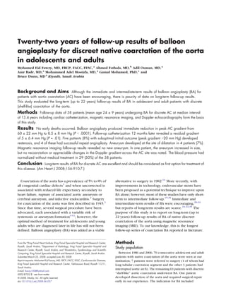
A, Magnetic resonance imaging scan of a patient with discrete coarctation (shell-like) (black arrow); notice remnant of the ductus arterious (white
arrow). B, One year after BA of the same patient, coarctation gradient decreased from 70 mm Hg to zero, notice remenant of the ductus arterious
(white arrow).
angiographic evidence of significant discrete aortic coarctation catheter balloon was inserted and inflated by hand for 5 to
with coarctation pressure gradient N20 mm Hg at cardiac 10 seconds until the stenotic waist disappeared. Hemodynamic
catheterization. All demographic, hemodynamic, echocardio- measurements and biplane aortic angiography were performed
graphic, and MRI follow-up data were encoded in prospective immediately before and after coarctation angioplasty, with
database program starting in 1986. Written informed consent special precaution to avoid manipulating the tip of the catheter
was obtained from all patients before BA. or guide wire over the area of the freshly dilated coarctation.
Definition Follow-up evaluation
Discrete coarctation of the aorta was defined as shelf-like Two patients who came from neighboring countries were lost
morphology in MRI (Figure 1) or angiographic images. Severe to follow-up. In addition, 3 other patients underwent surgical
isthmus or transverse aortic arch hypoplasia was defined as a repair within 1 year after BA. The remaining 58 of the 63 patients
ratio of the diameter of this structure to descending aorta at the were reassessed 6 months after the procedure and yearly
level of diaphragm of b0.6.19,30 Successful outcome was defined thereafter. Their clinical evaluation was accomplished by direct
as peak systolic gradient after BA of ≤20 mm Hg. Aneurysm was interview of the patients at clinic visits and included assessment
defined as an area of dilatation that was 150% of the aortic of peripheral pulse, evidence of radiofemoral delay, and supine
diameter at the level of the diaphragm or a discrete secular BP measurement in the right arm. Patients on antihypertensive
dilatation at that site that was not present before medication were given a trial off treatment for 1 month.
the intervention.26 Fifty patients underwent repeat catheterization and biplane
aortography with pressure measurement across the coarctation
Initial evaluation segment, 1 year after BA. The remaining 8 patients refused
Clinical evaluation before angioplasty included right arm repeat catheterization. All 58 patients had MRI and echocardio-
blood pressure (BP) measurement, chest radiograph, 12-lead graphic examination annually for the first 10 years, followed by
electrocardiogram, echocardiographic examination with mea- repeat MRI, and echocardiographic examination at 2-year
surement of the Doppler gradient across the coarctation using intervals thereafter. Their follow-up was concluded in
the precoarctation velocity (ie, ΔP (pressure difference) = December 2007.
4v22 − 4v12), and MRI.
Statistical analysis
Balloon angioplasty technique Data are presented as the mean ± SD. The paired Student t test
The technique used for BA has been previously reported.31 An was used to compare data before and after angioplasty and at
angioplasty balloon was selected with a diameter equal to that of follow-up. Multiple logistic regression analysis was used to
the isthmus or 1 to 2 mm smaller than the diameter of the identify variables associated with persistence hypertension. The
descending thoracic aorta at the level of the diaphragm. After variables included were age, gender, baseline coarctation
2,000 IU of heparin was given intravenously, the angioplasty gradient, baseline BP, and residual coarctation gradient.
3. American Heart Journal
912 Fawzy et al November 2008
Table I. Immediate and intermediate follow-up result
Parameters Before BA Immediately after P 12-month later P
Aortic pressure above Co (mm Hg) 170 ± 20 134 ± 16 b.0001 130 ± 12.8 .18
Catheter Co gradient (mm Hg) 60 ± 22 8.5 ± 8 b.0001 5 ± 6.4 .01
Doppler gradient (mm Hg) 61.9 ± 17.8 16.0 ± 8.4 b.0001
Co, Coarctation.
Freedom from reintervention was studied using Kaplan-Meier Complications
test. The analysis was performed with SAS Statistical Software There were no immediate deaths; one patient devel-
(SAS,
oped dissection of the aorta and underwent surgical
V 9.1, SAS Institute Inc, Cary, NC). A P value b.05 was
considered statistically significant.
repair. Thrombosis of the femoral artery developed in one
patient and required surgical thromboembolectomy.
Results Intermediate follow-up
Study subjects Catheter coarctation gradient. Follow-up catheter-
Sixty-three adolescent and adult patients (16 females) ization and angiography were performed 1 year after
underwent BA for native discrete coarctation of the aorta dilatation in 50 patients. Eight patients refused repeat
during a 22-year period. Their ages ranged from 14 to 55 catheterization. The gradient across the coarctation site
(mean 24 ± 9) years. In one patient, aortic dissection was further decreased to 5 ± 6.4 mm Hg (P = .01) (Table I).
developed early in our experience and required immedi- In comparison to values immediately after dilatation, there
ate surgical repair without sequelae. Apart from bicuspid was no further change in the systolic pressure in the aorta
aortic valve found in 26 patients (41%), additional above the coarctation site (134 ± 16 mm Hg to 130 ±
congenital heart defects were present in 7 patients (small 12.8 mm Hg, respectively; P = .18) (Table I).
ventricular septal defect 2 patients, a subaortic mem- Doppler coarctation gradient. The Doppler gradi-
brane in two, valvular aortic stenosis in one, and ent across the coarctation site decreased from 61.9 ±
moderate mitral regurgitation in one). All patients were 17.8 mm Hg before angioplasty to 16.0 ± 8.4 mm Hg
hypertensive (systolic BP 150-260 mm Hg). 1 year after angioplasty (P b .0001).
Restenosis. Restonosis is defined as residual gradient
Immediate results N20 mm Hg at rest on follow-up catheterization. The
The peak catheter coarctation gradient decreased from restenosis occurred in 5 patients (8%) and was mainly
60 ± 22 mm Hg to 8.5 ± 8 mm Hg (P b .0001) in because of suboptimal initial outcome. In 4 of these
57 patients (90%), and notably, gradient decreased to patients the anatomy of the coarctation was discrete, the
≤20 mm Hg at first dilation. In 49 of the 57 patients aortic arch and isthmus were of reasonable size, and the
(78%), the immediate coarctation gradient decreased to initial suboptimal relief of obstruction was due to the
≤10 mm Hg, and in 25 patients, the gradient was zero. small size balloon used in the first attempt early in our
In the remaining 9 patients, the gradient was N10 mm experience. Repeat dilatation with appropriately sized
Hg. Six patients had gradient of 12 to 15 mm Hg and in balloon catheter was carried out 6 to 12 months later. In
three, the gradient was 18 to 20 mm Hg. These 3 all four, the gradient decreased to 0 to 15 mm Hg and
patients had moderate degree of hypoplasia of the remained low at repeat catheterization 12 months later.
isthmus. Neither paradoxical hypertension nor mesen- The fifth patient, in whom the morphology of coarctation
teric vasculitis was encountered after angioplasty. The in a biplane aortogram at restudy 1 year later was deemed
systolic pressure in the aorta above the coarctation site unsuitable for angioplasty (Figure 2), underwent surgical
decreased from a mean of 170 ± 20 mm Hg to 134 ± repair. This was the only patient who had single-plane
16.0 mm Hg (P b .0001) (Table I). aortogram at the initial dilatation.
Aneurysm. The follow-up angiogram and MRI at
Suboptimal initial outcome 1 year after dilatation were scrutinized for aneurysm at
Suboptimal initial outcome, defined as immediate the site of BA. A total of 4 aneurysms were observed
residual coarctation systolic gradient N20 mm Hg, both on angiography and MRI giving an incidence of
was noted in 5 (8%) of the 63 patients. In four, an 7%. In 3 of these patients, the aneurysms were small
undersized balloon catheter was used in the absence of bulge measuring 2.0 to 2.3 cm in diameter. The fourth
an appropriate size balloon catheter early in our patient who had a 4-cm aneurysm underwent surgical
experiences, and one patient had narrow tortuous repair. None of the aneurysms could be detected on
coarctation segment. chest x-ray film.
4. American Heart Journal
Volume 156, Number 5
Fawzy et al 913
Figure 2
A, Aortogram in left arterior oblique view showing apparently discrete coarctation (arrow). B, Aortogram of the same patient in posterior-anterior
view showing tortuous coarctation (arrow) not suitable for BA.
Long-term follow-up results. Two patients living the remaining 29 patients, the BP was controlled with one
abroad were lost to follow-up and 3 required surgery medication in 4, 2 medications in 18, and 3 medications
within the first year after dilatation. The remaining in 7 patients.
58 patients were followed up for a median of 13.4 (mean Reintervention. Seven patients underwent repeat
12 ± 7) years (range 1-22 years), 23 of those were intervention, 4 patients with recoarctation responded to
followed up for a median of 18.3 (mean 18.5 ± 1.6) years. repeat BA, whereas one patient required surgery for
One patient who underwent BA at age 55 years died recoarctation. One patient with aneurysm underwent
15 years later from stroke. surgery 1 year after BA. Aortic dissection at the time of BA
Magnetic resonance imaging. The site of previous also necessitated surgical repair. Freedom from interven-
coarctation is shown to be well dilated (Figures 3 and 4). tion was 89% at 1 year, and this level was maintained
Aneurysms. Follow-up MRI studies revealed no new throughout the follow-up period (Figure 5).
aneurysm at the site of angioplasty. Of the 4 aneurysms
that were recognized during the first year of follow-up,
1 patient underwent surgery within 1 year after BA, Discussion
1 patient had aneurysm that increased in diameter from This study has demonstrated excellent long-term (up to
23 to 30 mm 20 years after dilatation, and the remaining 22 years) results of BA of discrete (shelf-like) aortic
2 patients had noted no appreciable changes in the size coarctation, and we propose that it should be used as first
of aneurysms. option for the treatment of discrete coarctation in the
Follow-up Doppler coarctation gradient. In com- absence of severe hypoplasia of the isthmus or transverse
parison to the value obtained 1 year after dilatation, the arch. Other investigators demonstrated that the outcome
Doppler coarctation gradient showed a small but in patients with discrete coarctation submitted to BA in
statistically insignificant further decrease at the last whom a residual gradient of ≤10 mm Hg was achieved
follow-up study (from 16.0 ± 8.4 to 13.7 ± 4.9 mm Hg, was not significantly different from cases with discrete
respectively; anatomy submitted to stent implantation.21 Surgery has a
P = .064). risk profile that encourages the pursuit of less invasive
Normalization of BP. The BP was normal (b140/ treatment options. Thus, an early surgical mortality rate of
90 mm Hg) without medication in 29 patients (50%). In 1.3% was reported in a study covering a wide age range
5. American Heart Journal
914 Fawzy et al November 2008
Figure 3
Serial MRI showing no restenosis or aneurysm formation up to 20 years of follow-up.
beyond infancy and recoarctation, and aneurysm forma- MRI, whereas the Doppler gradient at the site of
tion were noted in 5.8%.11,36 Cowley et al35 demonstrated coarctation decreased slightly at last follow-up compared
equivalent relief of obstruction and an equivalent need with 1 year after dilation probably because of remodeling
for repeat intervention for both of the aorta. Suarez de Lezo41 described neointimal
surgery and BA, but the risk of aneurysm formation was proliferation in 27% of patients after 2 to 3 years of follow-
higher among patients treated with BA. In addition, up, only 3 patients developed restenosis secondary to
surgery carries a small risk of recurrent laryngeal nerve neointimal proliferation, and multiple stents were used in
injury, phrenic nerve injury, chylothorax, wound infec- infancy in each of these 3 patients. The recoarctation rate
tion, postcoarctectomy syndrome, and paradoxical after surgery in the adult population is unclear and is
hypertension.36 The incidence of paraplegia is approxi- likely to be higher than the reoperation rate, as detection
mately 0.5%, despite various techniques for spinal of recurrence is dependent on the thoroughness of
cord protection.37 follow-up using imaging techniques.11
Coarctation restenosis Aneurysm formation
Recoarctation is a common complication after both The presence of cystic medial necrosis observed in two
angioplasty and surgical repair in infants and children, thirds of the resected aortic coarctation segments42
in whom recoarctation rate after angioplasty may range may provide a pathologic basis for the developments
from 15% to 30%.30,38 Recoarctation is uncommon of aneurysms associated with native coarctation, after
in adults, with incidence varying from 0% to BA and stent implantation or even after surgery. Over-
9%27,29,33,34,39,40—a finding that is corroborated by our stretching the coarctation is thought to increase the
study where recoarctation was encountered in 5 patients risk of the aneurysm, rupture, and dissection. Of our
(8%) all of whom had suboptimal initial outcome. No 4 patients who had aneurysms, 2 were treated with larger
recoarctation was observed on long-term follow-up using balloon catheter. However, one patient developed a 4-cm
6. American Heart Journal
Volume 156, Number 5
Fawzy et al 915
Figure 4 Figure 5
Kaplan-Meier curve showing freedom from intervention.
BA at mean follow-up 66 ± 37 (range 12-123 months).46
Multiple logistic regression analysis conducted in all
58 patients failed to identify a positive relation between
persistent hypertension and residual coarctation gradient
Magnetic resonance imaging scan of the aorta 22 years after BA of or baseline BP or age. Schräder et al47 reported a 79% rate
discrete coarctation in a patient. The coarctation segment is nicely of normalization of BP after BA in adolescent and adults
dilated (arrow); notice complete regression of the poststenotic with coarctation of the aorta and Walhout et al33
dilatation of the descending aorta.
encountered hypertension requiring medication in 6 of
18 adult patients (33%). We previously demonstrated that
aneurysm despite an appropriate-sized balloon catheter patients in whom BP became normal after BA also had
was used for angioplasty. Early studies by Cooper et al43 normal response of BP to exercise and regression of left
and Brandt et al44 reported high incidence of aneurysm ventricular hypertrophy.46 Hypertension in the absence
formation after BA; most subsequent investigators have of residual coarctation appears to be related to the
reported an incidence varying between 1.8% and duration of preangioplasty hypertension, possibly related
15%,24,27,28,31 concurring with our results (7%). No to insufficient resetting of the baroreceptors after BA.33
aneurysms were encountered by Koerselman et al40 and Pedra et al23 demonstrated stenting, and BA were
Walhout et al.33 Aneurysm were also encountered after similarly effective to normalize BP levels, which allowed
the use of stent with an incidence varying between 1.4% either discontinuation or dosage reduction of antihyper-
and 17%16-18,23-25 and also after CP covered stent tension medications. The incidence of later hypertension
implantation.23 The natural history of a small aneurysm after surgical repair of coarctation in adults varies
after BA is unknown. In our series, the aneurysm between 33% and N50%.4,10,48
increased in size in one patient on follow-up MRI 20 years
later, and in the other 2 patients, no appreciable changes Reintervention
in size were noted. Although development of aneurysm Freedom from reintervention was 90% at 1 year and
after BA is of concern, aneurysms are also known to 87% at 5 years as reported by Ovaert et al.32 This concurs
develop after surgical repair of coarctation especially with our findings, in which we noted freedom from
after patch aortoplasty, with incidence varying from 9% to reintervention to be 89% at 1 year and maintained
30%.5,9,12 Close follow-up is required for patient with or throughout the follow-up period.
without aneurysm, and we found that MRI is valuable
noninvasive imaging modality for follow-up of patients
who underwent coarctation angioplasty.45 Conclusion
This study demonstrated excellent long-term (up to
Normalization of the BP 22 years) results of BA for native discrete (shelf-like)
Blood pressure reverted to normal without medication coarctation in adolescent and adult patients. When
in 29 patients (50%). We have previously reported that compared against historical control subjects, the results
74% of patients have normal BP without medication after of BA compare favorably with reported results of surgical
7. American Heart Journal
916 Fawzy et al November 2008
repair or stenting. Accordingly, we recommend BA as the 20. Tyagi S, Singh S, Mukhopadhyay S, et al. Self- and
first option for treatment of discrete coarctation in balloon-expandable stent implantation for severe native coarcta-
adolescent and adult patients. tion of aorta in adults. Am Heart J 2003;146:920-8.
21. Zabal C, Attie F, Rosas M, et al. The adult patient with native
coarctation of the aorta balloon angioplasty or primary stenting.
We thank Suzanne Tobias and Jovett Lopez for typing Heart 2003;89:77-83.
the manuscript. 22. Chessa M, Carrozza M, Butera G, et al. Results and mid-long-term
follow-up of stent implantation for native and recurrent coarctation of
the aorta. Eur Heart J 2005;26:2728-32.
References 23. Pedra CAC, Fontes VF, Esteves CA, et al. Stenting vs balloon
1. Rao PS. Coarctation of the aorta. Semin Nephrol 1995;15:87-105. angioplasty for discreet unoperated coarctation of the aorta in
2. Campbell M. Natural history of coarctation of the aorta. Br Heart J adolescents and adults. Catheter Cardiovasc Internv 2005;64:
1970;32:633-40. 495-506.
3. Craaford C, Nylin G. Congenital coarctation of the aorta and its 24. Suarez de Lezo J, Pan M, Romero M, et al. Percutaneous intervention
surgical treatment. J Thorac Surg 1945;14:347-61. on severe coarctation of the aorta: a 21 year experience. Pediatr
4. Pennington DG, Liberthson RR, Jacobs M, et al. Critical review of Cardiol 2005;26:176-89.
experience with surgical repair of coarctation of the aorta. J Thorac 25. Forbes TJ, Moore P, Pedra CA, et al. Intermediate follow-up following
Cardiovasc Surg 1979;2:217-29. intravascular stenting for treatment of coarctation of the aorta.
5. Bromberg BI, Beekman RH, Rocchini AP, et al. Aortic aneurysm after Catheter Cardiovasc Interv 2007;70:569-77.
patch aortoplasty repair of coarctation: prospective analysis of 26. Beekman RH, Rocchini AP, Dick II M, et al. Percutaneous balloon
prevalence, screening tests and risks. J Am Coll Cardiol 1989;14: angioplasty for native coarctation of the aorta. J Am Coll Cardiol
734-41. 1987;10:1078-84.
6. Beekman RH, Rocchini AP, Behrendt DM, et al. Long-term outcome 27. Fletcher SE, Nihill MR, Grifka RG, et al. Balloon angioplasty of native
after repair of coarctation in infancy: subclavian angioplasty does not coarctation of the aorta: midterm follow-up and prognostic factors.
reduce the need for reoperation. J Am Coll Cardiol 1986;8:1406-11. J Am Coll Cardiol 1995;25:730-4.
7. Martin MM, Beekman RH, Rocchini AP, et al. Aortic aneurysms after 28. Mendelsohn AM, Lloyd TR, Crowley DC, et al. Late follow-up of
subclavian flap angioplasty repair of coarctation of the aorta. Am J balloon angioplasty in children with a native coarctation of the aorta.
Cardiol 1988;61:951-3. Am J Cardiol 1994;74:696-700.
8. Pinzon JL, Burrows PE, Benson IN, et al. Repair of coarctation of the 29. de Giavanni JV, Lip GYH, Osman K, et al. Percutaneous balloon
aorta in children: postoperative morphology. Radiology 1991;180: dilatation of aortic coarctation in adults. Am J Cardiol 1996;77:
199-203. 455-9.
9. Knyshaw GV, Sitar LL, Glagola MD, et al. Aortic aneurysms at the site 30. Rao PS, Galal O, Smith PA, et al. Five to nine year follow-up results
of the repair of coarctation: a review of 48 patients. Ann Thorac Surg of balloon angioplasty of native coarctation in infants and children.
1996;61:935-9. J Am Coll Cardiol 1996;27:462-70.
10. Cohen M, Fuster V, Steele PM, et al. Coarctation of the aorta 31. Fawzy ME, Sivanandam V, Galal O, et al. One to ten-year
long-term follow-up and prediction of outcome after surgical follow-up results of balloon angioplasty of native coarctation of
correction. Circulation 1989;80:840-5. the aorta in adolescents and adults. J Am Coll Cardiol 1997;30:
11. Kappetein AP, Zwinderman AH, Bogers AJ, et al. More than 1542-6.
thirty-five years of coarctation repair: an unexpected high relapse 32. Ovaert C, McCrindle BW, Nykanen D, et al. Balloon angioplasty of
rate. J Thorac Cardiovasc Surg 1994;107:87-95. native coarctation: clinical outcomes and predictors of success. J Am
12. Kodolitsch YV, Aydin MA, Koschyk DH, et al. Predictors of aneurysm Coll Cardiol 2000;35:988-96.
formation after surgical correction of aortic coarctation. J Am Coll 33. Walhout RJ, Lekkerker JE, Ernst SP, et al. Angioplasty for coarctation
Cardiol 2002;39:617-24. in different aged patients. Am Heart J 2002;144:180-6.
13. Bouchart F, Dubar A, Tabley A, et al. Coarctation of the aorta in 34. Paddon AJ, Nicholson AA, Ettles DF, et al. Long-term follow-up of
adults: surgical results and long-term follow-up. Ann Thorac Surg percutaneous balloon angioplasty in adult aortic coarctation.
2000;70:1483-8. Cardiovasc Intervent Radiol 2000;23:364-7.
14. Singer MI, Rowen M, Dorsey TJ. Transluminal aortic balloon 35. Cowley CG, Orsmond GS, Feola P, et al. Long-term, randomized
angioplasty for coarctation of the aorta in the newborn. Am Heart J comparison of balloon angioplasty and surgery for native
1982;103:131-2. coarctation of the aorta in childhood. Circulation 2005;111:
15. Marshall AC, Perry SB, Keane JF, et al. Early results and medium-term 3453-6.
follow-up of stent implantation for mild residual or recurrent aortic 36. Kirklin JW, Barratt-Boyes BC. Cardiac surgery. 3rd ed. Philadelphia:
coarctation. Am Heart J 2000;139:1054-60. Churchill Livingstone; 2003. p. 1315-75.
16. Harrison DA, Mc Laughlin PR, Lazzam E, et al. Endovascular stents in 37. Connolly JE. Hume memorial lecture, prevention of spinal cord
the management of coarctation of the aorta in adolescent and adult: complications in aortic surgery. Am J Surg 1998;176:92-101.
one year follow-up. Heart 2001;85:561-6. 38. Park Y, Lucas VW, Sklansky MS, et al. Balloon Angioplasty of native
17. Cheatham JP. Stenting of coarctation of the aorta. Catheter aortic coarctation in infants 3 month of age and younger. Am Heart J
Cardiovasc Intervn 2001;54:112-25. 1997;134:917-23.
18. Duke C, Qureshi SA. Aortic coarctation and recoarctation: to stent or 39. Tyagi S, Arora R, Kaul UA, et al. Balloon angioplasty of native
not to stent?J Interv Cardiol 2001;14:283-98. coarctation of the aorta in adolescents and young adults. Am Heart J
19. Hamdan MA, Maheshwari S, Fahey JT, et al. Endovascular stents for 1992;123:674-8.
coarctation of the aorta: initial results and intermediate-term 40. Koerselman J, de Vries H, Joarsma W, et al. Balloon angioplasty
follow-up. J Am Coll Cardiol 2001;38:1518-23. of coarctation of the aorta: a safe alternative for surgery in
8. American Heart Journal
Volume 156, Number 5
Fawzy et al 917
adults: immediate and mid-term results. Catheter Cardiovasc Interv 45. Fawzy ME, Sinner WV, Rifai A, et al. Magnetic resonance imaging
2000;50:28-33. compared with angiography in the evaluation of intermediate-term
41. Suárez de Lezo J, Pan M, Romero M, et al. Immediate and follow-up result of coarctation balloon angioplasty. Am Heart J 1993;126:
findings after stent treatment for severe coarctation of aorta. Am J 1380-4.
Cardiol 1999;83:400-6. 46. Fawzy ME, Sivanandam V, Peiters F, et al. Long-term effects of
42. Isner JM, Donaldson RF, Fulton D, et al. Cystic medial necrosis in balloon angioplasty on systemic hypertension in adolescent and
coarctation of the aorta: a potential factor contributing to adverse adult patients with coarctation of the aorta. Eur Heart J 1999;20:
consequences observed after percutaneous balloon angioplasty of 827-32.
coarctation sites. Circulation 1987;75:689-95. 47. Schräder R, Bussmann WD, Jacobi V, et al. Long-term effects of
43. Cooper RS, Ritter SB, Rothe WB, et al. Angioplasty for balloon coarctation angioplasyt on arterial blood pressure in
coarctation of the aorta: long-term results. Circulation 1987;75: adolescent and adult patients catheter. Cardiovasc Diagn 1995;36:
600-4. 220-5.
44. Brandt III B, Marvin Jr WR, Rose EF, et al. Surgical treatment of 48. Wells WJ, Prndergast TW, Berdjs F, et al. Repair of coarctation of the
coarctation of the aorta after balloon angioplasty. J Thorac aorta in adults. The fate of systolic hypertension. Am Thorac Surg
Cardiovasc Surg 1987;94:715-9. 1996;61:1168-71.
The following article is an AHJ Online Exclusive.
Full text of this article is available at no charge at our website:
www.ahjonline.com
Transcatheter closure of the patent ductus arteriosus using the new
Amplatzer duct occluder: Initial clinical applications in children
Basil Thanopoulos, MD, PhD, a Nikolaos Eleftherakis, MD, a Konstantinos Tzannos, MD, b and Christodoulos Stefanadis, MD, PhD b Athens, Greece
Background In spite of recent advances in transcatheter manage- Results The mean PDA diameter was 3.6 ± 1.3 mm (range 0.6-5
ment, the occlusion of certain anatomic types of patent ductus arteriosus (PDA), mm). The mean device diameter (waist diameter) was 4.3 ± 1.4 mm
especially in infants and small children, remains a challenge. The aim of the (range 3-6 mm). Complete echocardiographic closure of the ductus at 1-
study was to report initial human experience with transcatheter closure of PDA month follow-up was observed in 24 (96%) of 25 patients. Immediately
in 25 patients using the new Amplatzer duct occluder (ADO II) (AGA Medical, after the procedure, there was a mild left pulmonary stenosis (Doppler
Golden Valley, MN). gradient of 15 mm Hg) in 2 of 25 patients. No other complications were
observed.
Methods The median age of the patients was 3.2 years (range 0.1-
5 years), and the median weight was 10.5 kg (range 3-18 kg). The device Conclusions The ADO II is a promising addition to our armamen-
used is a modified ADO II made of fabric-free fine Nitinol wire net into 2 very- tarium for PDA closure. Further studies are required to document its efficacy,
low-profile disks with an articulated connecting waist. Both disks are 6 mm safety, and long-term results. (Am Heart J 2008;156:917.e1-917.e6.)
larger than the diameter of the connecting waist. Connecting waist diameters
range from 3 to 6 mm.