17.occlusal schemes anatomic and semiamatomic occlusion
•Télécharger en tant que PPT, PDF•
9 j'aime•2,488 vues


Recommandé
Recommandé
Contenu connexe
Tendances
Tendances (20)
Similaire à 17.occlusal schemes anatomic and semiamatomic occlusion
Similaire à 17.occlusal schemes anatomic and semiamatomic occlusion (20)
Plus de www.ffofr.org - Foundation for Oral Facial Rehabilitiation
Plus de www.ffofr.org - Foundation for Oral Facial Rehabilitiation (20)
Dernier
Dernier (20)
17.occlusal schemes anatomic and semiamatomic occlusion
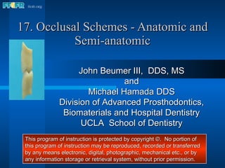
- 1. 17. Occlusal Schemes - Anatomic and Semi-anatomic John Beumer III, DDS, MS and Michael Hamada DDS Division of Advanced Prosthodontics, Biomaterials and Hospital Dentistry UCLA School of Dentistry This program of instruction is protected by copyright ©. No portion of this program of instruction may be reproduced, recorded or transferred by any means electronic, digital, photographic, mechanical etc., or by any information storage or retrieval system, without prior permission.
- 2. Semi-anatomic Denture Teeth Begin by positioning the appropriate protrusive insert, and check to ensure that the incisal guide pin is set at zero and in contact with the incisal guide table. Protrusive insert Protrusive Inserts Zero setting
- 4. Mark the casts indicating midline, crest of the ridge, and the retromolar pad . These landmarks will be used to check your denture setup. Maxilla Midline Anterior land Mandible Ridge Retromolar pad Cast Landmarks
- 5. Anterior land Cast Landmarks - Maxilla Midline Incisive papilla
- 6. Lines indicating the crest of the ridge Cast Landmarks -Mandible Midpoint of retromolar pad Land Mark on land indicating the midpoint of the retromolar pad
- 8. To set the remaining maxillary anterior teeth a clear glass or plastic slab is positioned on the mandibular record base to represent the plane of occlusion. When setting anatomic posterior teeth we recommend setting the maxillary posterior teeth before the mandibular posterior teeth. To aid in positioning the maxillary teeth, a line is inscribed on the slab indicating the crest of the mandibular ridge. Setting the Maxillary Anterior Teeth Mark indicating midpoint of the retromolar pad
- 9. Setting the Maxillary Anterior Teeth These two lines, inscribed on the plastic plane, indicate the crest of the alveolar ridge. These lines will be used to position the maxillary posterior denture teeth to insure that the mandibular posterior teeth are centered over the ridge. The lingual cusp tips of the posterior maxillary teeth should contact these lines. Lines indicating the crest of the ridge
- 10. Setting the Maxillary Anterior Teeth Soften some baseplate wax and attach some to the ridge lap portion of the other maxillary central incisor and attach it to the record base as shown. Set the lateral incisors and cuspids as shown previously (Section 13c, 1a Lingualized occlusion).
- 11. Setting the Maxillary Anterior Teeth Note the angulations of the anterior teeth in relation to the occlusal plane when viewed in profile. Occlusal plane
- 12. Setting the Maxillary Anterior Teeth “ Toed-in” Position Note how the cervical and incisal edges of the cuspid are aligned vertically (yellow line). The facial surface of the cuspid however, is canted inward and appears “toed in” (red line) due to the prominence of the cervical area of the tooth (yellow arrow). The centrals and laterals are inclined slightly towards the distal.
- 13. The long axis of the premolars should be perpendicular to the occlusal plane and the buccal and lingual cusp tips should touch the occlusal plane. Arranging the premolars in this way insures that the adjacent marginal ridges will be on the same level. This is an important factor when setting the opposing premolars. Setting the Maxillary Posterior Teeth Occlusal plane
- 15. Setting the Maxillary Posterior Teeth The curve of Wilson and the curve of Spee begin in the molar region. The mesial lingual cusp tip of the 1 st molar contacts the occlusal plane but the buccal cusp tips and the distal lingual cusp are elevated about .5mm off the occlusal plane (yellow line) . The Maxillary 1 st Molar
- 35. Setting the Mandibular Central Incisors In most patients the labial surface of the mandibular incisors should be roughly perpendicular to the occlusal plane. Occlusal plane The Central Incisors
- 37. Setting the Mandibular Anterior Teeth Horizontal overlap This practice will idealize the amount of horizontal and vertical overlap and ensure that anterior guidance is not introduced into the setup. Horizontal overlap Vertical overlap
- 41. Anatomic Denture Teeth (30 degree)
- 42. Anatomic Denture Teeth (30 degree) These teeth are arranged in the same fashion as the semi-anatomic teeth shown previously. Use the same sequence of steps as we have just shown.
- 43. Anatomic Denture Teeth (30 degree) Begin by positioning the appropriate protrusive insert, and check to ensure that the incisal guide pin is set at zero and in contact with the incisal guide table. Protrusive insert Protrusive Inserts Zero setting
- 44. Setting Anatomic Teeth (30 degree) When you are finished check to see that the posterior teeth are on plane and the posterior teeth centered over the mandibular ridge. Make corrections as necessary.