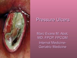
2. Definition
• Any lesion caused by unrelieved
pressure leading damage of underlying
tissue
– Synonymous to decubitus ulcer and
bedsores but the above term denotes the
primary pathophysiologic factor
3. Staging
• Stage I
– Nonblanchable erythema of intact skin;
may also be other discoloration, warmth,
edema and induration
– 10-fold increase in risk of developing
higher-staged ulcers
4. • Stage II
– Partial-thickness skin loss involving the
epidermis or also the dermis
• Stage III
– Extend to the subcutaneous tissues and
deep fascia
– Typically show undermining
• Stage IV
– Involve muscle and bone
11. • Eschar formation
– Full-thickness injury
– Has to be removed
prior to staging
• Pressure-related
blister formation
– Cannot be staged
clinically
12. Epidemiology
• Acute care setting
– Stage II and higher prevalence 3-11%,
incidence 1-3%
– After 1 week of confinement, incidence
28%, prevalence 8-30%
– >50% occur in patients >70 years old
13. • In nursing homes
– Prevalence of 20-33% and incidence of 11-
14%
• Sepsis
– Most serious complication of pressure
ulcers
– In-house mortality of 60% when the ulcer is
the source of bacteremia
14. • Infected pressure ulcers
– Most common infection in skilled nursing
facilities (6% of residents)
• Osteomyelitis
– In 26% of non-healing pressure ulcers
15. • Associated with prolonged and
expensive hospitalizations
• Associated with pain
– 59-85% of those who can communicated
describe pain
– 45% report ulcer pain as “horrible”
16. • Increased mortality
– 60% at 1 year after discharged for those
who develop a pressure ulcer
17. Pathophysiology
• 4 factors implicated: pressure, shearing
forces, friction and moisture
• Muscle and subcutaneous
tissuemore sensitive to pressure
injury
18.
19. • Pressure on bony prominences
– 100-150 mmHg on regular mattress while
lying down
– 300 mmHg on ischial tuberosities while
sitting
– Enough to decrease transcutaneous
oxygen tension to 0
• Other factors may lower the time or
pressure needed to cause full-thickness
injuries
20. • Shearing forces-tangential forces on the
skin when the patient slides while sitting
or lying down in an elevated position
– Lowers the pressure needed to cause
ulcers
• Friction leads to intraepidermal
blistersunroofed, leading to
superficial erosions
• Moisturemay lead to maceration
21.
22.
23. • Effect of pressure
– Ischemia and accumulation of cellular
toxins
– Damage begins in deeper tissues
– Persistent pressurevascular leakage and
interstitial edemaeventual hemorrhage
(stage I)
• Superimposed bacterial infection (both
deep and superficial)
24.
25. Presentation
• Any disease that leads to immobility
predisposes to pressure ulcers
• Risk factors other than immobility
– Incontinence (particularly fecal)
– Nutritional factors (decreased lymphocyte
count, hypoalbuminemia, inadequate
intake, decreased body weight, depleted
triceps skin fold)
26. • Other factors
– Dry skin
– Increased body temperature
– Decreased blood pressure
– Age
– Age-related skin changes
27. Assessment
• Includes assessment of risk factors,
including nutritional assessment
• Location, size, stage and wound
characteristics of ulcer at onset
– Includes assessment of tracts,
undermining, tunneling, exudate, necrotic
tissue, granulation and epithelialization
28.
29. • Follow-up assessment using above
parameters at least weekly
– PUSH score
– Decrease in ulcer size over 2 weeks usually
predicts healing
• Sinograms to assess tract extent
• Cultures using needle aspiration or biopsy
speciments; may include bone biopsys
– Culture of swabs not helpful as bacterial
colonization in eventual
– Important if ulcer does not heal after 4 weeks or if
with obvious infection
30.
31.
32. • Osteomyelitis diagnosis may be difficult
due to similarity of pressure-induced
bone changes
– Presence of abnormal plain radiograph,
WBC count of 15,000 and ESR >120 mm
has probability of osteomyelitis of 70%
33. • Most common bacterial isolates
– Gram (-) aerobic rods (45% of isolates)
– Gram (+) aerobic cocci (39%)
– Bacteroides species, most common
anaerobic isolate
34. Management
Pharmacologic
• Vitamin and mineral supplementation for
those with deficiencies
• Systemic antibiotics indicated for patients with
– Sepsis
– Cellulitis
– Osteomyelitis
– Prevention of bacterial endocarditis in those with
VHD and requiring debridement
35. • Broad spectrum antibiotics for those
with suspected bacteremia, pending
culture results
– Ampi-sulbactam
– Carbapenems
– Pip-tazo
– Clindamycin/metronidazole + quinolones
36. • Vancomycin for methicillin-resistent
Staphylococcus aureus
• Deeper ulcers may have some benefit
for topical antibiotics
– Silver sulfadiazine x 2 weeks
– Avoid iodophors, sodium hypochlorite or
acetic acid (toxic to fibroblast)
37. Nonpharmacologic
• Adequate dietary, especially protein
intake
– Target 30-35 kcal/kg BW/day with 1.25-
1.50 g CHON/kg BW
– May use alternative feeding methods if oral
intake is inadequate
– Vitamin and mineral supplementation
38. • Use of pressure-relieving devices
– Regular air/foam mattresses
– Egg-crate foam mattresses
– Static mattresses (should not bottom out
and provide at least 2.5 cm of support)
• Usually appropriate for those who can still
assume different positions
– Dynamic mattresses
• Air-fluidized mattresses
• Low-air loss mattresses
39.
40.
41.
42. • Debridement
– Sharp debridement
– Mechanical approaches (wet-to-dry
dressing, irrigation, hydrotherapy)
• Irrigation pressure 4-15 psi using a 30-cc
syringe with a 18G needle
– Enzymatic approaches (collagenases)
– Autolytic approaches (contraindicated in
infected ulcers)
43. • Occlusive dressings for clean wound
– Not proven to me more effective for stage
III or IV ulcers but reduces the nursing time
needed
• Moist gauze dressing using normal
saline for the ulcer base
• The aim of dressing the ulcer is to
maintain a moist environment for would
healing and autolytic debridement
44.
45.
46. • Skin sealants
– Prevents friction and
protects from adhesives
– Contains alcohol and
should not be used under
most hydrocolloids
47. • Impregnated gauze
– Gauze impregnated with
saline or other
substances
– Make sure that
impregnating substance
is not harmful to wound
healing
– Limited absorbent
capacity
48. • Composite dressings
– Combination of
different dressing
groups
– Properties depend on
the components
49. • Transparent film dressing
– Polyurethane and
polyethylene membrane
coated with a layer of
acrylic, hypoallergenic
adhesive
– Promotes epithelialization,
moist wound healing
– Bacterial barrier, autolysis
– May reinjure wound on
removal
– Can lead to wound edge
maceration
– Not for wounds with
moderate to heavy
exudation
50. • Hydrocolloid
– Gelatin or
carboxymethycellulose in a
polyisobutylene adhesive
base
– Moist would healing with
absorption of light to
moderate wound fluid
– Increased wear time
– Reduces pain, promotes
autolysis
– Not for those withg heavy
exudate
– Odor on removal
– Limited absorption
51. • Hydrogels
– May or may not have
supporting fabric net
– High water content with
varying gel forming material
– Moist wound healing with low
to moderate drainage
– Promotes autolysis
– Reduces pain and rehydrates
dry wounds; cooling effect
– Does not cause reinjury on
removal
– Can dry out or may macerate
surrounding tissues
– Candidiasis may occur with
inappropriate use
52. • Wound fillers
– Made of copolymer
starch or dextranomer
beads which absorb
wound fluid to form a gel
– Moderate to large
absorption and fills up
dead space
– Moisture retentive and
promotes autolysis
– Requires another
dressing to hold it in
– May have an odor
– Requires wound irrigation
to remove
53. • Enzyme debriding
agent
– Can debride necrotic
tissue
– Hard eschar chould be
removed first
– Discontinued when
granulation appears
– Require secondary
dressing
– May be inhibited by
irrigation solutions
54. • Alginates
– Calcium or sodium salts
of alginic acid
– Moisture retentive and
promotes autolysis
– Moderate to large
moisture absorption
– Reduces pain and can fill
dead space
– Should not be used in
low-exudate wounds and
may dry out
55. • Lubricating agents
– Promotes moist wound
healing
– Limited autolysis
– Reduces pain
– Requires secondary
dressing
– Non-absorptive
– May be used to
impregnate gauze
56. • Foams
– Hydrophilic and non-
adherent modified
polyurethane foam
– In wafers, pillows; with
film covers
– Surfactant impregnated
or with a charcoal layer
– Moderate to large
absorption
– Moist wound healing
– Can be used with topical
medications and
infected wounds
– Requires taping
57. • Collagen
– Bovine collagen attached to
nylon mesh, or powder or
paste
– Also comes in 90% collagen
and 10% alginate
– Absorbs small to moderate
exudate
– Non-adherent
– For contaminated, infecteed
wounds
– Can be used with topical
agents
– Requires secondary dressing
– Sensitivity to bovine material
58. • Surgical correction (attempted only in
clean wounds)
– Primary closure
– Skin grafting
– Myocutaneous flaps
• 30% complication rate
• Complications included necrosis, dehiscence,
flap infection, hematoma
• 70% healing rate by time of discharge
– Removal of underlying bony prominences
59. • Other modalities
– Hyperbaric oxygen therapy
• Effects not statistically significant
– Growth factors
• For ulcers that do not heal with a
comprehensive approach
– Larvae therapy
– Vacuum-assisted closure
• Reduces bacterial load and improves perfusion
and granulation
– Electrical stimulation
• Improves healing in small trials; dose and type
of wound to be applied with not yet determined
60. Prevention
• Systematic risks assessment
– Braden scale
• A score of 18 or less in any patient indicates
risk for pressure ulceration
– Norton scale
61.
62.
63. • Appropriate skin care
– Systematic skin inspection
– Skin cleaning with mild cleansing agent at
time of soiling and at regular intervals
– Minimize skin dryinguse moisturizers
– Minimize excessive moisture
– Minimize friction and shear forces
– Ensure adequate dietary intake
64. • Frequent repositioning every 2 hours for supine
patients
– The back should be at a 30° angle with the support
surface; avoid a 90° angle
– Minimize head elevation to compelling indications like
postfeeding or if in respiratory distress
• If patient needs to be seated, should not be for
more than 1 hour; positions are shifted every 15-
30 minutes
– May use pillows behind the knees, back or neck to
provide more support
– Avoid doughnut rings (increases venous congestion)
65. • Off-loading devices of extremities in the
supine or seated position
• Sheepskin and foam egg crate mattress
(or other foam overlays)
– Inexpensive but do not have the capability
of reducing pressure enough to reduce
injury
• Use of pressure-relieving mattresses
– 60% reduction in incidence of pressure
ulcers
66. • Treatment of infections distant from
clean pressure ulcers
– Bacteremia from distant infections may
seed in the clean ulcer due to least
resistance