
Recommandé
Contenu connexe
Tendances
Tendances (20)
En vedette
Similaire à Pulp protection
Similaire à Pulp protection (20)
Dernier
Dernier (20)
Pulp protection
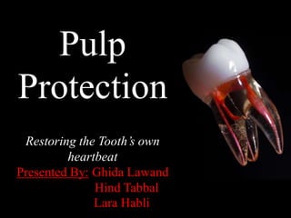
- 1. Pulp Protection Restoring the Tooth’s own heartbeat Presented By: Ghida Lawand Hind Tabbal Lara Habli
- 2. Outline: Pulp Irritants Protection in shallow and moderate cavities Pulp protection in deep cavities Indirect pulp capping direct pulp capping Materials used for pulp protection New Materials and mesthods to protect pulp
- 3. The dental pulp is a soft connective tissue of mesenchymal origin present within the pulp chamber and root canals of teeth. It is not considered an external tissue, yet its exposure to external stimuli is unceasing due to several factors that make the pulp extremely sensitive to environment outside. We have to protect it from any type of irritant
- 4. Pulpal irritants A) Bacterial irritants (Most common cause for pulpal irritation) B) Traumatic 1-Caries Tooth fracture Luxation Avulsion Parafunctional habits like bruxism 2- Periodontal pocket and abscess 1-Acute trauma 2- Chronic Trauma
- 5. C) Iatrogenic: 1. During tooth preparation a) Heat production during cutting procedures: Pulp temperature 11°F Destructive reaction Revolution per minute (RPM) of the bur: As RPM increases,heat production increases. Speed must not exceed 3,000 rpm. Pressure: It is directly proportional to heat generation. Surface area of contact: The more the contact between the tooth structure and revolving tool, the more is the heat generation
- 6. Excessive heat generation leads to change in dentin color due to vascular stasis and hemorrage in the subodontoblastic vascular plexus present in the pulp
- 7. b) Pressure exerted: Pressure of hand or rotary instruments Nuclear aspiration of odontoblasts or nerve endings from pulp tissues into the dentinal tubulesDisturb odontoblasts metabolism leading to their complete degeneration and disintegration. c) Remaining Dentin Thickness (RDT)
- 8. 2. Orthodontic movement 3. Periodontal and periapical curettage
- 9. 4. Use of chemicals Temporary & permanent fillings, bases, liners, and use of alcohol that leads to pulpal injury due to its cytotoxicity, acidity, heat formed and marginal leakage Chemical irritants applied to dentin can result in damage and disorganization in the subadjacent pulp
- 11. Because of these various irritants pulp needs protection…
- 13. Conventional methods Direct pulp capping is placing a biocompatible material over the exposed pulp to maintain vitality and promote healing. Direct Pulp Capping WHY? 1) To maintain the vitality of the remaining pulp tissue 2) To prevent root canal treatment 3) To help conserve tooth structure
- 14. Indications Recent small mechanical exposure of pulp during (< 24 hours): a) Tooth preparation b) Traumatic injury. No or minimal bleeding at the exposure site.
- 15. Contraindications Wide pulp exposure Pre-operative history of Spontaneous pain Presence of bleeding at exposure site Radiograph doesn’t show any pulp pathology
- 16. Clinical Procedure 3.When vital & healthy pulp is exposed, check fresh bleeding 2. Isolate the tooth with rubber dam 1. Administer local anesthesia 4. Clean the area with saline solution 5. Dry it with a cotton pellet 6. Apply calcium hydroxide (preferably Dycal) over the exposed area
- 17. 7. Give interim restoration such as zinc oxide eugenol for 6 to 8 weeks b) If not pulpotomy or pulpectomy is requested a) Remove the cement to inspect the exposure site. If secondary dentin formation takes place over the exposed site restore the tooth permanently with protective cement base and restorative material.
- 18. In indirect pulp capping, all caries are removed except the ones that lie adjacent to the pulp. Caries near the pulp is left in place to prevent pulp exposure and preparation is enclosed with a biocompatible material. Indirect Pulp Capping
- 19. Indications 1. Deep carious lesion near the pulp tissue but not involving it 2. No mobility of tooth 3. No history of spontaneous toothache 4. No tenderness to percussion 5. No radiographic evidence of pulp pathology 6. No root resorption or radicular disease should be present radiographically. Root resorption
- 20. Clinical Procedure It’s the same procedure as the direct pulp capping except that the pulp is not exposed. A thin layer of dentin and some amount of caries is left to avoid exposure. Placement of calcuim hydroxide and zinc oxide eugenol dressing after excavation of soft caries
- 21. Factors affecting Pulp Capping success 1) Age of the patient: Due to vascularity of the pulp, young patients have greater potential for success than older ones Young patient Old patient 2) Type of exposure: Mechanically done pulpal exposure has better prognosis than exposure caused by caries, due to less pulpal inflammation and deleterious effect of bacterial toxins on the pulp
- 22. 3) Size of the exposure: In large exposures, it is difficult to control the hemorrhage and tissue seepage. Small pinpoint exposures are easy to manage and have a greater potential for success 4) History of pain: If previously pain has not occurred in the tooth, the potential for success is more
- 23. Recent methods Laser in pulp capping Mechanism: CO2 laser emits an infrared beam Stimulates mineralization in dental pulp cells Therapeutic benefit for direct pulp capping and pulpotomy in clinical practice
- 24. Conventional Materials Materials used for Pulp Protection Recent Materials Varnish Base Sealer Liner 1) Zinc oxide eugenol liners 2) Calcium hydroxide 3) Flowable composites 4) Glass ionomers 1) Zinc Oxide Eugenol 2) Zinc phosphate cement 3) Polycarboxylate cement 4) Glass ionomer cement Growth Factors Cements Stem Cells Enzymes
- 25. Ether or chloroform Organic copalResin gum Solvent evaporates Definition: It is an organic copal or resin gum suspended in solutions of ether or chloroform. When we put it on the tooth surface the organic solvent evaporates leaving a protective film Two coats of varnish should be applied using a small cotton pellet to ensure sufficient wetting of cavity walls A) Varnish
- 26. Indications To seal the dentinal tubules Dentinal tubules Open Dentinal tubules Sealing dentinal tubules with varnish Dentinal tubules blocked by varnish 2. Protects the tooth from chemical irritants from cements reducing postoperative pain 3. Reduces microleakage around restorations 1. Prevents discoloration of tooth with an amalgam restoration by preventing migration of ions into the dentin
- 27. Under Composite Resin Varnishes dissolve in the monomer of the resin & also interfere with their polymerization of resins With Glass Ionomer Restorations It interferes the bonding of tooth to these cements Contraindications
- 28. B) Sealer Indications • To seal dentinal tubules • To treat dentin hypersensitivity. An adhesive sealer is commonly used under indirect restorations. For application, cotton tip applicator is used to apply sealer on all areas of exposed dentin. C) Liners • Fluid materials that can adapt more readily to all aspects of a tooth preparation • Used to create a uniform, even surface that aids in adaptation of more viscous filling materials (amalgams, composites) • Do not have sufficient thickness, hardness and strength not used alone in deep preparations 1. Protect pulp from chemical irritants by sealing ability 2. Stimulate formation of reparative dentin. Indications
- 30. 1. Zinc oxide eugenol liners • Used to alleviate pain from mild-to- moderate inflammation of pulp. In low concentration it acts as obtundant In high concentration it acts as chemical irritant Contraindication: It inhibits polymerization Should not be used under bonding agents & composite restorations
- 31. 2- Calcium hydroxide Most common agent considered as the “gold standard” of direct pulp capping materials against which new materials should be tested Advantages: 1. Causes dentin mineralization by activating the enzyme ATPase 2. Stimulates reparative dentin formation 3. Biocompatible 4. High pH (12.5) neutralizes acidity of silicate and zinc phosphate cements Disadvantages: 1. Low strength 2. High solubility Dissolves rapidlyUsed over small areas requiring pulp protection / Applying glass ionomer or zinc phosphate base to prevent its dissolution.
- 33. 3- Flowable composites Composites with a lower amount of filler more fluid consistency, less strength and lower modulus
- 34. 4- Glass ionomers Renewable source of fluoride under restorations Reduce the incidence of caries Fluoride Glass ionomer cements (GIC): Bond to tooth structure Act as a thermal barrier Ability to bond in a moist environment Easy to use. Anticariogenic.
- 35. Light-cured resin-modified glass ionomers (RMGIs) Provide good adhesion to both tooth structure and restorative materials High strength Flexible (low modulus of elasticity) Dual-setting reaction: 1) Light-activated, methacrylate crosslinking reaction 2) Slower, delayed, acid-base reaction Which gives RMGIs an additional period of maximum flexibility to absorb stress from the adjacent shrinking composite.
- 36. Classification of bases Protective bases Sedative bases Insulating bases They protect the pulp before restoration is placed They help in calming the pulp which has been irritated by mechanical, chemical or other means They protect the tooth from thermal shock. D) Bases Bases should have sufficient strength so that they can withstand forces of mastication and condensation of permanent restorations.
- 37. Excellent sealing quality. Bacteriostatic in nature. Anodyne effect. Reduces the thermal conductivity of metallic restorations Blocks undercuts in the preparation wall in case of cast restorations. Chemically bonds to tooth Antibacterial properties Fluoride release Anticariogenic property Chemical bond to tooth Well tolerated by the pulp. Materials used as bases Zinc oxide eugenol Zinc phosphate cement Polycarboylate cement Glass ionomer cement
- 38. Pulp Protection according with depth of tooth preparation
- 39. Recent Materials used for Pulp Protection
- 40. Biodentin Biodentine is a calcium-silicate based material, it has been used in various clinical applications: Advantages: Biocompatible so no pulp inflammatory responses Can be used wherever dentin is damaged Outstanding sealing properties Used as base or liner under composite restorations Adequate compressive and flexural strength Creates faster dentin bridges Better properties than glass ionomer and calcium hydroxide Radio opacity for following up
- 42. (a & b) Pre-operative photograph showing in 11 with pulp exposure (c) Preoperative radiograph (d and e) A 3mm layer of Biodentine located over the uncovered pulp (f) Immediate post-operative radiograph showing 3mm barrier of Biodentine (g) Post-operative radiograph after 18 months showing a well-formed radio- opaque barrier (h) Post-operative recall photograph after 18 months Clinical Procedure:
- 43. Dental pulp engineering and regeneration
- 44. Mineral Trioxide Aggregate (MTA) 1) Characteristics: Non-toxic material Low or no solubility Stimulate reparative dentin development by a normal defending process of an early pulpal wound healing (evidence was the presence of odontoblast like cells) Minimal inflammation at early healing stage 2) Composition: a. Tricalcium silicate b. Tricalcium aluminate c. Tricalcium oxide d. Silicate oxide
- 45. 3) Manipulation: Mixed with sterile water in a 3:1 powder to liquid ratio Setting time: MTA sets in 5 minutes 4) How does MTA work? Tricalcium oxide Tissue fluids Calcium hydroxide Hard tissue formation
- 46. 5) Clinical procedure a) Radiograph before performing the operative procedure b) A Photograph that shows the uncovered pulp tissue c) Photograph showing settlement of MTA above the pulp tissue d) Radiograph after restoring the tooth permenantly e) Six months follow up radiograph
- 47. Why is MTA better than Calcium Hydroxide? MTA Calcium hydroxide VS. 1. Rapid cell growth promotion in vitro 2. Greater ability to maintain the integrity of pulp tissue 3. Thicker and rapidly formed dentinal bridge 4. Less hyperemia 5. Lower level of necrosis
- 48. Caster Oil Bean (COB) Cement Histological sections comparing the rate of regeneration between calcium hydroxide and COB indicating that the regeneration is faster with COB The castor oil bean (COB) (Ricinus communis) is a polyester formed by an amino radical which was initially developed as a biomaterial for bone repair and regeneration after local bone injury. Advantages: Confers bactericidal effect Has biocompatibility with living tissues. It has great potential to facilitate tissue healing Excellent structural properties, Low cost Good physicochemical properties
- 49. Thercal 2) Composition: Tricalcium silicate particles in a hydrophilic monomer that provides significant calcium release making it a uniquely stable and durable material as a liner or base. 3) Mechanism: Calcium release stimulates hydroxyapatite and secondary dentin bridge formation 4) Indications: Any pulpal exposures (carious exposures, mechanical exposures or traumatic exposures ) 1) Characteristics: TheraCal is a light cured, resin modified calcium silicate filled liner designed for use in direct and indirect pulp capping, as a protective base/liner under composites, amalgams, cements, and other base materials.
- 51. Why is Thercal better than MTA & Calcium Hydroxide? MTA Calcium hydroxideThercal VS. Higher calcium releasing ability Lower solubility than either MTA or Calcium Hydroxide due to the capability of TheraCal to be cured to a depth of 1.7 mm which avoids the risk of dissolution.
- 52. Clarity on the biology of caries, comprehension of technological advances and conviction about enhanced restorative products has initiated pulp preservation that indeed is a benefit to the clinician and the patient. Science is a mystery that we won’t ever stop trying to reveal its secrets so what’s the next material we’ll discover?
- 53. References: - N. G. (2015). Textbook of Operative Dentistry (3rd ed.). London: The Health Sciences Publisher page 213- 223 - A. (2014, January 12). Recent Advances in Pulp Capping Materials: An Overview. Retrieved January 8, 2014 -M. (n.d.). High-Tech Pulp Capping Using Laser and CAD/CAM.
- 54. Thank You