1. Ultrasound Obstet Gynecol 2012; 40: 418–425
Published online 17 September 2012 in Wiley Online Library (wileyonlinelibrary.com). DOI: 10.1002/uog.10116
Barriers to prenatal detection of congenital heart disease:
a population-based study
N. M. PINTO*, H. T. KEENAN†, L. L. MINICH*, M. D. PUCHALSKI*, M. HEYWOOD‡
and L. D. BOTTO§
*Division of Cardiology, Department of Pediatrics, University of Utah School of Medicine, Salt Lake City, UT, USA; †Division of Critical
Care Medicine, Department of Pediatrics, University of Utah School of Medicine, Salt Lake City, UT, USA; ‡University of Utah School of
Medicine, Salt Lake City, UT, USA; §Division of Medical Genetics, Department of Pediatrics, University of Utah School of Medicine, Salt
Lake City, UT, USA
KEYWORDS: congenital heart disease; fetal; prenatal; ultrasound
ABSTRACT
Objective To evaluate the extent and determinants of
missed prenatal detection of congenital heart disease
(CHD) in a population-based setting.
Methods This was a retrospective cohort study of cases
with CHD, excluding minor defects, identified between
1997 and 2007 by a statewide surveillance program.
We examined a comprehensive list of potential risk fac-
tors for which data were available in the surveillance
database from abstracted medical charts. We analyzed
the association of fetal, maternal and encounter factors
with 1) whether a prenatal ultrasound was performed and
2) prenatal detection of CHD.
Results CHD was detected prenatally in only 39% of
1474 cases, with no improvement in detection rate over
the 10-year period. Among the 97% (n = 1431) of moth-
ers who underwent one or more ultrasound examinations,
35% were interpreted as abnormal; fetal echocardiogra-
phy was performed in 27% of the entire cohort. Maternal
and encounter factors increasing the adjusted odds of
prenatal detection included: family history of CHD (OR,
4.3 (95% CI, 1.9–9.9)), presence of extracardiac defects
(OR, 2.7 (95% CI, 1.9–3.9)) and ultrasound location i.e.
high risk clinic vs clinic (OR, 2.1 (95% CI, 1.3–3.1)).
Defects that would be expected to have an abnormal
outflow-tract view were missed more often (64%) than
were those that would be expected to have an abnormal
four-chamber view (42%).
Conclusion The majority of CHD cases over the 10-
year study period were missed prenatally and detection
rates did not increase materially during that time. The
failure to detect CHD prenatally was related to encounter
characteristics, specifically involving screening ultrasound
examinations, which may be targeted for improvement.
Copyright 2012 ISUOG. Published by John Wiley &
Sons, Ltd.
INTRODUCTION
Congenital heart disease (CHD) is one of the most com-
mon and lethal birth defects1
. Approximately 1% of
liveborn infants have CHD. Of these, 18% die within
a year2
and 40% require some type of intervention3
.
CHD diagnosis prior to delivery allows for early parental
counseling. Although data regarding the impact of prena-
tal diagnosis of CHD on mortality are conflicting4–8
, it
is widely accepted that for prenatally diagnosed infants
requiring intervention, planned delivery and appropri-
ate postnatal care improve preoperative hemodynamic
stability, decreasing perioperative morbidity5,9
.
Efficient screening for fetal CHD is challenging,
requiring a population-based approach, as most cases
occur in mothers without known risk factors10,11
.
Consensus recommendations in the USA advocate CHD
screening during standard second-trimester ultrasound
examination, using a four-chamber view of the fetal
heart, plus, if ‘technically feasible’, an outflow-tract
view12,13
. Published studies report detection rates for
CHD as high as 55–65% with the four-chamber view
alone and 80–84% with the addition of the outflow-
tract view14,15
. However, current screening practices in
most developed countries detect only 30–50% of CHD
cases2,11,16,17
. While low rates of prenatal detection are
well documented16–18
, the reasons for failed detection
have not been well studied.
Several studies cite low rates of prenatal CHD detection
even when > 90% of women in the population undergo
fetal ultrasound examination14,16,17,19–21
. Therefore, fac-
tors other than low use of prenatal ultrasound, including
gestational age at the time of ultrasound, maternal habi-
tus, technical ability to obtain appropriate views, CHD
Correspondence to: Dr N. M. Pinto, 100 N. Mario Capecchi Drive, Salt Lake City, UT 84113, USA (e-mail: Nelangi.Pinto@imail.org)
Accepted: 30 September 2011
Copyright 2012 ISUOG. Published by John Wiley & Sons, Ltd. ORIGINAL PAPER
2. Prenatal detection of CHD 419
diagnosis and the ultrasound operator’s and reader’s expe-
rience, likely play a greater role in CHD detection. Studies
of predictors of failed CHD detection have been per-
formed in select cohorts20,22
. However, we are unaware of
any systematic population-based study of the potentially
modifiable factors related to failed detection.
We used population data from the Utah Birth Defect
Network (UBDN) to: 1) determine the rate of failed
prenatal detection of CHD, 2) determine when during
pregnancy the opportunity to detect CHD is missed, and
3) identify maternal and encounter-related risk factors for
failed prenatal detection.
METHODS
Cases
This retrospective cohort study included all cases of major
CHD identified by the UBDN from 1997 to 2007 for all
live births, stillbirths and terminations at > 20 weeks’
gestation. We excluded cases with only isolated septal
defects (except for inlet-type ventricular septal defects) or
mild valve abnormalities (isolated stenosis or regurgita-
tion without associated ventricular chamber hypoplasia).
Inlet ventricular septal defects were included as most can
be seen on an appropriate four-chamber screening view
at the level of the atrioventricular valves (while outflow
tract or perimembranous defects may be missed) and most
will require postnatal intervention. Cases of severe val-
var stenosis with associated ventricular hypoplasia were
included as these again should be seen on a four-chamber
screening view and in these cases intervention is almost
always required. Cases were reviewed and then coded
using the Center for Disease Control recommended mod-
ified ICD-9-DM codes23
. If the case had multiple CHD
codes, it was assigned a primary diagnosis based on
the most significant defect. For each case we determined
which, if any, ultrasound view would be expected to be
abnormal at screening, according to the particular defects
present. If a case had multiple defects, all defects and
their expected abnormalities on screening images were
used to designate them as either an ‘expected abnor-
mal four-chamber screening view’, ‘expected abnormal
outflow-tract view’, ‘expected abnormal both views’ or
‘expected abnormal neither view’.
Data source
The UBDN is a well-established, robust population-based
statewide surveillance system that meets the requirements
of the Centers for Birth Defects Research and Preven-
tion methodology and participates in the National Birth
Defects Prevention Study. The UBDN, under the auspices
of the Utah Department of Health, prospectively monitors
all births (live births, stillbirths and pregnancy termina-
tions) of mothers who reside in Utah to identify major
birth defects. Age at first diagnosis is up to 24 months.
The UBDN has over 100 data sources, resulting in a high
level of case ascertainment. Potential cases are reviewed
by three medical geneticists (including one who is also
board-certified in maternal–fetal medicine (MFM)). Most
CHD cases are also reviewed by a pediatric cardiolo-
gist. The UBDN began collecting CHD data in 1997 for
conotruncal and left-sided obstructive lesions. In 1999,
ascertainment expanded to include all heart defects with
the exception of isolated ventricular septal defects, which
were included from 2003. The database includes detailed
information regarding maternal characteristics, prenatal
care and imaging and postnatal diagnosis and imaging.
Data collection
Maternal and encounter characteristics were collected
from the UBDN database. A positive family history was
defined as a history of CHD in a first-degree relative.
Ultrasound reader was defined in a hierarchical fashion
in the order in which referrals would typically be made.
Thus, cases in which multiple ultrasound examinations
had been performed and interpreted by obstetricians
and/or radiologists and MFM specialists were coded as
read by a MFM; those in which ultrasound examinations
had been interpreted by obstetricians and radiologists
were coded as read by a radiologist; those in which
they were interpreted only by obstetricians were coded
as read by an obstetrician. Location of ultrasound
examination was treated in a similar hierarchical fashion,
with high-risk clinics, followed by hospitals and then
general clinics. We defined a screening ultrasound as
the first ultrasound examination performed between
16 and 24 weeks’ gestation, as this is when anomaly
screening is performed. Cases delivered in 2003–2007
were reviewed for available prenatal ultrasound reports.
Though data from ultrasound reports, including timing,
location, reader and diagnoses, had been abstracted for
all cases, paper reports were not retained prior to 2003.
Reports were reviewed solely for detailed documentation
regarding the cardiac screening views obtained and
whether they were read as normal or abnormal. Paper
reports were not used as a source for other study variables.
Additional socioeconomic variables and measures
of distance were obtained from the 2000 census
data using the University of Utah’s Department of
Geography’s Digitally Integrated Geographic Information
Technologies (DIGIT) lab. Using the maternal address
at delivery, the DIGIT lab provided census-tract level
measures of socioeconomic status, including education,
median income and population below the poverty level
(defined by the Census bureau for family size and number
of dependents24
). Census-tract rural-urban commuting
areas were used to define residence as ‘urban’ (codes
1–3) or ‘rural’ (codes 4–10)25
. Travel time to the nearest
pediatric hospital with a fetal cardiology program was
calculated using distance and road speed data, with a
maximum speed of 55 mph.
Statistical analysis
The cohort was described using frequencies and propor-
tions. Odds ratios were used to examine the association of
Copyright 2012 ISUOG. Published by John Wiley & Sons, Ltd. Ultrasound Obstet Gynecol 2012; 40: 418–425.
3. 420 Pinto et al.
fetal, maternal and encounter factors with documentation
of having undergone a prenatal ultrasound examination
and prenatal diagnosis of CHD. Logistic regression was
used to model risk factors for undergoing a prenatal
ultrasound examination and detection of CHD. Covari-
ates were included in the model if on univariate analysis
P < 0.2. Models were examined for collinearity and, if
found, the variable with the strongest association was
retained. Log likelihood ratios were used to backwards
eliminate covariates. All analyses were conducted using
Stata 11.0 (StataCorp, College Station, TX, USA).
The study was approved by the institutional review
boards of the University of Utah and the Utah Department
of Health.
RESULTS
There were 1474 cases of CHD ascertained by the UBDN
in 1997–2007 that met our study inclusion criteria; their
characteristics are given in Table S1. The number of cases
of CHD was lower in 1997–1998, when only conotruncal
and left-sided obstructive heart lesions were collected
by the UBDN, but stable through the rest of the study
period. Most mothers were white and had an education
at high-school level or lower. A family history of CHD
was reported in 3% of cases. Extracardiac malformations
were present in 38% of cases and 1% had heterotaxy.
The majority of mothers (87%) had their first prenatal
visit in the first trimester. About half (53%) of the cohort
had prenatal ultrasound examinations performed only in
a clinic (family practice or obstetric), 32% had one or
more ultrasound examinations performed in a hospital
and 15% had one or more performed in a MFM clinic.
The interpreting physician’s specialty could be identified in
690 (47%) cases. For screening ultrasound examinations,
62% were read by an obstetrician, 12% by a radiologist
and 25% by a MFM.
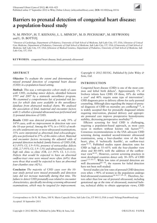
Rate of prenatal detection
The proportion of CHD cases detected prenatally in this
cohort was 39% (574/1474), with no significant differ-
ences according to year of delivery (Figure 1, P = 0.10).
The lowest detection rates (Figure 2) were for aortopul-
monary windows (0%) and total anomalous pulmonary
venous return (6%). Detection was also low for conotrun-
cal or outflow tract anomalies, including truncus arterio-
sus (24%), tetralogy of Fallot with pulmonary stenosis
(26%) and transposition of the great arteries (14%).
Missed opportunity for CHD detection
Almost all (97%) mothers of CHD cases underwent at
least one prenatal ultrasound examination and 77%
had an ultrasound exam between 16 and 24 weeks’
gestation. However, 60% of CHD cases in which a
prenatal ultrasound examination had been performed
were missed. Fetal echocardiograms were performed in
180
160
140
120
100
80
60
40
20
0
CHDcases(n)
1997∗1998∗ 1999 2000 2001 2002 2003 2004 2005 2006 2007
Birth cohort year
Figure 1 Rate of prenatal detection of congenital heart defects
(CHD) from 1997 to 2007 in the state of Utah. There was no
significant difference in detection according to birth cohort year
(P = 0.10). *Figures in 1997 and 1998 represent only conotruncal
and left obstructive types of CHD. , defects detected; , defects
missed.
27% of CHD cases. Family history of CHD was associated
with having undergone a fetal echocardiogram (65% vs
47%, P = 0.02). However, 35% of mothers with a family
history of CHD did not receive a fetal echocardiogram.
Although most (89%) cases with an abnormal ultrasound
were seen by a MFM, 42% of these cases never had a fetal
echocardiogram. Of those with a fetal echocardiogram,
3% had a missed CHD diagnosis, predominantly
coarctation of the aorta (n = 8), with one case each of
double outlet right ventricle and double inlet left ventricle.
Factors related to undergoing an ultrasound
examination
Factors associated with failure to receive a prenatal
ultrasound examination included later initiation of
prenatal care, higher number of previous pregnancies and
maternal residence in a census tract in which 10–20%
of the population were below the poverty level; they did
not include maternal age, education or race (Table 1). In
the multivariate model, only late initiation of prenatal
care (in second trimester: odds ratio (OR), 0.35 (95%
CI, 0.12–0.976) and in third trimester: OR, 0.1 (95%
CI, 0.0–0.4)) was associated independently with failure
to undergo prenatal ultrasound.
Risk factors related to missed CHD diagnosis
Among mothers who underwent ultrasound examination,
maternal factors associated with lower prenatal detection
of CHD included younger age, fewer years of education,
excessive weight gain during pregnancy (> 16 kg (c. 35
lb)) and rural residence (Table 1). In contrast, a family
history of CHD increased the odds of prenatal detection
(OR, 2.1 (95% CI, 1.2–3.7)).
Encounter factors associated with lower prenatal
detection of CHD included ultrasound examinations
performed solely at general clinics compared with one
or more performed at a hospital or MFM clinic (Table 2).
Nevertheless, 67% of CHD cases which underwent
Copyright 2012 ISUOG. Published by John Wiley & Sons, Ltd. Ultrasound Obstet Gynecol 2012; 40: 418–425.
4. Prenatal detection of CHD 421
Single
ventricle
N
O
S
D
O
R
V
N
O
S
H
ypoplastic
rightventricle∗
Single
ventricle
D
ouble
inletleftventricle
Situsinversus
D
O
R
V
w
ith
norm
ally
related
greatarteries
A
bnorm
alsitus
Situsam
biguous
H
ypoplastic
leftheartsyndrom
e
H
ypoplastic
leftventricle∗
D
O
R
V
w
ith
transposed
greatarteries
C
ongenitally
corrected
TG
A
(L-TG
A
)
Tricuspid
atresia
TO
F
w
ith
pulm
onary
atresia
C
om
plete
com
m
on
atrioventricularcanal
C
om
m
on
atrium
Ebstein
anom
aly
Interrupted
aortic
arch
type-A
Inletventricularseptaldefect
TO
F
w
ith
pulm
onary
stenosis
Truncusarteriosus
Prim
um
atrialseptaldefect
C
oarctation
D
-TG
A
w
ith
ventricularseptaldefect
D
-TG
A
w
ith
intactventricularseptum
Totalanom
alouspulm
onary
venousreturn
A
ortopulm
onary
w
indow
Interrupted
arch
type-B
Pulm
onary
atresia
w
ith
intactventricularseptum
0
20
Type of CHD
40
60
80
100
Prenatallydiagnosed(%)
100
88.9
78.6
75
72.2
71.4
70.8
70.6
70.5
70
60
58.1
54.2
52.1
50
48.5
46.7
42.9
42.9
28.6
26.3
26.3
23.7
22.2
18.5
15.2
13
5.9
0
0
Figure 2 Prenatal detection of congenital heart defects (CHD) according to type of defect and expected abnormal cardiac screening view
(expected abnormal four-chamber view ( ), expected abnormal outflow-tract view ( ), expected neither abnormal four-chamber nor
abnormal outflow-tract views ( ), expected varying abnormal views ( )). Expected abnormal view based on primary diagnosis only, i.e.
additional defects that may have led to additional abnormal views not taken into account. *Hypoplastic right and left ventricles were cases
with severe pulmonary or aortic stenosis with significant associated ventricular hypoplasia noted on pre- or postnatal echocardiogram.
DORV, double outlet right ventricle; D-TGA, dextro transposition of the great arteries; L-TGA, levo transposition of the great arteries;
NOS, not otherwise specified; TOF, tetralogy of Fallot.
ultrasound examination in a hospital and 25% in a
MFM clinic were missed. Another factor related to lower
detection was travel time to the nearest fetal cardiology
program (per additional hour of travel: OR, 0.9 (95% CI,
0.78–0.97)). CHD cases without additional non-cardiac
defects were less likely to be diagnosed prenatally (56%
vs 29%, P < 0.001).
Defects were categorized based on the screening view(s)
expected to be abnormal (Figure 2). If a case had multiple
defects, all expected abnormal views (in addition to their
primary diagnoses) were considered. Compared with
defects for which neither view would be expected to
be abnormal, those with an expected abnormal four-
chamber view had the highest chance of being detected
prenatally (OR, 4.6 (95% CI, 3.6–5.7)), while those
with an isolated expected abnormal outflow-tract view
had a slightly increased likelihood of detection (OR, 1.8
(95% CI, 1.4–2.4)). However, 42% of cases with an
Copyright 2012 ISUOG. Published by John Wiley & Sons, Ltd. Ultrasound Obstet Gynecol 2012; 40: 418–425.
5. 422 Pinto et al.
Table 1 Maternal characteristics associated with undergoing ultrasound examination (US exam) and prenatal detection of congenital heart
disease (CHD) in Utah between 1997 and 2007
US exams received Prenatal detection of CHD
Characteristic n (%)* OR (95% CI) n (%)† OR (95% CI)
Maternal age
≥ 35 years 197 (96) 1 96 (49) 1
21–34 years 1096 (97) 1.61 (0.76–3.44) 430 (39) 0.51 (0.32–0.80)
< 21 years 138 (98) 2.10 (0.56–7.90) 45 (33) 0.68 (0.50–0.92)
Plurality Collinear (—) 1.45 (0.93–2.41)
Singleton gestation 1360 (97) 536 (39)
Multiple gestation 71 (100) 35 (49)
Initiation of prenatal care
First trimester 1400 (98) 1 488 (35) 1
Second trimester 135 (96) 0.35 (0.13–0.98) 55 (41) 1.04 (0.73–1.49)
Third trimester 38 (90) 0.12 (0.04–0.39) 11 (29) 1.03 (0.63–1.69)
Gravidity
Per additional pregnancy 0.89 (0.79–1.00) 1.10 (1.04–1.15)
Maternal BMI at first visit
< 25 kg/m2 838 (97) 1 340 (41) 1
≥ 25 kg/m2 320 (98) 0.95 (0.73–1.24) 126 (39) 0.95 (0.73–1.23)
≥ 30 kg/m2 273 (96) 0.91 (0.69–1.20) 105 (38) 0.90 (0.69–1.19)
Weight gain
≤ 16 kg (c. 35 lb) (normal) 419 (42) 1
> 16 kg (c. 35 lb) (excessive) 152 (35) 0.72 (0.58–0.91)
Maternal education
College graduate 169 (95) 1 87 (51) 1
High school 633 (98) 2.14 (0.88–5.19) 249 (39) 0.56 (0.40–0.78)
< High school 629 (97) 1.41 (0.62–3.28) 235 (37) 0.62 (0.44–0.86)
Family history 49 (98) 1.49 (0.20–11.04) 28 (57) 2.06 (1.16–3.66)
Maternal race
White 1191 (97) 1 464 (39) 1
Non-white 234 (97) 0.93 (0.40–2.12) 103 (44) 1.23 (0.93–1.64)
Census-tract level % of adults ≥ 25 years
with < high school education
0.0–14.9% 952 (97) 1 387 (41) 1
15.0–24.9% 314 (97) 0.86 (0.41–1.79) 111 (35) 0.79 (0.61–1.02)
25.0–39.9% 99 (96) 0.68 (0.23–1.98) 41 (41) 1.00 (0.66–1.51)
40.0–100.0% 24 (96) 0.66 (0.09–5.03) 13 (54) 1.64 (0.74–3.63)
Census-tract level % of adults ≥ 25 years
with college degree
40.0–100.0% 176 (98) 1 74 (42) 1
25.0–39.9% 387 (96) 0.41 (0.12–1.43) 171 (44) 1.02 (0.72–1.46)
15.0–24.9% 484 (98) 0.69 (0.19–2.46) 173 (36) 0.75 (0.53–1.06)
0.0–14.9% 342 (97) 0.58 (0.16–2.14) 134 (39) 0.85 (0.59–1.23)
Census-tract level % below poverty level‡
0.0–4.9% 516 (98) 1 211 (41) 1
5.0–9.9% 437 (98) 0.93 (0.39–2.21) 169 (39) 0.90 (0.69–1.17)
10.0–19.9% 277 (94) 0.32 (0.15–0.70) 112 (40) 0.92 (0.69–1.23)
20.0–100.0% 159 (99) 1.7 (0.37–7.72) 60 (38) 0.88 (0.61–1.27)
Census-tract level rural residence§
Urban 1247 (97) 1 506 (41) 1
Rural 142 (96) 0.66 (0.27–1.61) 46 (32) 0.69 (0.78–0.99)
*Proportion of the cohort who underwent an ultrasound examination. †Proportion of cases detected out of those who underwent an
ultrasound examination. ‡Defined by the Census bureau for family size and number of dependents24. §Census-tract rural-urban commuting
areas were used to define residence as ‘urban’ (codes 1–3) or ‘rural’ (codes 4–10)25. BMI, body mass index.
expected abnormal four-chamber view, 64% with an
expected abnormal outflow-tract view and 30% with
both views expected to be abnormal were not detected
prenatally.
On multivariate analysis, after adjusting for maternal
race and rural residence, prenatal detection was related
independently to several encounter factors, including
total number of fetal ultrasound examinations, location
of ultrasound examination and an abnormal screening
ultrasound result (Table 3). Cases with a family history of
CHD and those with an additional non-cardiac congenital
defect had higher odds of prenatal detection. Maternal
age, education and weight gain during pregnancy were
not retained in the final model.
Copyright 2012 ISUOG. Published by John Wiley & Sons, Ltd. Ultrasound Obstet Gynecol 2012; 40: 418–425.
6. Prenatal detection of CHD 423
Table 2 Encounter factors related to prenatal detection of
congenital heart disease (CHD) identified in Utah between 1997
and 2007 when a prenatal ultrasound (US) examination had been
performed
Characteristic
CHD
detected
(n (%)) Odds ratio (95% CI)
US location
Clinic 164 (22) 1
Hospital 65 (31) 1.60 (1.14–2.25)
MFM/high-risk clinic 341 (74) 10.00 (7.64–13.10)
US interpreter
Obstetrician 18 (6) 1
Radiologist 10 (16) 2.77 (1.21–6.32)
MFM 268 (79) 56.41 (32.7–97.2)
US outcome
Normal 93 (11) 1
Suspected abnormality 74 (75) 24.38 (14.75–40.27)
Abnormal 398 (86) 52.03 (36.96–73.24)
Screening US location*
Clinic 191 (28) 1
Hospital 59 (38) 1.60 (1.14–2.25)
MFM/high-risk clinic 211 (71) 10.00 (7.64–13.10)
Screening US interpreter*
Obstetrician 116 (32) 1
Radiologist 36 (50) 2.77 (1.21–6.32)
MFM 110 (75) 56.41 (32.7–97.2)
Number of US exams 1.79 (1.65–1.93)
(per additional exam)
Presence of additional
congenital defects
311 (57) 3.18 (2.55–3.98)
Presence of heterotaxy 12 (60) 2.29 (0.92–5.63)
Travel time to fetal
cardiology program
0.90 (0.78–0.97)
(per additional hour)
*Screening US is first US exam performed between 16 and 24
weeks. MFM, maternal–fetal medicine specialist.
Table 3 Multivariate regression model for prenatal detection of
congenital heart disease
Odds ratio (95% CI) P
Maternal characteristics
Plurality 0.5 (0.2–0.9) 0.04
Family history 4.3 (1.9–9.9) < 0.01
Maternal rural residence 0.6 (0.3–1.2) 0.14
Encounter characteristics
Each additional US exam 1.6 (1.4–1.7) < 0.01
Suspected abnormality on US 17.3 (9.8–30.5) < 0.01
Abnormal US 31.3 (20.7–47.6) < 0.01
Additional congenital defect 2.7 (1.9–3.9) < 0.01
US performed at high-risk clinic 2.1 (1.3–3.1) < 0.01
US performed at hospital 0.8 (0.4–1.3) 0.31
US, ultrasound.
Subset analysis
Of the 705 cases delivered after 2003, 297 (42%) had
ultrasound reports available for review. Compared with
the whole cohort (n = 1474), this subset of patients had
a higher rate of prenatal detection (79%). The review of
their reports showed that 95% had documented cardiac
screening, including 65% with specific documentation
of a four-chamber view and 57% with documentation
of both four-chamber and outflow-tract views. A fetal
echocardiogram was never performed in 10% of cases
that documented an abnormal cardiac screen.
DISCUSSION
This study is the first in 15 years to provide data on
longitudinal trends in prenatal detection of CHD in the
USA. It also provides novel population-based findings on
potential predictors of missed prenatal detection of CHD.
Utilizing a statewide birth defect surveillance system, we
examined a comprehensive list of potential risk factors
not readily available in previous population studies of
prenatal CHD detection. We found that the majority of
CHD cases were missed prenatally and that detection
rates did not increase materially over the 10-year study
period. Although discouraging, the findings also suggest
missed opportunities in the screening process that could
be targeted to improve CHD detection.
The low rate of prenatal detection (39%) in our
cohort of approximately 1500 patients is consistent with
previous national and European publications14,16,17,19–21
.
The finding that CHD detection rates did not improve
significantly over the 10-year study period (1997–2007)
is in contrast to an earlier population study in Atlanta26
.
However, in that study the initial rate of detection was
very low (2.6%), and the study period overlapped with the
period of rapid evolution of fetal ultrasound technology
in the 1990s. Nevertheless, the lack of improvement in
prenatal detection rates in our more recent time period is
concerning and emphasizes the importance of identifying
modifiable factors that, if appropriately targeted, could
increase the sensitivity of current screening approaches.
Similar to previous studies14,16,17,19–21
, we found that
fetal ultrasound use was nearly universal. Thus, increasing
the rates of screening alone is unlikely to improve CHD
detection. The major risk factor for missed CHD detection
was failure to detect a cardiac abnormality on routine
ultrasound, particularly for defects expected to have only
an abnormal outflow-tract view. Potentially, interventions
aimed at improving the skills of those performing and
reviewing prenatal screening ultrasound examinations
could increase the detection of serious cardiac anomalies,
including many conotruncal malformations.
Opportunities for CHD detection may also have been
missed because some mothers did not receive higher level
imaging. Thirty-five percent of mothers with a family
history of CHD and 10% of those with abnormal cardiac
findings on screening ultrasound did not receive a fetal
echocardiogram. While some patients may have been
referred to MFM clinics first, evidence suggests that
evaluation by a MFM specialist alone is insufficient when
suspicion for CHD is increased by risk group or an
abnormal screen27
. In our study, 25% of patients scanned
at a MFM office had a CHD that went undetected.
Potential risk factors for failed prenatal CHD detec-
tion include sociodemographic factors and factors that
Copyright 2012 ISUOG. Published by John Wiley & Sons, Ltd. Ultrasound Obstet Gynecol 2012; 40: 418–425.
7. 424 Pinto et al.
affect image quality, such as maternal body habitus.
Studies examining sociodemographic factors show con-
flicting results20,22
. In our study, neither individual nor
geographic measures of socioeconomic status were associ-
ated with prenatal CHD detection. This lack of association
may be related to the current nearly universal use of
ultrasound. We also did not observe an independent asso-
ciation of any maternal factor, such as body habitus, with
CHD detection.
The primary risk factors for missed prenatal CHD
detection were fetal ultrasound location, ultrasound
interpreter and absence of extracardiac malformations.
Most heart defects are isolated, with no extracardiac
anomalies to increase suspicion. Additionally, most
patients received their screening ultrasound in low-risk
outpatient settings, where detection rates are lower
compared to tertiary or university-based settings20,22
.
The effects of location and interpreter are likely due
to variations in experience, training, and equipment. This
study was not designed to explore these factors. However,
even in best-case scenarios, among mothers who had one
of more ultrasound examinations performed at a MFM
clinic and interpreted by a MFM, a quarter of significant
CHD was missed prenatally.
Prenatal CHD detection was lower for those cases with
an expected abnormal outflow-tract view than for those
with an expected abnormal four-chamber view (of which
40% were still missed). This is consistent with other
studies16,18,20,22,28,29
. The addition of an outflow view
significantly enhances prenatal CHD detection, yet its
successful visualization is inconsistent and not uniformly
required30,31
. Currently, these views are not mandatory in
recommendations for cardiac screening in the USA12,13
,
although standardizing cardiac screening protocols and
enhancing training could improve the ability to obtain
this difficult view and improve detection rates28,29,32–35
.
In the UK and Canada, the routine assessment of outflow
tracts is already part of the guidelines for screening36,37
.
This study had some limitations. It used data obtained
for surveillance purposes; miscoding or data entry errors
could have been present. Miscoding is unlikely, since
all UBDN CHD cases undergo clinical review by a
pediatric geneticist specializing in CHD or a pediatric
cardiologist. Utah is predominately Caucasian, which
decreases our study’s generalizability, although our results
confirm findings reported in other regions of the country.
We were also unable to determine whether the lack
of indicated fetal echocardiograms was due to a lack
of referral or due to referred patients not actually
having a fetal echocardiogram. The analysis of ultrasound
reports must be interpreted with caution since only a
minority of patients (those with a higher rate of prenatal
detection) had reports available for review. Finally, in
using secondary data, we were unable to review screening
images directly. This will be important in future research
to generate additional insights into prenatal detection.
In conclusion, in spite of nearly universal ultrasound
screening, most (61%) significant CHD in this cohort
was missed prenatally. Our study identified multiple
points in the screening process amenable to improvement.
The one likely to have the largest population effect is
improvement to the initial screen done in the low-risk
outpatient setting. The scarce improvement in detection
rates over the recent decade suggests that initiatives to
enhance the screening process have been neither effective
nor widely disseminated. Since the primary risk factors for
failed detection of CHD appear to be related to screening
methods, targeted strategies amenable to widespread
adoption in clinical practice may improve detection. The
failure to detect these defects prenatally represents a
missed opportunity to provide counseling and timely care
for infants with CHD.
ACKNOWLEDGMENTS
We would like to thank Professor Paula Woodward,
MD, of the Department of Radiology, University of Utah
School of Medicine and Professor Michael Varner, MD,
of Maternal Fetal Medicine, Department of Obstetrics
and Gynecology, University of Utah School of Medicine
for their review and input on this paper. This study was
supported in part by the Children’s Health Research
Center, University of Utah as well as by an NIH
Institutional Career Enhancement Award for Dr Pinto
(1KM1CA156723-01).
REFERENCES
1. Hoffman JI. Incidence of congenital heart disease: II. Prenatal
incidence. Pediatr Cardiol 1995; 16: 155–165.
2. Kuehl KS, Loffredo CA, Ferencz C. Failure to diagnose congen-
ital heart disease in infancy. Pediatrics 1999; 103: 743–747.
3. Moller JH, Allen HD, Clark EB, Dajani AS, Golden A, Hay-
man LL, Lauer RM, Marmer EL, McAnulty JH, Oparil S, et al.
Report of the task force on children and youth. American Heart
Association. Circulation 1993; 88: 2479–2486.
4. Mahle WT, Clancy RR, McGaurn SP, Goin JE, Clark BJ.
Impact of prenatal diagnosis on survival and early neurologic
morbidity in neonates with the hypoplastic left heart syndrome.
Pediatrics 2001; 107: 1277–1282.
5. Kumar RK, Newburger JW, Gauvreau K, Kamenir SA, Horn-
berger LK. Comparison of outcome when hypoplastic left heart
syndrome and transposition of the great arteries are diagnosed
prenatally versus when diagnosis of these two conditions is
made only postnatally. Am J Cardiol 1999; 83: 1649–1653.
6. Tworetzky W, McElhinney DB, Reddy VM, Brook MM, Han-
ley FL, Silverman NH. Improved surgical outcome after fetal
diagnosis of hypoplastic left heart syndrome. Circulation 2001;
103: 1269–1273.
7. Bonnet D, Coltri A, Butera G, Fermont L, Le Bidois J,
Kachaner J, Sidi D. Detection of transposition of the great
arteries in fetuses reduces neonatal morbidity and mortality.
Circulation 1999; 99: 916–918.
8. Ewigman BG, Crane JP, Frigoletto FD, LeFevre ML, Bain RP,
McNellis D. Effect of prenatal ultrasound screening on perinatal
outcome. RADIUS Study Group. N Engl J Med 1993; 329:
821–827.
9. Satomi G, Yasukochi S, Shimizu T, Takigiku K, Ishii T. Has
fetal echocardiography improved the prognosis of congenital
heart disease? Comparison of patients with hypoplastic left
heart syndrome with and without prenatal diagnosis. Pediatr
Int 1999; 41: 728–732.
10. Kleinert S. Routine prenatal screening for congenital heart
disease. Lancet 1996; 348: 836.
Copyright 2012 ISUOG. Published by John Wiley & Sons, Ltd. Ultrasound Obstet Gynecol 2012; 40: 418–425.
8. Prenatal detection of CHD 425
11. Stumpflen I, Stumpflen A, Wimmer M, Bernaschek G. Effect of
detailed fetal echocardiography as part of routine prenatal
ultrasonographic screening on detection of congenital heart
disease. Lancet 1996; 348: 854–857.
12. AIUM Practice Guideline for the Performance of Obsteric
Ultrasound Examinations, 2007; http://aium.org/resources/
guidelines/obstetric.pdf [Accessed May 2011].
13. American College of Radiology Practice Guideline for the Per-
formance of Obstetrical Ultrasound. 2007; http://www.nice.org.
uk/nicemedia/live/11947/40145/40145.pdf [Accessed June
2011].
14. Chew C, Halliday JL, Riley MM, Penny DJ. Population-based
study of antenatal detection of congenital heart disease by
ultrasound examination. Ultrasound Obstet Gynecol 2007; 29:
619–624.
15. Bromley B, Estroff JA, Sanders SP, Parad R, Roberts D, Frigo-
letto FD, Jr, Benacerraf BR. Fetal echocardiography: accuracy
and limitations in a population at high and low risk for heart
defects. Am J Obstet Gynecol 1992; 166: 1473–1481.
16. Khoo NS, Van Essen P, Richardson M, Robertson T. Effective-
ness of prenatal diagnosis of congenital heart defects in South
Australia: a population analysis 1999–2003. Aust N Z J Obstet
Gynaecol 2008; 48: 559–563.
17. Stoll C, Garne E, Clementi M. Evaluation of prenatal diagnosis
of associated congenital heart diseases by fetal ultrasonographic
examination in Europe. Prenat Diagn 2001; 21: 243–252.
18. Acherman RJ, Evans WN, Luna CF, Rollins R, Kip KT, Colla-
zos JC, Restrepo H, Adasheck J, Iriye BK, Roberts D, Sacks AJ.
Prenatal detection of congenital heart disease in southern
Nevada: the need for universal fetal cardiac evaluation. J Ultra-
sound Med 2007; 26: 1715–1719; quiz 1720–1721.
19. Randall P, Brealey S, Hahn S, Khan KS, Parsons JM. Accuracy
of fetal echocardiography in the routine detection of congenital
heart disease among unselected and low risk populations: a
systematic review. BJOG 2005; 112: 24–30.
20. Friedberg MK, Silverman NH, Moon-Grady AJ, Tong E, Nourse
J, Sorenson B, Lee J, Hornberger LK. Prenatal detection of
congenital heart disease. J Pediatr 2009; 155: 26–31.
21. Buskens E, Grobbee DE, Frohn-Mulder IM, Stewart PA,
Juttmann RE, Wladimiroff JW, Hess J. Efficacy of routine fetal
ultrasound screening for congenital heart disease in normal
pregnancy. Circulation 1996; 94: 67–72.
22. Wong SF, Chan FY, Cincotta RB, Lee-Tannock A, Ward C.
Factors influencing the prenatal detection of structural
congenital heart diseases. Ultrasound Obstet Gynecol 2003;
21: 19–25.
23. Feldkamp M, MacLeod L, Young LP, Lecheminant K, Carey JC.
The Methodology of the Utah birth defect network: congenital
heart defects as an illustration. Birth Defects Res A Clin Mol
Teratol 2005; 73: 693–699.
24. http://www.census.gov/hhes/www/poverty/data/threshld/index.
html
25. United States Department of Agriculture. Rural-Urban Com-
muting Area Codes Overview. http://www.ers.usda.gov/data-
products/rural-urban-commuting-area-codes.aspx [Accessed
May 2011].
26. Montana E, Khoury MJ, Cragan JD, Sharma S, Dhar P, Fyfe D.
Trends and outcomes after prenatal diagnosis of congenital
cardiac malformations by fetal echocardiography in a well
defined birth population, Atlanta, Georgia, 1990–1994. J Am
Coll Cardiol 1996; 28: 1805–1809.
27. Meyer-Wittkopf M, Cooper S, Sholler G. Correlation between
fetal cardiac diagnosis by obstetric and pediatric cardiologist
sonographers and comparison with postnatal findings. Ultra-
sound Obstet Gynecol 2001; 17: 392–397.
28. Garne E, Stoll C, Clementi M. Evaluation of prenatal diagnosis
of congenital heart diseases by ultrasound: experience from
20 European registries. Ultrasound Obstet Gynecol 2001; 17:
386–391.
29. Hunter S, Heads A, Wyllie J, Robson S. Prenatal diagnosis
of congenital heart disease in the northern region of
England: benefits of a training programme for obstetric
ultrasonographers. Heart 2000; 84: 294–298.
30. Wigton TR, Sabbagha RE, Tamura RK, Cohen L, Minogue JP,
Strasburger JF. Sonographic diagnosis of congenital heart
disease: comparison between the four-chamber view and
multiple cardiac views. Obstet Gynecol 1993; 82: 219–224.
31. Kirk JS, Riggs TW, Comstock CH, Lee W, Yang SS, Wein-
house E. Prenatal screening for cardiac anomalies: the value
of routine addition of the aortic root to the four-chamber view.
Obstet Gynecol 1994; 84: 427–431.
32. Carvalho JS, Mavrides E, Shinebourne EA, Campbell S, Thila-
ganathan B. Improving the effectiveness of routine prenatal
screening for major congenital heart defects. Heart 2002; 88:
387–391.
33. Tegnander E, Eik-Nes SH. The examiner’s ultrasound experi-
ence has a significant impact on the detection rate of congenital
heart defects at the second-trimester fetal examination. Ultra-
sound Obstet Gynecol 2006; 28: 8–14.
34. Ogge G, Gaglioti P, Maccanti S, Faggiano F, Todros T. Prenatal
screening for congenital heart disease with four-chamber and
outflow-tract views: a multicenter study. Ultrasound Obstet
Gynecol 2006; 28: 779–784.
35. Galindo A, Herraiz I, Escribano D, Lora D, Melchor JC, de
la Cruz J. Prenatal detection of congenital heart defects: a
survey on clinical practice in Spain. Fetal Diagn Ther 2011;
29: 287–295.
36. NHS National Institute for Health and Clinical Excellence.
Antenatal care: routine care for the healthy pregnant woman.
http://www.nice.org.uk/CG062 [Accessed June 2011].
37. Cargill Y, Morin L, Bly S, Butt K, Denis N, Gangnon R,
Hietala-Coyle M, Lim KH, Ouellet A, Racicot M, Salem S.
Content of a complete routine second trimester obstetrical
ultrasond examination and report. J Obstet Gynecol Canada
2009; 31: 272–275.
SUPPORTING INFORMATION ON THE INTERNET
The following supporting information may be found in the online version of this article:
Table S1 Demographics and descriptors of cases of congenital heart disease identified in Utah between
1997 and 2007
Copyright 2012 ISUOG. Published by John Wiley & Sons, Ltd. Ultrasound Obstet Gynecol 2012; 40: 418–425.