Use of ConeBeam Computed Tomography to determine cochlear length and implant array positioning
•
1 j'aime•291 vues

This study aimed to develop a technique for calculating cochlear length and determining cochlear implant electrode position using cone-beam computed tomography (CBCT) scans. The researchers examined CBCT scans of 100 subjects who received cochlear implants with either straight or contour electrode arrays. They calculated cochlear length based on the number of electrodes inserted at 360 degrees. For straight arrays, cochlear length ranged from 27.44 to 35.91 mm. For contour arrays, length ranged from 17.8 to 22.24 mm. The study concluded that CBCT can be used to precisely map electrode position to the cochlear's tonotopic map, which may improve outcomes for cochlear implantation.

Recommandé
Contenu connexe
Tendances
Tendances (20)
En vedette
En vedette (20)
Similaire à Use of ConeBeam Computed Tomography to determine cochlear length and implant array positioning
Similaire à Use of ConeBeam Computed Tomography to determine cochlear length and implant array positioning (20)
Plus de HEARnet _
Plus de HEARnet _ (20)
Dernier
Dernier (20)
Use of ConeBeam Computed Tomography to determine cochlear length and implant array positioning
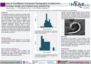
- 1. Use of ConeBeam Computed Tomography to determine cochlear length and implant array positioning Fadwa Alnafjan 1,2,3, A.Prof Melville Da Cruz 3, A.Prof Catherine McMahon 1,2 1 The HEARing Cooperative Research Centre, ² Macquarie University, Linguistics Department & ³ Westmead Hospital, Department of Otolaryngology Abstract A validated technique to calculate cochlear length and cochlear implant electrode position using pre- and post-implantation Computed Tomography (CT) has previously been reported. However, CT exposures subjects to high levels of radiation twice (once per scan). More recently, Cone Beam Computed Tomography (CBCT) has been used for cochlear implantees, exposing them to lower radiation, but providing similarly high quality images. The aim of this study was to develop a technique to calculate the length of the cochlea within individuals and, from this, to determine whether the distribution of the length was normal. Study Design 100 temporal bone CBCT images of in vivo subjects implanted with Cochlear straight or contour arrays by one surgeon. Main Outcome Measure Main Outcome Measure: CBCT images were examined by two independent examiners who calculated the length of the cochlea based on the number of electrodes inserted at 360°. Results The length of the organ of Corti based on the position of the straight array within the cochlea was 27.44 to 35.91 mm (mean = 32.24 mm). The length of the spiral ganglion based on the position of the contour array within the cochlea ranged from 17.8 to 22.24 mm (mean = 19.43 mm). Conclusion Using an evidence-based paradigm, we have developed a novel method of calculating cochlear length that can be used with CBCT. This enables a more precise mapping of the electrode position to the tonotopic map, which may result in improved outcomes of cochlear implantation. creating sound value www.hearingcrc.org Cone-beam computed tomography (CBCT) image of an implanted electrode array (Slim Straight Electrode) showing the three landmarks needed to measure the cochlear length; (i) bony lip of round window (RW), (ii) modiolus, and (iii) most apical electrode (#22). These are used to estimate the number of electrodes inserted at 360 degrees (solid line) and the length between this point and the bony lip of the RW (dashed line). The distribution of cochlear length for 77 ears with a straight array (CI422 and CI522). The dashed curve shows a normal distribution. The distribution of cochlear length for 23 ears with a contour array (CI24RE and CI512). The dashed curve shows a normal distribution.