

Recommandé
Contenu connexe
Tendances
Tendances (20)
En vedette
En vedette (20)
Similaire à Urinary system
Similaire à Urinary system (20)
Dernier
Dernier (20)
Urinary system
- 1. The Urinary System City Campus, L1.10, Cova Square, Jalan Teknologi, Kota Damansara, PJU 5, 47810 Petaling Jaya, Selangor Phone: 03-6142 6666 Fax: 03-6142 6655 Website: www.iucn.edu.my Email: info@iucn.edu.my
- 2. The Urinary System Learning Outcomes: • List the parts of the Urinary System. • State the function of the kidneys. • Outline the gross structure of the kidneys. • Describe the structure of a nephron. • Name the four processes involved in urine formation and describe the action of each. • Describe the process of urine formation. • State the composition of urine. PN111: INTRODUCTION TO HUMAN BODY 2 2
- 3. The Urinary System • Also called the excretory system. • Excrete, remove and eliminate metabolic waste products from the blood. • Regulate volume, acid-base balance and electrolyte composition of body fluids. • Blood supply – renal artery and renal vein. PN111: INTRODUCTION TO HUMAN BODY 2 3
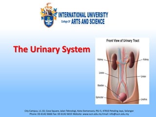
- 4. The Urinary System The main excretory system and consists of the following structures: • 2 kidneys (secrete urine) • 2 ureters (convey urine from the kidneys to the bladder) • 1 urinary bladder (collects urine) • 1 urethra (through which urine is discharged) PN111: INTRODUCTION TO HUMAN BODY 2 4
- 5. The Kidneys Main functions of kidneys are: • Removal of nitrogenous waste material, certain salts and excess water from the blood. • Secretion of waste products in the form of urine. • Production and secretion of erythropoietin, the hormone that controls formation of red blood cells. • Production and secretion of renin, an enzyme that helps in the control of blood pressure. PN111: INTRODUCTION TO HUMAN BODY 2 5
- 6. Structure of the Kidney • A bean shaped organ app. 10cm long and 5 cm wide, 2.5 cm thick and weigh 150 g. • Extend from the 12th thoracic vertebra to the 3rd lumbar vertebra. • Rt. Kidney is slightly lower than Lt. kidney. • A protective layer of fat (adipose capsule) around the organ. • An outermost layer of fascia (connective tissue) anchors the kidney to the peritoneum and abdominal wall. • Medial border – hilum, where the renal artery, renal vein and ureter connect with the kidney. PN111: INTRODUCTION TO HUMAN BODY 2 6
- 7. Gross structure of the Kidney Divided into 3 areas of tissues: • A fibrous capsule, surrounding the kidney. • The cortex, a reddish brown layer of tissue, below the capsule and outside the pyramids. • The medulla, the innermost layer, consisting of pale conical-shaped striations, the renal pyramids. • The hilum – concave medial border of the kidney where blood and lymph vessels, the ureter and nerves enter. PN111: INTRODUCTION TO HUMAN BODY 2 7
- 8. Gross structure of the Kidney • The renal pelvis – the funnel-shaped structure that acts as a receptacle for the urine formed by the kidney. • Calyces – cuplike extensions of renal pelvis surrounding the tips of the pyramids and collects urine. • Urine in pelvis passes down the ureters to the bladder. PN111: INTRODUCTION TO HUMAN BODY 2 8
- 9. Structure of the Kidney PN111: INTRODUCTION TO HUMAN BODY 2 9
- 10. The Nephron • The nephron is the structural and functional unit of the kidney. • Consists of a specialized tubular structure and closely associated blood vessels. • Responsible for the formation of urine. • Each kidney contains 1,000,000 nephrons that filter the blood and form urine. • About 99% of the initial filtrate from the glomerulus is reabsorbed by the nephron and returned to the blood in the peritubular capillaries. • When nephrons are damaged they are not replaced or regenerated. PN111: INTRODUCTION TO HUMAN BODY 2 10
- 11. The Nephron • About one third of the nephrons in a kidney must be functional to ensure survival of the organ. The parts of the nephron are as follows: • Bowman's capsule - This closed end at the beginning of the nephron is located in the cortex. • Proximal convoluted tubule or proximal tubule - The first twisted region after the Bowman's capsule; it is in the cortex. PN111: INTRODUCTION TO HUMAN BODY 2 11
- 12. The Nephron • Loop of Henle - A long, hairpin loop after the proximal tubule, it extends from the cortex down into the medulla and back. • Distal convoluted tubule or distal tubule - This second twisted portion of the nephron after the loop of Henle is located in the cortex. • Collecting duct - This long straight portion after the distal tubule that is the open end of the nephron extends from the cortex down through the medulla. PN111: INTRODUCTION TO HUMAN BODY 2 12
- 13. The Nephron PN111: INTRODUCTION TO HUMAN BODY 2 13
- 14. Formation of Urine • A complete process, involving many exchanges between blood stream and kidney tubules. • There is ample time for exchange to take place as fluid filtered from the blood travels through the twists and turns of the nephron. • The processes allow the kidney to “fine tune” body fluids as they adjust the composition of the urine. PN111: INTRODUCTION TO HUMAN BODY 2 14
- 15. Formation of Urine Four processes occurring in successive portions of the nephron accomplish the function of urine formation. • Glomerular filtration allows diffusible materials (water and small molecules) to pass from the blood into the nephron. • Tubular reabsorption moves useful substances (glucose, amino acids,sodium, calcium, potassium, phosphate and chloride) back into the blood while keeping waste products (urea and uric acid) in the nephron for elimination. PN111: INTRODUCTION TO HUMAN BODY 2 15
- 16. Formation of Urine • Tubular secretion moves additional substances from the blood into the nephron for elimination. Movement of hydrogen ions is one means by which the pH of body fluids is balanced. • The countercurrent mechanism concentrates the urine and reduces the volume excreted. The pituitary hormone ADH allows more water to be reabsorbed from the nephron. PN111: INTRODUCTION TO HUMAN BODY 2 16
- 17. The Path of the Formation of Urine • Blood enters the afferent arteriole, passes through the glomerulus, to Bowman’s capsule. • In Bowman’s capsule, it becomes filtrate (blood minus RBC and plasma proteins) continues through the proximal convoluted tubule to the collecting tubule (by now, 99% of filtrate has been reabsorbed) • App. 1 ml of urine is formed per minute and travels all the way down to bladder for excretion. PN111: INTRODUCTION TO HUMAN BODY 2 17
- 18. PN111: INTRODUCTION TO HUMAN BODY 2 18
- 19. PN111: INTRODUCTION TO HUMAN BODY 2 19
- 20. Normal Constituents of Urine • Nitrogenous waste products – urea, uric acid, and creatinine. • Electrolytes – sodium chloride, sulfates and phosphates. • Pigment – from bile compounds, foods and drugs. PN111: INTRODUCTION TO HUMAN BODY 2 20
- 21. Composition of Urine • Water 96% • Urea 2% • Uric acid • Creatinine • Ammonia • Sodium • Potassium 2% • Chlorides • Phosphates • Sulphates • Oxalates PN111: INTRODUCTION TO HUMAN BODY 2 21
- 22. Ureter, Bladder, Urethra Learning Outcomes: • Describe the Ureter, Bladder and Urethra. • State the functions of ureter, bladder and urethra. • Describe the physiological processes of micturition. PN111: INTRODUCTION TO HUMAN BODY 2 22
- 23. Ureters • Two long, narrow tube (1/4 inch wide and 10 to 12 inches long) carrying urine from the kidneys to the bladder. • When the muscles contract, peristalsis is initiated, pushing urine down the ureter into the urinary bladder. • When urine accumulates and the pressure in the bladder rises, the ureters are compressed and the openings occluded to prevent reflux of urine back into the kidney. PN111: INTRODUCTION TO HUMAN BODY 2 23
- 24. Cross Section of Ureter PN111: INTRODUCTION TO HUMAN BODY 2 24
- 25. Structure of the Ureters Consists of 3 layers of tissue: • An outer covering of fibrous tissue. • A middle muscular layer consisting of interlacing smooth muscle fibres. • An inner layer, the mucosa, composed of transitional epithelium. PN111: INTRODUCTION TO HUMAN BODY 2 25
- 26. Function of Ureter • Propel urine from the kidneys into the bladder by peristaltic contraction of the smooth muscle layer. • Occurs several times per minute, increasing in frequency with the volume produced. PN111: INTRODUCTION TO HUMAN BODY 2 26
- 27. Urinary Bladder • A hollow, muscular, pear shaped organ for storage of urine. • Can stored up to 500 mls of urine. • Lies in the pelvic cavity. • When distended, the bladder rises into the abdominal cavity. • Voiding takes place by contractions of the bladder, which forces urine through the urethra and to the outside opening, the urinary meatus. PN111: INTRODUCTION TO HUMAN BODY 2 27
- 28. Structure of the Urinary Bladder The bladder wall is composed of three layers: • An outer layer of loose connective tissue, containing blood and lymphatic vessels and nerves, covered on the upper surface by the peritoneum. • A middle layer of interlacing smooth muscle fibres and elastic tissue called the detrusor muscle. • A inner layer of mucosa, composed of transitional epithelium. PN111: INTRODUCTION TO HUMAN BODY 2 28
- 29. Cross Section of Urinary Bladder PN111: INTRODUCTION TO HUMAN BODY 2 29
- 30. Structure of the Urinary Bladder • The trigone is a triangular-shaped region in floor of bladder. • Marked by the openings of the two ureters and urethra. • As the bladder fills with urine, it expands upward, leaving the trigone at the base stationary. • This prevents stretching of the ureteral openings and possible backflow. PN111: INTRODUCTION TO HUMAN BODY 2 30
- 31. The Urethra • A canal extending from neck of bladder to external urethral orifice. • Longer in the male (8-10 inches) than in female (4 inches). • External urethral orifice is guarded by the external urethral sphincter (voluntary control). PN111: INTRODUCTION TO HUMAN BODY 2 31
- 32. Structure of the Urethra The walls consist of three layers of tissue. • The muscle layer is continuous with that of the bladder. It is mainly elastic tissue and smooth muscle fibres, under autonomic nerve control. • The submucosa is a spongy layer containing blood vessels and nerves. • The mucosa is continuous with that of the bladder in the upper part of the urethra. The lower part consists of stratified squamous epithelium, continuous externally with the skin of vulva. PN111: INTRODUCTION TO HUMAN BODY 2 32
- 33. Micturition • Urination (micturition) involves physiological processes within the urinary tract and the brain. • The slight need to urinate is sensed when urine volume reaches about one-half of the bladder's capacity. • The brain suppresses this need until a person initiates urination. • Neurons in the brain and in smooth muscle of the bladder govern the detrusor muscle; it is not controlled voluntarily. PN111: INTRODUCTION TO HUMAN BODY 2 33
- 34. Micturition • The nervous system stimulates the detrusor muscle to contract into a funnel shape and expel urine, once a person initiates urination. • Pressure in the bladder increases and the detrusor remains contracted until the bladder empties. • Once empty, pressure falls and the bladder relaxes and resumes its normal shape. • Normally, the detrusor muscle contracts and relaxes according to the volume of urine in the bladder and the initiation of urination. PN111: INTRODUCTION TO HUMAN BODY 2 34
- 35. CLINICAL CONNECTION • Nephroptosis • Kidney transplant • Loss of plasma proteins in urine causes edema • Glucosuria • Diuretics • Dialysis • Cystoscopy • Urinary incontinence PN111: INTRODUCTION TO HUMAN BODY 2 35
- 36. PN111: INTRODUCTION TO HUMAN BODY 2 36