
Recommandé
Contenu connexe
Tendances
Tendances (20)
En vedette
En vedette (20)
Similaire à Electron Microscope Shanthakumar
Similaire à Electron Microscope Shanthakumar (20)
Dernier
Dernier (20)
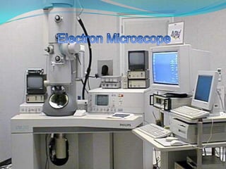
Electron Microscope Shanthakumar
- 2. Comparison b/w light and electron microscopes LIGHT MICROSCOPE ELECTRON MICROSCOPE Magnification can be done upto 2000 times Resolving power is less Photons are involved Magnificationcan be done upto 2 million times Have much greater resolving power than ordinary microscope Electrons are involved
- 3. Scale 3
- 4. Reason for greater resolution and magnification of electron microscope Wavelength of an electron[de Broglie] is very much smaller than that of a light photon Wavelength of an electron = λ=h/(√2mE) Wavelength of a light photon = λ=h/E
- 6. Year-1933
- 8. Construction of EM [TEM]
- 10. WORKING OF AN ELECTRON MICROSCOPE
- 11. Types of ELECTRON MICROSCOPE
- 16. TEM ELECTRON GUN ELECTROMAGNETIC LENSES VACUUM PUMPS OPENING TO INSERT SAMPLES OPERATION PANEL DISPLAY SCREEN WATER SUPPLY TO COOL THE INSTRUMENT
- 17. ELECTRON GUN The electron gun produces a stream of monochromatic electrons of energy 100-400keV . The extraction of electrons is of two types 1. Thermionic emission using thermal energy 2. Field emission by applying very large electric field 1010 A/m FE gun is more expensive and must be used in high vacuum conditions.
- 19. The beam of electrons is focused using condenser lenses [1&2] The beam is restricted by the condensor aperture Then it strikes the specimen and transmitted The transmitted portion is focused by Objective Lens into an image Intermediate and projector lenses enlarge the image Image is formed on phosphor screen Darker area – few electrons – thick region Lighter area – more electrons – thin region.
- 22. very high resolution
- 23. Resolving power is
- 24. Magnification is 1,000,000 times greater than the size of the object
- 27. To get 2-D Image of biological cells , virus , bacteria etc.
- 28. In fields such as thin film technology , metallurgy , microbiology etc.
- 31. It captures the images of the specimen surface by scanning it with a high-energy beam of electrons , in a scan pattern
- 32. produces a 3-dimensional image of specimen’s surface features24
- 34. SEM electron gun electromagnetic lenses vacuum pumps opening to insert specimen operation panel screen for display cryo – unit for cryosem electronic instruments
- 36. If a non conductive material has to be viewed , then it has to be coated by a thin layer of electrically conductive material
- 38. SPUTTERING
- 39. WORKING SEM produces signals in the form of secondary electrons , backscattered electrons, characteristic x-rays , light , specimen current, and transmitted electrons These signals are formed by the interaction of the electron beam with the surface of the specimen and require specialized detectors for their detection Depth of the specimen can be expressed in the image Back scattering of electrons help to detect the distribution of elements in the specimen
- 40. CHARACTERISTIC X-RAY SIGNAL Characteristic x-ray signals are formed when the electron beam removes an inner shell electron of the sample causing a high energy electron to fill the space and release energy Help to identify the composition of elements in sample
- 43. Image can be directly viewed
- 44. 3-Dimensional image can be obtained
- 45. has large depth of focus
- 47. preparation of sample is difficult and tedious
- 49. wide application in medical , science and engineering fields
- 50. To find the structural composition of paper pulps, ceramic materials , polymers etc.
- 52. SEM AND TEM PHOTOS SEM TEM
- 53. Comparison of images SEM of the compound eye of a fly! TEM of bacteria
- 54. Some more images obtained using SEM SEM of Yersiniapestis – which causes plague SEM image of Streptococcus pyogenes , which causes scarlet fever
- 55. EM Image of the chloroplast of spinach
- 56. EM Image of RBC
- 57. SEM Image of a mesh
- 58. RBC OF MAN
- 59. NEURON CELL
- 60. MOSQUITO
- 61. CULTURED CELLS OF HUMAN BEING
- 62. HUMAN CELLS
- 63. HEAD OF A BLACK ANT
- 64. Websites for further info: www.nobelprize.org www.wikipedia.com www.pbrc.hawaii.edu/microangela www.mos.org/sln/SEM/ www.britannica.com/EBchecked/topic-art/183561/110970/Scanning-electron-microscope
- 65. Presentation done by… SHANTHA KUMAR . T EEE RMD ENGG COLLEGE
- 66. THANK YOU!