Myoglobin1
•Télécharger en tant que PPT, PDF•
2 j'aime•4,005 vues

MYOGLOBIN IS THE STRUCTURAL SIMILAR PROTEIN OF HEAMOGLOBIN.

Recommandé
Contenu connexe
Tendances
Tendances (20)
En vedette
En vedette (20)
Plus de HARIS.P
Dernier
Dernier (20)
Myoglobin1
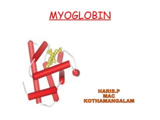
- 1. MYOGLOBIN HARIS.P MAC KOTHAMANGALAM
- 2. Myoglobin 16,700 153 1
- 4. highlights regions of secondary structure A “mesh” image emphasizes the protein surface. visualizing pockets in the protein where other molecules might bind
- 5. A ribbon representation, including side chains (blue) for the hydrophobic residues Leu, Ile, Val, and Phe. A space-filling model with all amino acid side chains.
- 8. PROTOPORPHYRIN IX HEME
- 10. Steric effects on the binding of ligands to the heme of myoglobin.