
![[object Object],[object Object],[object Object],[object Object],[object Object],[object Object],[object Object],[object Object],[object Object],[object Object],Cerebral Cortex](data:image/gif;base64,R0lGODlhAQABAIAAAAAAAP///yH5BAEAAAAALAAAAAABAAEAAAIBRAA7)
Recommandé
Contenu connexe
Tendances
Tendances (20)
En vedette
En vedette (20)
Similaire à Cbr cortex2k1
Similaire à Cbr cortex2k1 (20)
Dernier
Dernier (20)
Cbr cortex2k1
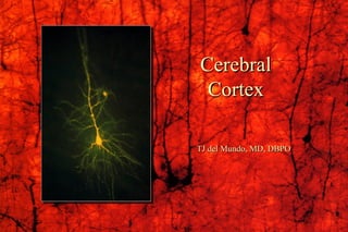
- 1. Cerebral Cortex TJ del Mundo, MD, DBPO
- 3. Numerical Data Total surface area : 2200 cm 2 (2.5 ft 2 ) about 1/3 ------ surface area about 2/3 ------ hidden in the sulci Thickness : 1.5 mm (V I) - 4.5 mm (M I) Generally, thickest over the crest of the convolution and, thinnest in the depth of sulci Weight : 600 gm (40 % of total brain weight) 180 gm --------- neurons 420 gm --------- glial cells Cerebral Cortex
- 4. Numerical Data Number of neuronal cells in cerebral cortex neurons ----------- 10-15 billion glial cells ---------- 50 billion Estimation of number of cortical neurons von Economo and Koskinas (1925) 14.0 billion Shariff (1953) 6.9 billion Sholl (1956) 5.0 billion Pakkenberg (1966) 2.6 billion Cerebral Cortex
- 5. Isocortex – typical 6 layered cortex I. Molecular Layer II. External Granular Layer III. External Pyramidal Layer IV. Internal Granular Layer V. Internal Pyramidal Layer VI. Polymorphic Layer
- 6. Histological Organization Cellular Elements 1. Pyramidal Cell - output neuron giant pyramidal cell of Betz 2. Fusiform Cell --- modified pyramidal cell 3. Granular (Stellate) Cell basket cell, double bouquet cell, bipolar cell, chandlier cell, neurogliform cell 4. Horizontal Cell of Cajal (Retzius-Cajal cell) 5. Cells of Martinotti Cerebral Cortex
- 7. 1. Pyramidal Cell 2. Fusiform Cell 3. Granular (Stellate) Cell 4. basket cell 5. double bouquet cell 6. chandlier cell 7. neurogliform cell 8. Horizontal Cell of Cajal 9. Cells of Martinotti a: axon Cerebral Cortex
- 8. I. Molecular Layer II. External Granular Layer III. External Pyramidal Layer Line of Kaes-Bechterew IV. Internal Granular Layer Outer band of Baillarger - Line of Gennari in area 17 V. Internal Pyramidal Layer Giant pyramidal cell of Betz Inner Band of Baillarger VI. Polymorphic Layer Golgi Nissl Weigert
- 9. 1. corticocortical fiber association fiber commissural fiber 2. thalamocortical fiber - specific and non-specific 3. extrathalamic subcortical fiber cholinergic fiber - acetylcholine basal nucleus of Meynert mesolimbic dopaminergic fiber - dopamine ventral tegmental area serotonergic fiber – serotonine - raphe nuclei norepinephrinergic fiber - norepinephrine nucleus locus ceruleus Cortical Afferent Fiber
- 10. Cortical Afferent Fiber 1. association fiber 2. commissural fiber 3. specific thalamocortical fiber 4. non-specific thalamocortical fiber
- 11. 1. Corticofugal Fiber - Projection Fiber corticostriate fiber corticothalamic fiber corticorubral fiber corticotectal fiber corticopontine fiber cortico-olivary fiber corticobulbar fiber corticospinal fiber 2. Corticocortical Fiber Association fiber Commissural fiber Cortical Efferent Fiber
- 12. 5. association fiber 6. commissural fiber 7. corticostriate fiber 8. corticorubral fiber corticopontine fiber corticobulbar fiber 9. corticospinal fiber corticotectal fiber 10. corticothalamic fiber Cortical Efferent Fiber
- 13. A. pyramidal neuron B. excitatory granular cell C. inhibitory granular cell 1. afferent fiber 2. efferent fiber 3. corticothalamic fiber Columnar Cortical Unit and Cortical Circuitary
- 14. A. Homotypical isocortex ------- association cortex B. Heterotypical isocortex 1. granular cortex --- primary sensory cortex V I (17), S I (3), A I (41) 2. agranular cortex --- motor cortex M I (4), PM (6) Regional Variation of Cortical Lamination
- 15. Von Economo’s classification of cortical types 1. agranular, 2. frontal, 3. parietal, 4. polar, 5. granular
- 16. 1. agranular, 2. frontal, 3. parietal, 4. polar, 5. granular
- 17. Phrenology of Gall and Spurzheim Clinical evidences Broca’s area (1861) Jacksonian epilepsy (1864) Experimental evidences Fritsch and Hitzig (1870) --- motor cortex von Gudden (1870) ---- visual cortex Ferrier (1873) ---- auditory cortex Functional Localization of Cerebral Cortex
- 18. Albertus Magnus (1206-1280) Phrenology of Gall (1758-1828) and Spurzheim (1776-1832)
- 19. PET (positron emission tomography) scan
- 20. based on cytoarchitectonic studies Campbell (1905) -------- about 20 areas Brodmann (1909) ------ 47 areas - most popular Vogt and Vogt (1919) - over 200 areas von Economo (1929) -- 109 areas Morphological Classification of Cortical Areas
- 21. Brodmann’s cytoarchitectorial map (Lateral surface)
- 22. Brodmann’s cytoarchitectorial map (Lateral surface)
- 23. Sensory area primary sensory area secondary sensory area Motor area primary motor area secondary motor area supplementary motor area Association area parietal, occipital and temporal cortex - conceptual elaboration of sensory data prefrontal (frontal) cortex - judgement, foresight Functional Localization of Cerebral Cortex
- 24. Somesthetic Area (Somesthesia) S I, S II Visual Area (vision) V I, V II Auditory Area (Hearing) A I, A II Vestibular Area (Equilibrium) Gustatory Area (Taste) Olfactory Area (Smell) Sensory Areas
- 25. S I ----- 3, 1, 2 (postcentral gyrus) afferernts: ventrobasal complex (VPLc, VPM) discrimination of position and intensity of sensation S II ---- superior bank of lateral fissure no clinical disorders Somesthetic Association Cortex ------- 5, 7 (parietal lobule, precuneus) afferents: S I, LP of thalamus integration of geneal sensation with past experience tactile agnosia, astereognosis Somesthetic Area
- 27. Thalmocortical connection (VPLc S I) Central region --- cutaneous (3b, 1) Peripheral region --- deep (3a, 2) Primary Somesthetic Area
- 28. Secondary Somesthetic Area (SII) superior bank of lateral fissure
- 29. V I ----- 17 (striate cortex - line of Gennari) greatly thickened outer band of Baillarger heterotypical isocortex afferent: LGd of thalamus visual field defect: homonymous quadranopsia and macular sparing V II ---- 18, 19 (visual association area) afferents: V I, pulvinar of thalamus integration of vision with past experience visual agnosia cf. occipital eye field Visual Cortex
- 30. Visual Areas
- 31. V4 (color) Face recognition Perceive Facial Expression Visual association areas
- 32. A I ----- 41, 42 (trannsverse temporal gyrus of Heschl) heterotypical isocortex afferents: MGv of thalamus - core projection slight diminution in auditory acuity A II ---- 22 (Wernike's area of original connotaion) not well-defined afferents: non-laminar part (MGm, MGd) – belt projection A I auditory agnosia - sensory aphasia Auditory Cortex
- 33. Auditory Areas Planum temporale
- 34. Auditory Areas A I ----- 41, 42 A II ---- 22
- 35. Vestibular Area Area 3a and 2v of S I afferents: VPLo [superior temporal gyrus anterior to A I] Gustatory Area Area 43 (inferior end of postcentral gyrus) afferents: VPMpc Olfactory Area Piriform Lobe - Limbic System Other Primary Sensory Areas
- 37. primary Motor Area (M I) Premotor Area (PM) Supplementary Motor Area (SMA) Frontal Eye Field Motor Areas
- 38. Motor Homunculus
- 39. M I ------- 4 precentral gyrus of lateral surface anterior part of paracentral lobule heterotypical agranular cortex giant pyramidal cell of Betz afferents: premotor area, SMA, S I VLc, VPLo of thalamus Motor Homunculus Upper Motor Neuron (UMN) syndrome Primary Motor Area
- 40. Premotor Area (PM) ------ lateral surface of 6 afferents: VLc, VPLo of thalamus from cerebellum Supplementary Motor Area (SMA) -------------------------- medial surface of 6 afferents: VLo, Vapc of thalamus from basal ganglia Frontal Eye Field ---------- 8 voluntary tracking movement Other Motor Areas
- 41. Brodman ’ s Map of Motor and Sensory Areas
- 42. Language Areas ----- 22, 39, 40, 44, 45 Posterior Parietal Association Area ------ 5, 7 (39, 40) body image Temporal Association Area ------ 20, 21, 37, 38 (22) multisensory integration, conceptual ideation Prefrontal Association Area ----- 9, 10, 11, 12, 46, 47 (44, 45) judgement, foresight, personality Association Areas
- 43. Order of Cortical Maturation 1 2 3 3 3 2 1 1
- 44. Agnosia Tactile agnosia Visual agnosia Alexia Auditory agnosia Apraxia Aphasia Wernicke’s (receptive) aphasia Broca’s (Motor) aphasia conduction aphasia global aphasia Disorders of Association Cortex
- 45. Apraxia The inability to execute a voluntary motor movement despite being able to demonstrate normal muscle function.
- 46. Sensory Language Area (Wernike's area) ---- 22, 39, 40 Receptive Aphasia - area 22 defect in comprehension, good spontaneous speech Anomic Aphasia - word finding difficulty Jargon aphasia - fluent, but unintelligiable jargon 39 (supramarginal gyrus), 40 (angular gyrus) Superior Longitudinal Fasciculus Conduction Aphasia good comprehension, good spontaneous speech poor repetition, poor response Motor Language Area (Broca’s area) --- 44, 45 Motor Apahsia good comprehension, no speech Language Areas
- 47. Language Areas (Geschwind Model)
- 48. Photograph of the brain of Paul Broca ’ s patient called “ Tan ” (real name is Leborgne). Broca ’ s Area Pars triangularis and pars opercularis of the inferior frontal gyrus of dominant hemisphere.
- 49. Paul Broca (1824-1880) Carl Wernicke (1848-1905)
- 50. PET (positron emission tomography) scan
- 51. Composite radioisotope brain scan
- 52. Cerebral Dominance (Lateralization, Asymmetry) Dominant Hemisphere Language – speech, writing Calculation Non-dominant Hemisphere Spatial Perception (3D subject) Singing Playing musical instrument
- 53. Language Speech Writing Calculation 3D perception Singing Playing Musical instrument
- 54. Roger Sperry (1913-1994) 1981 Nobel Laureate Split Brain Commissuratomy (split corpus callosum) Two minds in one brain?
- 56. Phineas Gage (1823-1861, accident in 1848)
- 57. Phineas Gage’s lesion reconstructed (H. Damasio and R. Frank, 1992)
- 58. Prefrontal Leucotomy (Frontal Lobotomy) Antonio Egas Moniz
- 59. Conceptual Framework of Cerebral Function