Development of an Automated Comet Assay for Genotoxicity Assessment on TK6 cell line
•
1 j'aime•605 vues

Quantifying DNA damage is mandatory to assess potential adverse effects of candidate drugs or molecules or extracts developed in the dermo-cosmetic industry, but also to assess the efficacy of therapeutic approaches with the aim of producing tumor cell genotoxicity in cancer treatment. The comet assay is a sensitive, well established technique for quantifying DNA damage in eukaryotic cells. Compatible with the detection of a wide range of DNA damaging agents, its principle consists in the migration of fragmented DNA in an electrophoresis gel (damaged DNA forming the tail of the comet), while intact DNA moves at a slower rate (head of the comet). The percentage of fragmented DNA in the comet tail is a direct measure of DNA damage.

Recommandé
Contenu connexe
Tendances
Tendances (20)
En vedette
En vedette (18)
Similaire à Development of an Automated Comet Assay for Genotoxicity Assessment on TK6 cell line
Similaire à Development of an Automated Comet Assay for Genotoxicity Assessment on TK6 cell line (20)
Plus de HCS Pharma
Plus de HCS Pharma (20)
Dernier
Dernier (20)
Development of an Automated Comet Assay for Genotoxicity Assessment on TK6 cell line
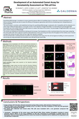
- 1. Development of an Automated Comet Assay for Genotoxicity Assessment on TK6 cell line M. ROUDAUT1, S. ASTRI2, N. ORSINI2, A.-P. LUZY2*, J. BURSZTYKA1* & N. MAUBON1 1 HCS Pharma, Biopôle, 6 rue Pierre Joseph Colin, 35000 Rennes 1* julian.bursztyka@hcs-pharma.com 2 Galderma R&D, Les Templiers, 2400 route des Colles, 06410 Biot ²* anne-pascale.luzy@galderma.com Abstract Quantifying DNA damage is mandatory to assess potential adverse effects of candidate drugs or molecules or extracts developed in the dermo-cosmetic industry, but also to assess the efficacy of therapeutic approaches with the aim of producing tumor cell genotoxicity in cancer treatment. The comet assay is a sensitive, well established technique for quantifying DNA damage in eukaryotic cells. Compatible with the detection of a wide range of DNA damaging agents, its principle consists in the migration of fragmented DNA in an electrophoresis gel (damaged DNA forming the tail of the comet), while intact DNA moves at a slower rate (head of the comet). The percentage of fragmented DNA in the comet tail is a direct measure of DNA damage. The comets are evaluated via fluorescence staining and microscopy. Two drawbacks are often mentioned about the comet assay: preparing the comet slides with cells in agarose to perform electrophoresis is time consuming and reduce the throughput of the method, and, if manual scoring of the comets are done, there can be a high inter-operator variability. Here we will describe a combined process to perform an automated comet assay, from the cell and agarose handling, to comet assessment and scoring, by using automated instrumentation and a 96-well format comet slide. Methods Results Conclusions & Perspectives Image analysis with Columbus allowed automated scoring of the comets, with exclusion of abnormal ones. Nice dose effects were observed when looking at the % of DNA in Tail after incubation of TK6 cells with genotoxic compounds like etoposide and 4NQO. No comet were observed nore scored when TK6 cells were incubated with a non genotoxic compound like cycloheximide. Conclusion : Automation of the comet assay using 96-well slides allows medium throughput screening to assess potential adverse effects of new drug candidates on Tk6 cells. Assessment of cytotoxicity in parallel need to be developed. These are encouraging preliminary results that should be completed with an additional list of genotoxic and non genotoxic compounds. Cell culture: human lymphoblast cells (TK6) were routinely maintained in RPMI supplemented with 10% serum, 1% non-essential aminoacids, 1% L- glutamine. For the comet assay, cells were seeded at 10 000 cells/well in 96-well Greiner polypropylen round bottom plates (100 µL/well). Cells treatments: the comet assay was performed by replacement of the medium in each well by fresh medium containing etoposide, 4- nitroquinoline 1-oxide (4NQO) or cycloheximide at different concentrations in duplicate. After a 3h incubation, cells were embedded into agarose (Trevigen), and immediately dispensed on the CometAssay® 96 well slide (Trevigen) using the Caliper Sciclone ALH3000 liquid handler (Perkin Elmer). The comet assay in alkalin condition has been carried following recommendations of Trevigen, provider of the consumables used during the experiment. Image acquisition: Images were acquired on the Operetta plateform (Perkin Elmer). Image analysis: Images analysis was done using the Columbus Software, allowing detection of the head of each comet (corresponding to the original nucleus) and detection of the tail (which extent and intensity are proportional to damages undergone by DNA). The % of DNA in Tail is calculated as follows: (Sum of intensity in the tail/sum of intensity in the whole comet)*100. A minimum of 52 comets were scored per well, except with the most cytotoxic incubated concentrations of 4NQO, at 10 & 30 µM). To get this poster, please flash the QR- code You can use the I-NIGMA application from your store CTRLEtoposide4NQO CTRL Cmax - - - - - - - - - - - - - - - - - - - - - - - - - - Cmin Etoposide Cycloheximide 4NQO Cycloheximide Heatmap of the 96-well plate Segmentation Calculated % DNA in Tail Mean of the duplicates, error bares show the two calculated values CTRL Cmax - - - - - - - - - - - - - - - - - - - - Cmin Etoposide Cycloheximide 4NQO Dose effect on nucleic count