
Recommandé
Contenu connexe
Similaire à A look at camels - نظرة إلى الإبل
Similaire à A look at camels - نظرة إلى الإبل (20)
Plus de hhalhaddad
Plus de hhalhaddad (20)
Dernier
Dernier (20)
A look at camels - نظرة إلى الإبل
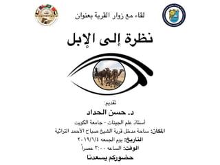
- 1. ا#بل إلى نظرة :تقديم الحداد حسن .د الكويت جامعة - الجينات علم أستاذ التراثية ا>حمد صباح الشيخ قرية مدخل ساحة :ا5كان ٢٠١٩/١/٤ الجمعه يوم :التاريخ ًاعصر ٣:٠٠ الساعه :الوقت يسعدنا حضوركم بعنوان القرية زوار مع لقاء
- 2. ا#بل إلى نظرة الحداد حسن .د الكويت جامعة - الجينات علم أستاذ الشعبي للموروث الكويت دولة مهرجان (٢٠١٨/١٩) @jamalid_reportJamalid ReportJamalidreport Jamalid Report Laboratory Kuwait University, College of Science Khaldiya Campus, 1Kh Lab No. 150 jamalidreport@gmail.com 900 camel 900 22635
- 3. ﷲ ﺣﯾﺎﻛم
- 5. SamplEase Jamalid Report Laboratory Kuwait University, College of Science Khaldiya Campus, 1Kh Lab No. 150 jamalidreport@gmail.com hagenetics.org bader.alhajeri@gmail.com hhalhaddad@gmail.com @jamalid_reportJamalid Report@jamalidreport العلوم كلية البيولوجية العلوم قسم (الجينات )علم دكتوراه ماجستير و بكالوريوس (دقيقه )بيولوجيا
- 6. بل؟Yا إلى نظرة اذاZ
- 7. اﻟﻨﻈﺮ ﻋﺪم ﻋﻠﻰ ﺗﻌﺎﺗﺒﻨﻲ اﻵﯾﺔ رأﯾﺖ ﺑﻌﺪم اﺳﺘﻤﺮاري ﻋﻦ وﺗﺴﺘﻔﮭﻢ أﻟﻢ ﻣﻦ ّﻲﻋﻠ ﺧﻔﻒ ﻣﺎ وﻟﻜﻦ ،اﻟﻨﻈﺮ اﻵﯾﺔ ﺑﺄن إﺣﺴﺎﺳﻲ ھﻮ اﻟﻌﺘﺎب .ﻟﻠﻨﻈﺮ أﯾﻀﺎ ﺗﺪﻋﻮﻧﻲ
- 8. اللغوي وتراثنا بلYا إلى نظرة
- 9. اﻟﻌرﺑﻲ ﺗراﺛﻧﺎ ﻓﻲ ﻣﺗﻐﻠﻐل اﻟﺟﻣل إن
- 10. وأقاربها بلYا إلى نظرة
- 12. ومنتجاتها بلYا إلى نظرة
- 14. وقدراتها بلYا إلى نظرة
- 15. terinaria 6 (4): 417-423, 2011 2-6197 Publications, 2011 ding Author: Adel M. Badawy, Department of Surgery,Radiology and Anaesthesiology, Faculty of Veterinary Medicine Benha Univeisity, Qalubia, Egypt. Tel: +202-42132555, Mob: +2011-8859378, E-mail: adelbadawybadawy@yahoo.com. 417 Computed Tomographic Anatomy of the Fore Foot in One-Humped Camel (Camelus dromedrus) Adel M. Badawy Department of Surgery, Anaesthesiology and Radiology, Faculty of Veterinary Medicine (Moshtohor), Benha University, Qalubia, Egypt act: The purposes of this study was to describe and identify , the complex anatomical structures of digits oot pad of the fore limb in one-humped camel by using the computed tomography scan (CT scan) and anatomy, which would be used in diagnosis of foot and footpad disorders. The study was performed ree pairs of camel's fore feet. Transverse and sagital CT images were obtained by using Hitachi-CXR ti-Slice CT Scanner. The different anatomical structures of the digits and footpad were identified in the n sections, the fixed slices and the dissected specimens. The CT images were compared with the ponding sections at the same levels and selected for their identity and photographed. The analogous mical structures were identified on the transverse and sagital slices and labeled with the corresponding ures on the CT images. Results revealed that, the frontal tip of each digit was covered by a characteristic nail. The distal and middle phalanges were horizontally situated, while the proximal phalanx obliquely oned. Their planter surfaces were separated from the ground by foot pad. Three digital cushions enclosed ommon capsule were found underneath each digit. On CT images, the inner structures of the foot and foot were appeared with various gray scales. It was concluded that, camel has unique feet morphological arities. CT was efficient imaging modality that provides a cross-sectional image with superior soft tissue entiation and no superimposition of the overlying structures, which can be used for better diagnosis of nd foot pad abnormalities. words: Camel CT scan Cross-section Digits Footpad INTRODUCTION value to evaluation of soft tissue, although ls are a fascinating and little studied animals. ligaments [8-10]. Alongside, ultrasonography provides a unique among artiodactyls in their regular a small view and each structure has to be separately nt of pacing gait and having a unique foot imaged and a cross sectional examination through the gy assumed to be an adaptation for this mode of entire digit is not possible. On the other hand, soft n [1-3]. The uniquely designed wide spread feet tissue is difficult to be evaluated by ultrasonography m to walk on shifting sand in the desert and in the digit [11]. CT, through its high spatial resolution ky terrain whereas the footpad is used to grip and moderate differentiation of tissue contrast is a and steep inclines. Their feet are secondarily fastened exceptionally useful technique for visualizing e, with a splayed -toed foot, loss of hooves, general anatomy [12]. f a broad foot pad and loss of the interdigital The use of CT in large animal medicine is currently allowing the divergence of the third and limited by logistical problems of acquiring CT images; its [3-5]. Imaging techniques play a major meanwhile a few CT studies on horse's foot have been e modern biomedical research [6,7]. Current done [13]. Computed tomography (CT) is anatomic cross- imaging techniques such as radiography and sectional imaging that uses x-rays and x-ray attenuation raphy provide limited information for to create the images [14, 15]. The CT gantry houses a row of the camel foot. Radiography has limited of x-rays detectors across from an x-ray generator. ultrasonography provides visualization of tendons and Global Veterinaria, 6 (4): 417-423, 2011 (A) (B) Fig. 1: (A) Dorsoplanter CT image and (B) Dorsal view of dissected foot. 1, metacarpal bone ;2, divided metacarpal bone; 3, fetlock joint; 4, proximal sesamoid bones; 5, proximal phalanx; 6, pastern joint; 7, middle phalanx; 8, distal phalanx; 9, sole; 10,nail or pes; 11, interdigital notch; 12, interdigital septum; a, tendon of muscle extensor digitorum lateralis; b and c , medial and lateral tendons of muscle extensor communis; d, true common extensor tendon ; e, proper extensor tendon of the third digit ; f, proper extensor tendon of the fourth digit. (A) (B) Fig. 2: (A) Sagital CT image and (B) cross section at the level of the middle of the nail. 1, distal exterimity of the metacarpal bone ;2, proximal phalanx; 3, middle phalanx ;4, distal phalanx; 5, proximal sesamoid bone; 6, deep digital flexure tendon; 7, fibrocartilagenous enlargement of the deep digital flexure tendon; 8, middle scutum; 9,yellow bed; 10, digital cushion; 11, common capsule of the digital cushions; 12 epidermal layer of the sole; 13 sole pad; 14, distal scutum;,N, nail. (A) (B) 419 lanx;9,sole;10,nailorpes;11,interdigitalnotch;12,interdigitalseptum;a,tendonofmuscleextensor torumlateralis;bandc,medialandlateraltendonsofmuscleextensorcommunis;d,truecommonextensor on;e,properextensortendonofthethirddigit;f,properextensortendonofthefourthdigit. (A)(B) SagitalCTimageand(B)crosssectionatthelevelofthemiddleofthenail.1,distalexterimityofthe acarpalbone;2,proximalphalanx;3,middlephalanx;4,distalphalanx;5,proximalsesamoidbone;6,deep talflexuretendon;7,fibrocartilagenousenlargementofthedeepdigitalflexuretendon;8,middlescutum; llowbed;10,digitalcushion;11,commoncapsuleofthedigitalcushions;12epidermallayerofthesole;13 pad;14,distalscutum;,N,nail. (A)(B) TransverseCTimageand(B)crosssectionatthelevelofthesolepad,showingsomewhatroundsoleand rdigitalnotch. الشحم خف خدةZا خف ّeالل الخف الرمال على شيZا خف
- 16. Body Temperature of the Camel and Its Relation to Water Economy KNUT SCHMIDT-NIELSEN,l BODIL SCHMIDT-NIELSEN,l p2S. A. JARNUM3 AND T. R. HOUPT4 From the Department of Zoology, Duke University, Durham, North Carolina, U.S.A. and Centre de Reclzerches Sahariennes, B&i A bbds, Algeria ABSTRACT SCHMIDT-NIELSEN, KNUT, BODIL SCHMIDT-NIELSEN, S. A. JARNUM AND T. R. HOUPT. (Duke U., Durham, N. C. and Centre de Recherches Sahari- ennes, Beni Abbes, Algeria.) Body temperature of the camel and its relation to water economy. Am. J. Physiol. 188(r): 103-112. Io57.-The rectal tem- perature of normal healthy camels at rest may vary from about 34OC to more than 4oOC. Diurnal variations in the winter are usually in the order of 2OC. In summer the diurnal variations in the camel deprived of drinking water may exceed 6°C but in animals with free access to water the varia- tions are similar to those found in the winter. The variations in temperature are of great significance in water conservation in two ways. a) The increase in body temperature means that heat is stored in the body instead of being dissipated by evaporation of water. At night the excess heat can be given off without expenditure of water. b) The high body temperature means that heat gain from the hot environment is reduced because the temperature gradient is reduced. The effect of the increased body temperature on heat gain from the environment has been calculated from data on water expendi- ture. These calculations show that under the given conditions the variations in body temperature effect a considerable economy of water expenditure. The evaporative heat regulation in the camel seems to rest exclusively on evaporation from the skin surface (sweating), and there is no apparent in- crease in respiratory rate or panting connected with heat regulation. The evaporation from isolated skin areas increases linearly with increased heat load. The critical temperature at which the increase sets in is around 35OC. The fur of the camel is an efficient barrier against heat gain from the en- vironment. Water expenditure is increased in camels that have been shorn. N A PREVIOUS PAPER (I) we described the unusualtolerance of the camel to water considerable economy with water could be achieved by changesin body temperature (2, BODY TEMPERATURE AND WATER ECONOMY IN THE CAMEL *OS FIG. 2. Variations in rectal temperature in 2 camels and I donkey for a 3-wk. period. Diurnal fluctuation in rectal temperatures as well as the increase in these fluctuations during water deprivation are discussed in text. Air temperature corresponds closely to standard meteorological observations. Dotted line refers to black bulb temperature. magnitude as observed by Sergent and Lheritier (IO). During this period (January) the air temperature varied between approxi- mately 0°C and I~OC. Under theseconditions of low air temperature andrelatively lowradia- tion the heat gain from the environment probably wasnegligible. In his paper Sergent observed that excep- tionally low rectal temperatures in the camel were always associatedwith rain. He found that temperatures between 34°C and 35OC were not unusual after rainy nights. We ex- pected a similar reactions in our experimental camels. However, on three occasionswhen What we see from this chart is that the donkey and the camel,in spiteof beingat rest, have rhythmical diurnal excursions in the temperature curve many times as great as thoseof man. The curves are alsomore irregu- lar, and the minima and maxima are rather variable by human standards. The most conspicuousfeature of the curves, however, is that the temperature fluctuations aremuch greater in the animalswhenthey are deprived of drinking water. To showin detail the effect of water intake on the temperature curve a portion of the complete graph is pre- sentedin figure 3. For both camelsthis section الحرارة تخزين حرارة درجة أعلى حرارة درجة أقل اءZا حفظ
- 17. Water Balance of the Camel BODIL SCHMIDT-NIELSEN,LF~ KNUT SCHMIDT-NIELSEN,I T. R. HOUPT3 AND S. A. JARNUM” From the Department of Zoology, Duke Vrt;versity, Dwham, North Carolina, ad Cetdre de Rechemhes Sahariennes, Beni Abbes, Algeria ABSTRACT Camels (Cuwzeltis dmmedarius) were exposed to prolonged periods of water deprivation during winter, spring and summer in the Sahara desert. Determinations were made of: weight changes, water and food intake, urine flow and concentrations, plasma concentrations, etc. It was found that the camel can tolerate a loss of water corresponding to 300/a of its body weight even when exposed to the severe desert heat. Other mammals de- hydrated in a hot environment may die from circulatory failure already when the water loss involves 12 y0 of the body weight. Unlike many other mammals the camel does not lose its appetite when deprived of water but continues to eat normally until the desiccation becomes very severe. It has a low urine output (0.5-r l/day when kept on a diet of dates and hay), a low water content in the feces, and, when dehydrated in the summer, a very low evaporative water loss.When offered water the camel drinks in IO minutes enough water for compIete rehydration. The Iongest period that we kept a camel on dry food without drinking water in the hot summer was 17 days. This camel was not working and it had its protective fur which decreased the heat gain from the environment. It is concluded that the ability of the camel to withstand prolonged dehydration is due to: a) toler- ance to an extremely high degree of desiccation of the body and b) low over- all water expenditure. Particularly effective as a water conserving mecha- nism is the low evaporative water loss during dehydration in the summer. THE CAMEL has a legendary reputation for ability to tolerate water deprivation for prolongedperiodsof time. It is sur- prising therefore that scientific literature about its physiology is quite inadequate. The litera- ture on the camel mainly consists of travel reports and veterinary handbooks. Informa- tion pertinent to the water balance of the camelhas beenreviewed recently (I). For a large animal like the camel the prob- lems of survival in a desert environment are quite different from those of, for example, a small rodent which has an underground bur- row. A large animal cannot escapethe heat of the day, and it will consequentlyhave to spend Received for publication August 8, 1955 l Dept. of Zoology, Duke Univ., Durham, N. C. 2 Established Investigator of the American Heart Association. 3 Dept. of Physiology, School of Veterinary Medi- cine, Univ. of Pennsylvania, Philadelphia, Pa. 4 Bispebjerg Hospital, Dept. A. Copenhagen, Den- mark. water for evaporative cooling in order to maintain constant body temperaturewhenever the environment is so warm that the heat transfer between body and environment re- sults in a net heat gain in the body. The small rodents can and must escapethe heat because evaporative cooling is too expensivean under- taking for them (2). Accordingly, smallrodents do not spendwater for heat regulation, and it is possible for certain of them to maintain water balance even when the water intake is restricted to the preformed water in dry seeds and the water formed by the oxidation of the food in the body. This is accomplishedby an extreme degree of water economy (3). How- ever, the large animal that sweats or pants can never become independent of intake of free water, becausethe amount of water neces- sary for heat regulation is large comparedwith the other needs. The adaptation of the camel to its hot en- vironment does not, therefore, involve inde- ded from www.physiology.org/journal/ajplegacy by ${individualUser.givenNames} ${individualUser.surname} (163.015.154.053) on August 6, 2018. Copyright © 1956 American Physiological Society. All rights reserved. OF THE CAMEL 193 TABLE 4. COMPARISON BETWEEN THE WATER LOSS DURING WATER DEPRIVATION AND THE AMOUNT OF WATER INGESTED AT THE TERMINATION OF THE DEHYDRATION PERIOD T- - DrinkingWeight Loss kg i%if. * Camel A =7*3 7*8 36.5 16.2 34*o 14.5 64.0 22.0 (124)” 37*4 72.5 26.5 Camel Q Days With- out Water ggf l3%e ing - De- hydr. 12/14 223 I/27 226 4119 234 5129 29= 6/22 332 7/4 274 6/I 2!474 6/22 452 7/4 447 kg To of de- hydr. b.wt. 16.3 7*9 40.0 21.1 44*o 22.0 56.0 24.9 61.7 29.7 66.5 33J 9 =7 =7 II 17 7 7 23.0 8 22.6 7 27.2 Rortkey B 103 28.1 103 29*4 104 31.8 in to e, y re n 12/d I I2/2I 12123 I2/27 I/2 d7 3/2I 3/27 619 6/17 11.8 14.2 IO.2 12.2 4*4 50 9*2 r1.3 9.7 11.9 9.0 11.6 154 19.1 =7*4 22.6 20,2 27.2 (23d (31*5H 20.3 27.8 (26.9H (36*8H 96-S 4 94*8 5 93.7 2 93-4 4 89.6 4 91.0 4 96.7 3 96*5 6 107 4 104 4 13.5 I4 11.3 11.9 4-7 5-0 12.4 13.3 8.5 9*5 9.0 9*9 15.7 16.2 “9.5 20.2 32*5 3044 31.1 29.9ey of ht ne ng he * This weight loss includes the loss of considerable edema produced by excessive administration of NaCl, t Figures in parentheses represent total amount of water consumed in the course of several hours. On تأثر دون الوزن من ٪٣٠ من أكثر ماء فقدان فقودZا الوزن وتعويض بسرعه اءZا شرب
- 18. وأعصاب تuعض يحوي أنف ختياريYا والفتح للغلق قابل
- 19. وأعمارها بلYا إلى نظرة
- 20. سنانzا نمو حالة ثم ً|أو السلوك على ًابناء بلYا أعمار تسمية
- 22. متفق صفات حسب نتاجYا لجودة عرض هي زاينZا مسابقات eربZا من عليها
- 23. الحيوانات مزاين عن ًاكثير تختلف | بلzا مزاين مسابقات خرىzا
- 24. زاينZا إبل إلى عامة نظرة
- 25. Shaele Majaheem Waddah Shageh Homor Sofor black Mezayen breeds Crow-black Majaheem Black Majaheem Light Majaheem Smoky-brown Sofor Syrupy Sofor Light Sofor Brown Shaele Milky Shaele Light Shaele Red Homor Blackened Homor Twilight Homor Wheat Shageh Light Shageh Rosy Waddah Blond Waddah White Waddah اﻟﻣزاﯾن إﺑل ﺗﻘﺳم ،ﻣﺟﻣوﻋﺗﯾن اﻟﻰ ًﻻاو ﻣﺑﻧﻲ اﻟﺗﻘﺳﯾم وھذا اﻟﺻﻔﺎت ﻋﻠﻰ .اﻟظﺎھرﯾﺔ
- 26. ()ﺣداد،ﺣراب اﻟرأس ﻋﻠﻰ وﻋﻣودﯾﺔ ﻣﺳﺗﻘﯾﻣﮫ ()اﻟﻣﺟﺎھﯾم اﻟﺳوداء اﻷﺑل أذن ()ﺧرع ﻟﻠﺧﻠف ﻣﺎﺋﻠﺔ ()اﻟﻣﻐﺎﺗﯾر اﻟﻣﻠوﻧﮫ اﻹﺑل أذن
- 27. a. b. c. اﻟﻣﺟﺎھﯾم ذﯾل ودﻗﯾق طوﯾل اﻟﻣﻧﺑت اﻟﻣﻐﺎﺗﯾر ذﯾل وﺳﻣﯾك ﻗﺻﯾر اﻟﻣﻧﺑت
- 28. زاينZا إبل إلى دقيقة نظرة
- 29. ًﺎﺗﺣﻣﯾﺻ اﻟﻣﺣﻣﺻﺔ اﻟﻘﮭوة ﻟون إﻟﻰ وأﻗرب ،اﻟﺳواد دون داﻛن ﻟوﻧﮭﺎ .ﺑﻌده أو إﻋدادھﺎ ﻗﺑل اﻟﺗرﻛﯾﺔ اﻟﻘﮭوة ﻟون ﯾﻘﺎرب ﻟوﻧﮭﺎ .ًﻼطوﯾ
- 30. ﻟون ﻓﺈن ،اﻟﻘﮭوة درﺟﺎت ﺑﺎﺳﺗﺧدام ﻟﻸﻟوان اﻟﻌرب وﺻف أﺧذﻧﺎ ﻣﺎ إذا .اﻻﺣﺗراق دون إﻧﻣﺎ اﻟﻘﮭوة ﺗﺣﻣﯾص ﻣن ﻣﺗﻘدﻣﺔ ﻣرﺣﻠﺔ اﻟﺻﻔر اﻹﺑل
- 31. ﺑﺎﺳﺗﺧدام اﻟﺷﻌل ﻓروع ﻣﺧﺗﻠف ﻓﻲ اﻟوﺑر ﻟون ﺗوﺻﯾف اﻟﻣﻣﻛن ﻣن .ﺑﺎﻟﺣﻠﯾب اﻟﺷﺎي ﻣﺷروب
- 32. وﻗت اﻟﺳﻣﺎء ﻛﻣﺎ ﻣﻧﮭﺎ اﻟﺷﻔﻘﺎء اﻟﺣﻣر اﻹﺑل ﻓﻲ ﻋﻠﻰ اﻟﻣﺻﻔر ﺑﺎﻟﺑﯾﺎض اﻟﺣﻣرة ﺗﻣﺗزج ،اﻟﻐروب أﺳﻔﻠﮭﺎ اﻟﺑﯾﺎض و اﻟﺳﻣﺎء أﻋﻠﻰ اﻟﺣﻣرة ﺗﻛون ٍﻧﺣو
- 33. "اﻟﮭرﯾس " ﯾﺳﻣﻰ ﺑﻣﮭﺎرة ﺟدﺗﻲ ﺗﺻﻧﻌﮫ ٍﺑطﺑق اﻟﺷﻘﺢ اﻟﻣﻐﺎﺗﯾر ﻟون ﯾذﻛرﻧﻲ
- 34. وھذا اﻷﺑﯾض اﻟوﺑر ﺑﻠون ﺗﺗﻣﯾز اﻟوﺿﺢ اﻟﻣﻐﺎﺗﯾر إﺑل ﻋﺎم ﺑﺷﻛل .ﻣﺎ ًﺎﺷﯾﺋ رَﻔْاﻟﻣﺻ اﻟدﺳم اﻟﺣﻠﯾب ﻟون ﻣن ﻗرﯾب اﻟﺑﯾﺎض
- 35. بلYا أبحاث إلى نظرة
- 36. واﻟﺻﻔﺎت (DNA) ال ﻋﯾﻧﺎت ﺑﻧك إﻧﺷﺎد اﻟذﻛﯾﺔ ﻟﻠﮭواﺗف (App) ﺑﺎﺳﺗﺧدام اﻟﻣﻌﻠوﻣﺎت ﺟﻣﻊ 10266 | Ecology and Evolution. 2018;8:10266–10271.www.ecolevol.org 1 | INTRODUCTION All research requires data. Data are often collected unmethodically and unsystematically. It is often the case that only after the com- pletion of the field season, is this data organized and transcribed to a format that can be more readily archived, analyzed, and shared (i.e., a spreadsheet). This two-step process of data collection is time- intensive (reducing the time spent collecting samples) and reduces data quality (via omissions, transcription errors, etc.). The usability of any dataset is directly related to its organiza- tion and formatting and the scientific value of the dataset increases as it is used by different research groups. Collaboration and data sharing across research groups are hindered by the use of disparate data collection and organization schemes—thus, a unified system- atic data collection approach could increase collaboration and data sharing. While many researchers developed software to address and serve various scientific objectives (see lists in http://brunalab.org/ apps/), none of these applications provide a simple solution to the collection of biological specimen data. The closest application to serve this purpose is EpiCollect (Aanensen, Huntley, Feil, al-Own, & Spratt, 2009). However, the complex application design and multiple features, although valued, may limit its wide-scale adoption and im- plementation across fields. In designing SamplEase, we adopted the recommendations of Borer et al. concerning data formats, storage, and sharing potential as well as technical issues with naming files and categories of data (Borer, Seabloom, Jones, & Schildhauer, 2009). SamplEase is available in both Android and iOS platforms, designed to expedite collection, management, and sharing of biological specimen data in the field. 2 | SAMPLEASE WORKFLOW The general idea behind developing SamplEase is to (1) rapidly and conveniently collect and store biological specimen data along with photographs for each biological sample, (2) export the data in a Received: 16 March 2018 | Revised: 4 August 2018 | Accepted: 7 August 2018 DOI: 10.1002/ece3.4503 O R I G I N A L R E S E A R C H SamplEase: a simple application for collection and organization of biological specimen data in the field Hasan Alhaddad | Bader H. Alhajeri This is an open access article under the terms of the Creative Commons Attribution License, which permits use, distribution and reproduction in any medium, provided the original work is properly cited. © 2018 The Authors. Ecology and Evolution published by John Wiley & Sons Ltd. Department of Biological Sciences, Kuwait University, Safat, Kuwait Correspondence Hasan Alhaddad, Department of Biological Sciences, Kuwait University, Safat 13060, Kuwait Email: hhalhaddad@gmail.com Abstract Careful collection and organization of biological specimens and their associated data are at the core of field research (e.g., ecology, genetics). Fieldwork data are often col- lected by handwriting or unsystematically via an electronic device (e.g., laptop), a process that is time-intensive, disorganized, and may lead to transcription errors, as data are copied to a more permanent repository. SamplEase is an iOS and Android application that is designed to ease the process of collecting biological specimen data in the field (data associated with biological samples, such as location, age, and sex). In addition to biological specimen data, SamplEase allows for the assignment of photo- graphs to each collected sample, which provides visual records of each specimen in its environment. SamplEase outputs biological specimen data in a tabular format, fa- cilitating subsequent analyses and dissemination. Despite the simplicity of SamplEase, no similar data management application is readily available for researchers. K E Y W O R D S animal sampling, biological specimen data, data management, fieldwork, images SamplEase and Cdrom archive: Windows to study the dromedary Camelus dromedarius (Artiodactyla: Camelidae) and related camelids Hasan Alhaddad and Bader H. Alhajeri Department of Biological Sciences, Kuwait University, Kuwait SamplEase 1. Search 2. Download 3. Register 4. Link to Dropbox 5. Collect data 6. Upload data 7. View data Acknowledgment: Jaafar Hussian assisted in designing SamplEase. Sample ID Date Nickname Sex Age Sire Dam Breed Bio Sample Other 1 Other 2 Other 3 GIS Country Region Notes Breeder Name Breeder Phone Breeder Email Photo 1st Photos Total S-Other 1 S-Other 2 S-Other 3 Collector Name Collector Phone Collector Email Collector Affiliation 201701151200_1 201701151200 Hasan Male 4 y ? ? Wadh Saliva 29.32491182 - 47.97026849 Kuwait Khaldiya KU 6199998888 h@gmail.com 201701151200KU_1_1 3 Hasan Alhaddad 123456789 jamalidreport@gmail.com Kuwait University 201701151200_2 201701151200 Bader Male 5 y ? ? Sufur Hair 29.32491182 - 47.97026849 Kuwait Khaldiya KU 6199998888 h@gmail.com 201701151200KU_2_1 5 Hasan Alhaddad 123456789 jamalidreport@gmail.com Kuwait University Figure 1: How to use SamplEase, an iOS sampling application. Cdrom archive 1. Register SamplEase • SamplEase registration ensures that each sample links to the collector’s contact information (Table 1). • Registration information records the contribution of each investigator working within a team. 2. Link to Dropbox • SamplEase needs to be linked to a Dropbox account, where sampling data and associated images can be deposited. • If need be, SamplEase can be unlinked from one Dropbox account and linked to another. 3. Session information • A session is defined as a sampling trip, often to a specific breeder, in a specific location. • All samples collected at a single locality share the same session information. 4. Sample information • Unique information for each sample can be easily entered and saved. • Samples can be continually added until the sampling session is complete. The session ends when sampling is done. 5. SamplEase output • For each session, data is stored as a zip file, which contains a table of SamplEase data (Table 1), along with the associated images. • Data can be uploaded to the linked Dropbox account (when connected to the internet), in a folder with the label: App/SamplEase. • When not connected to the internet, the session data will be stored on the device temporarily, so that it can be uploaded to the linked Dropbox account when internet is restored. • Images are automatically assigned a unique name, based on the sample ID. Why use SamplEase? 1. Search 2. Download 3. Register 4. Link to Dropbox 5. Collect data 6. Upload data 7. View data Introduction • Collection, organization, and dissemination of biological data, in the form of specimens and associated information is essential for long and short-term research projects. Manual data collection (i.e. by hand) is time-intensive, hard to share, and is more susceptible to loss. • SamplEase is an application designed to ease the process of biological specimen sampling, particularly in a fieldwork setting. The application allows systematic assignment of basic data to each collected specimen. • SamplEase can be utilized by ecologists, geneticists, plant and animal breeders, and researchers from other fields. • It is easy to use. • Paperless data collection. • Data can be inputted in any language. • Data output is organized in a table. • Data is automatically backed up in Dropbox. • Data is uploaded as a zip file to save space. • Each collected sample is automatically assigned an ID, a date and time, and GIS coordinates. • Unlimited number of photos for each sample. • Assignment of sample IDs is based on the sampling date & time (yyyy-mm-dd-hh- mm_#). • Rapid collection of phenotypic, behavioral, and environmental data. • Images are named according to the sample ID. • The name of the output file corresponds to the date & time of the sampling session. • File names are arranged in chronological order (yyyy-mm-dd-hh-mm_name). Table 1: An example of a table outputted from SamplEase. • Cdrom archive is a bank of Camelus dromedarious biological specimens, used to explore its molecular and morphological variation. • To investigate the morphological variation and the genetic causes behind such variation, a large number of camels need to be screened molecularly. • Cdrom archive links the detailed morphological data obtained from SamplEase with the collected DNA samples (Tail hair), in order to verify molecular variants associated with a particular phenotype. • Blood, buccal swabs (saliva), and tail hair follicles are possible biological specimen sources that can be collected from camels in order to extract DNA for the Cdrom archive. • Blood samples offer sufficient quantities of high quality DNA but poses problems related to sample storage and collection (many breeders are resistant to providing blood samples). • Saliva samples are relatively easier collect and store compared to blood samples. However, the quality and quantity of DNA varies across samples, and may be contaminated with feeding material. • Hair samples are the easiest to collect and store and camel breeders do not mind providing such samples. We found that hair can provide sufficient good quality DNA for molecular analyses.
- 37. Al Sohi Al-Shahab (white + black tint) Al-Shiabeen (light color) Asail (pure) Awadi (gentle) Azaouad (black) Benadir (white) Brela Cajeh (red) Dkhan (smokey white) Grain (tawny) Homor (red) Khader (green) Kimta(h) Labied (white) Lahmami (color) Lakhdar (green)Lasfar (yellow)Lazrag (blue)Maghateer (light) Majaheem (black) Ramli (sandy) Shaele (brown) Shageh (wheat) Sofor (somkey yellow) Waddah (white) Zargeh (blue) JabaliRiverine Saharan SahelSaheli Sahelian Sahraoui Fellahi M aya M ehara A(i)rAdrar des Iforas Aftout AjjerAl M ahaliyat Alsertaw eya Altebestee Arak Awarik Azaouak Azawak Baladia Batinah Bikaneri Butana Campbellpuri Chami Danakil DeraIsmailKhan Deshi Dhatti Dhofar Fleuve Gandiol Guban HoratAlMadenia Jaisalmeri Jalore Jebel Judi Kachchi Kala Kala-Chitta Kanem Kharai Kharani Khawar Kohi Kubule Larri M ajorero M akraniM alvi Mewari Mewati MudughMultanOgadenPishinRakaRodbariSakarchagin SakraiShekhawati Shemelia Sifdaar Sindhi Sirohi Sowari Tibesi Tibesti Turkana Yerbent Afar Ait Khabbach Al-Shorarat Anafi Arvana Bagri Bandari Berabish Bim al Bishari Borena Brahvi Chaam bi Dhibian Dolbahanta GabbraGaddiGarre GhulmaniGoraneHelai JebliKabbashiKalkooi Kenani Lahwee Maalia Maganeen Mahamid Mareecha Rashaidi Reguibi Rendille ShanbaliTajakantTarguiUrfillaOuladSidiCheikh Camel Groups (138) Ecotype Function Phenotype (27) Geographic Region (63) Tribe's Name (38) وﻣﻣﯾزات ﺧﺻوﺻﯾﺔ ﻟدراﺳﺔ اﻟﻣزاﯾن أﺑل وإﻛﺗﺷﺎف اﻟوراﺛﺔ اﻹﺑل ﺻﻔﺎت ﺟﯾﻧﺎت
- 38. Shaele Majaheem Waddah Shageh Homor Sofor black Mezayen breeds Crow-black Majaheem Black Majaheem Light Majaheem Smoky-brown Sofor Syrupy Sofor Light Sofor Brown Shaele Milky Shaele Light Shaele Red Homor Blackened Homor Twilight Homor Wheat Shageh Light Shageh Rosy Waddah Blond Waddah White Waddah ﺻﻔﺎت وﻋرض ﺗرﺟﻣﺔ ﯾراھﺎ ﻛﻣﺎ اﻟﻣزاﯾن إﺑل ﻟدراﺳﺗﮭﺎ أﺻﺣﺎﺑﮭﺎ ًﺎوﺟﯾﻧﯾ ًﺎﺷﻛﻠﯾ
- 39. اﻟﺻﻔر اﻹﺑل ﻟﻠون اﻟﺟﯾﻧﻲ اﻟﺳﺑب دراﺳﺔ ()اﻟدﺑﺎﺳﻲ
- 40. Curly (47) Straight (66) Unknown (50) a. b. Unknown (163) اﻟﺳﺑب دراﺳﺔ ﻟطول اﻟﺟﯾﻧﻲ اﻟوﺑر وﻗﺻر اﻟﺳﺑب دراﺳﺔ اﻟوﺑر ﺷﻛل اﻟﺟﯾﻧﻲ ﻧﺎﻋم ()ﻣﻠﺳﺎء،ھﻠﺔ ﻣﻔﻠﻔل ()ﻣﻌﻛرﺷﮫ،ﻣﺣﻠق
- 41. اﻹﺑل ﻗﯾﺎﺳﺎت
- 42. 1 2 3 4 56 7 8 9 10 Mejaheem 1 2 3 4 56 7 8 9 10 1 2 3 4 56 7 8 9 10 1 2 3 4 56 7 8 9 10 1 2 3 4 56 7 8 9 10 Maghatir Omaniat ShaeleSofor وﻣﻛﺎن ﺷﻛل دراﺳﺔ اﻟﻣزاﯾن إﺑل ﻓﻲ اﻟﺳﻧﺎم
- 43. 3 2 9 5 6 1 4 8 7 10 11 12 Hijin (n = 69) Mezayen (n = 322) Beauty and the beast: A comparison of torso shapes of beauty-contest camels vs. racing camels Alhajeriand Bader H.,AlhaddadHasan,AlaqeelyRanda Department of Biological Sciences, Kuwait University, Kuwait Introduction • Dromedary camels (Camelus dromadarius) permeate Arab culture. • Perhaps the two most well-known camel types are those used for beauty-contests (Mezayen) and the ones used for racing (Hijin). • Torso morphology is one of the most important features that characterize these two camel types. Perhaps the most distinctive torso feature relate to the humps, which tend to be posteriorly- positioned in camels bred for beauty-contests, and anteriorly- positioned in racing camels. • In this study, we assess the exact discrepancy in overall torso shape between Mezayen and Hijin using geometric morphometric analysis—a technique that uses two-dimensional landmarks digitized on images of lateral views of camels collected from various social media accounts. @jamalid_report jamalidreport@gmail.com@jamalidreport Objectives 1. Determine if there are significant differences in the geometric shape of the torso of camels bred for beauty-contests (Mezayen) vs. those that are bred for racing (Hijin). 2. Assuming that there are significant differences in torso shape, determine the exact locations, and extent of the differences in the shape among Mezayen and Hijin camels. Materials and Methods • Camel images (n = 391) were acquired from public social media accounts (Figure 1). Only photographs in which the torso was positioned parallel to the photographic plane were used (Figure 2). • Twelve torso landmarks were digitized in a standardized manner using ImageJ software (Figure 2). • Landmark coordinates were subjected to generalized procrustes analysis (GPA) to obtain size-independent shape variables (procrustes coordinates), that were used to compare camel torso shapes. • A principal components analysis was conducted on the procrustes coordinates, in order to summarize the shape variation—the first three principal components were retained for subsequent analysis, as recommended by the broken-stick criterion. • A multivariate analysis of variance (MANOVA) was conducted on the retained principal components in order to test if the difference in torso shape among the camel types is significant. • In order to quantify the magnitude and the direction of the differences in torso shape among the two camel types, (1) the mean shape of each type was determined (Figure 3), and (2) the differences in the positions of each landmark in the corresponding type was visualized using a vector plot (Figure 4). Figure 2: Positions of the 12, two- dimensional landmarks used in this study. Circles represent positions on a Cartesian coordinate system. Conclusions • The geometry of torso shape of Mezayen and Hijin types were significantly different—the two most variable landmarks were associated with the posterior of the hump (landmark 2), which is more posteriorly positioned in the Mezayen, and the apex of the hump (landmark 3), which is more dorsally positioned in the Mezayen (Figure 4). • The flatter and the more anteriorly positioned humps of Hijin could be selected for in breeders for racing, since jockeys tend to mount camels from the posterior side of the humps. • Landmarks associated with the hindlimb (1, 11, and 12) also seem to be variable in the two breeds, with all of them being shifted anteriorly in the Mezayen relative to the Hijin—the more posteriorly placed hindlimb in Hijin could be another functionally useful trait selected for racing. Acknowledgments: We thank the following social media accounts for making their camel images publically available (images used in this study): ime1111, hjn_uae, alotaibi_654, smsrbywshr, djekv493hf, rashed1209, ahmedreshidi, aljhaam, 3lag_al7lal, camel.kw, theking2050, al_nahab, alsultan38, nsas669, zmool_alarab, o.77_, osaamah23, _a_qatar_9033, shr2222, swaihaan. Figure 3: Positions of landmark coordinates of all the Mezayen (n = 322) and the Hijin (n = 69) specimens used in this study. Grey circles represent the each specimen after being aligned using GPA, and the blue numbered circles represent the average location of each of the 12 landmarks based on the whole sample, in each of the two camel types. The numbers match the numbers in Figure 2. MANOVA indicates that the torso shapes of Mezayen and Hijin are significantly different (F=8.58, P<0.0001). Figure 1: Sample description. Are there significant differences in torso shape between camel types? How are Mezayen and Hijin torsos different? Figure 4: A vector plot that shows the magnitude and the direction of torso shape differences between the Mezayen and the Hijin types. This vector plot shows the corresponding coordinates of the reference mean Hijin shape being displaced towards the target mean Mezayen shape. The numbers match the numbers in Figure 2. For illustrative purposes, vector displacements have been magnified four times. −0.4 −0.2 0.0 0.2 0.4 −0.4−0.20.00.20.4 x y Mezayen 3 7 45 8 6 2 1 9 10 11 12 −0.4 −0.2 0.0 0.2 0.4 −0.4−0.20.00.20.4 x y Hijn 3 7 45 8 6 2 1 9 10 11 12 -0.4 -0.2 0.0 0.2 0.4 -0.4 -0.2 0.0 0.2 0.4 x y 3 7 4 5 8 6 2 1 9 10 11 12 es of sexes mong near mpling st @jamalid_report jamalidreport@gmail.com Conclusion • Male and female camels are significantly different in limb ratios, confirming our prediction of sexual dimorphism in limb morphology. • The limb ratios most important in distinguishing among the sexes were associated with the following structures: (6, 8) forearm, (5) forelimb, (7) front cannon bone, (14) stifle and hock, and (18) hindlimb. (Figures 1-4). length 22 24 Mid forearm width/ front cannon bone length (8/7) Length between the stifle and hock/ hindlimb length (14/18) Forearm length/ forelimb length (6/5) 5 6 7 8 14 18 Figure 4: The three ratios with the largest absolute values of the discriminant function coefficient (based on the DFA of the 11 limb ratios), indicating that they contributed to the sole discriminant function the most. The colors match those in Table 1. Figure 3: Percentage of correct/ incorrect sex classifications based on the linear discriminant function of the 11 limb ratios using data cross-validated via jackknife resampling. Figure 2: Bubble plot showing the separation of males and females in the 3D morphospace represented by the three limb ratios that contributed to the sole discriminant function the most (see Table 1; Figure 4). Forearm length/forelimb length (6/5) Midforearmwidth/frontcannonbonelength(8/7 6/5 -18.3 7/5 9.0 8/7 29.7 21/20 -6.8 20/18 -10.0 17/19 12.9 1/2 0.8 3/4 0.4 18/26 9.5 14/18 16.8 22/18 -0.3 mel breeders on their social media accounts, including: ime1111, hjn_uae, alotaibi_654, smsrbywshr, djekv493hf, ahab, alsultan38, nsas669, zmool_alarab, o.77_, osaamah23, _a_qatar_9033, shr2222, swaihaan). he 11 26) Female Male ↓ value for the length between the stifle and hock/hindlimb length (14/18) Overall correct assignments = 73%. %correct/incorrect classifications correctmales correctfemales incorrect males+females Are area ratios useful in distinguishing among camel types? A preliminary study indicates that they are Huda AlAskar, Hasan Alhaddad, and Bader H. Alhajeri Department of Biological Sciences, Kuwait University, Kuwait Introduction • Arabian camels (Camelus dromadarius) are greatly variable in external morphology. • Different types (which consist of different breeds) are bred for different purposes, which includes those used for racing and others used for milk and meat production. • Traditionally, camel types and breeds are distinguished based on discrete features, including coat color and binary/categorical morphological characteristics. • Recently, some studies have attempted to examine the morphometric structure of various camel breeds based on both linear distances and geometric landmarks, with varying degrees of success. • In this study, we propose a novel method of distinguishing among camel types and breeds, based on photographs of the lateral view of camels— area ratios, which examine the relative contribution of the area of different body regions (head, neck, torso, limbs) to the overall area of the lateral plane of the camels. Objectives 1. Examine the utility of area ratios in distinguishing among camels. 2. Determine at what level (type vs. breed) are area ratios useful at discovering morphometric structure in camels. 3. Discern the discriminating ability of different area ratios (best vs. worst). 4. Determine which camel breeds show the most distinct area ratios. Head ratio Neck ratio Torso ratio Limbs ratio Figure 1: Summary of the camel types/breeds sampled in this study and their subdivisions. ‘Mezayen’ camel types are those most commonly used for milk and meat production as well as in ‘beauty’ contests. ‘Other types’ represent a large sample of camel types/breeds including Omani, Sudani, Kenani, and Onafi. Are there differences in area ratios between Mezayen and other camel types? Figure 2: Subdivisions of different camel body areas used in calculating area ratios. Colors in the figure are the same as the colors used in the results below (boxplots, etc). ● ● ● ● 0.040.050.060.070.080.090.10 ● ● ● ● 0.140.160.180.200.220.24 ● 0.450.500.55 ● .250.300.350.40 F=4.74 P= 0.0300 F= 0.06 P= 0.8080 F= 9.32 P= 0.0024 F= 14.72 P= 0.0001 Mejaheem Sufer Shael ShaghWadh Other types n=141 n=101 n=44 n=23 n=21n=72 Figure 4: Differences in area ratios among Mejaheem and Maghateer (Mezayen subtypes). See Figure 3. Are area ratios useful in distinguishing among camel types? A preliminary study indicates that they are Huda AlAskar, Hasan Alhaddad, and Bader H. Alhajeri Department of Biological Sciences, Kuwait University, Kuwait Introduction • Arabian camels (Camelus dromadarius) are greatly variable in external morphology. • Different types (which consist of different breeds) are bred for different purposes, which includes those used for racing and others used for milk and meat production. • Traditionally, camel types and breeds are distinguished based on discrete features, including coat color and binary/categorical morphological characteristics. • Recently, some studies have attempted to examine the morphometric structure of various camel breeds based on both linear distances and geometric landmarks, with varying degrees of success. • In this study, we propose a novel method of distinguishing among camel types and breeds, based on photographs of the lateral view of camels— area ratios, which examine the relative contribution of the area of different body regions (head, neck, torso, limbs) to the overall area of the lateral plane of the camels. Are there differences in area ratios between Maghateer breeds? Materials and Methods • Camel images (n = 402) were acquired from publically available social media accounts—images come from various camel types and breeds (Figure 1). • Only photographs in which the lateral views of the camels were positioned parallel to the photographic plane were chosen (Figure 2). • Photographs were excluded if the following ‘natural’ standing posture was not displayed—neck up, the snout parallel to the ground, and the limbs parallel to each other and perpendicular to both the torso and the ground (Figure 2). • Area (in pixels) of the head, neck, torso, and limbs were estimated from the photographs using ImageJ. • Because there were no scale bars in the retrieved photographs, absolute size could not be compared, and only relative sizes of each body region were compared by calculating unit-less area ratios (shape variables). • Different area ratios were calculated by dividing the area of each region (head, neck, torso, limbs) by the total area (head + neck + torso + limbs)—these area ratios were then used as shape variables to compare among camels. • Differences in area ratios among camel types (Mezayen vs. other types), Mezayen subtypes (Mejaheem vs. Maghateer), and Maghateer breeds (Sufer, Shael, Shagh, vs. Wadh) were determined using analyses of variance (ANOVA). Results were visualized using boxplots. • The significance of the pairwise differences in the area ratios of camel breeds were determined via a post-hoc Tukey honest significant difference (HSD) test. Objectives 1. Examine the utility of area ratios in distinguishing among camels. 2. Determine at what level (type vs. breed) are area ratios useful at discovering morphometric structure in camels. 3. Discern the discriminating ability of different area ratios (best vs. worst). 4. Determine which camel breeds show the most distinct area ratios. Head ratio Neck ratio Torso ratio Limbs ratio Figure 3: Differences in area ratios among Mezayen and a group containing a sample of other camel types (‘Other types’). Inner boxplot lines are median values, box margins are 25th and the 75th percentiles, whiskers are 5th and 95th percentiles, and points beyond the whiskers are outliers. ANOVA results are also indicated. Figure 1: Summary of the camel types/breeds sampled in this study and their subdivisions. ‘Mezayen’ camel types are those most commonly used for milk and meat production as well as in ‘beauty’ contests. ‘Other types’ represent a large sample of camel types/breeds including Omani, Sudani, Kenani, and Onafi. Figure 5: Differences in area ratios among Maghateer breeds. See Figure 3. Are there differences in area ratios between Mezayen and other camel types? Within Mezayen types, are there differences in area ratios between Mejaheem and Maghateer (subtypes)? Acknowledgments: This project uses images posted in social media accounts of the following users: hjn_uae, shr2222, u02mda.7, aljhaam, mzayn5, osaamah23, abl_aljezra, saif22220, 3bdallah_1918, rashed1209, alsarem_777, saif050200, mjahim10, abu_faisal.3, ime1111, shr_2222, o.77_, b.mzain, alotaibi_654, smsrbywshr, djekv493hf, ahmedreshidi, nawader_almgahim, sagr_511_, 3lag_al7lal, camel.kw, 1faisalroqi, theking2050, fheed6666, Ahmedreshidi, al_nahab, 5aled_189, alsultan38, nsas669, zmool_alarab swaihaan, al_hjamh_911, otubu.511. Conclusions • Based on our results, area ratios seem to be useful in distinguishing among camels—at both the broad level (types and subtypes) and the narrow level (breeds) (Figures 3-5). • Our results indicate that head ratio is the most distinguishing feature across all levels (types, subtypes, and breeds). (Figures 3-5). • We find that both torso ratio and neck ratio is the least distinguishing features among camels—torso ratio only seems to distinguish the relatively large-bodied Mezayen camels from other camel types; while neck ratio differentiates only the two Maghateer breeds (long-necked Shagh vs. short-necked Wadh). (Figures 3-5). • At the narrowest level, the most disparate Maghateer breeds in area ratios are the Shagh and the Wadh—the prior having relatively larger heads and necks, and shorter limbs than the latter. (Figure 5). Figure 2: Subdivisions of different camel body areas used in calculating area ratios. Colors in the figure are the same as the colors used in the results below (boxplots, etc). ● ● ● ● ● ● 0.030.040.050.060.070.080.090.10 Mezayen Other types ● ● ● ● 0.120.140.160.180.200.220.24 Mezayen Other types ● 0.400.450.500.55 Mezayen Other types ● 0.250.300.350.40 Mezayen Other types F=4.74 P= 0.0300 F= 0.06 P= 0.8080 F= 9.32 P= 0.0024 F= 14.72 P= 0.0001 F=4.75 P= 0.0311 F= 0.11 P= 0.7400 F= 0.00 P= 0.9770 F= 0.17 P= 0.6780 ● 0.040.050.060.070.08 Mejaheem Maghateer 0.120.140.160.180.200.22 Mejaheem Maghateer ● 0.450.500.55 Mejaheem Maghateer 0.240.260.280.300.320.340.36 Mejaheem Maghateer F=5.85 P= 0.0008 F= 3.13 P= 0.0276 F= 0.87 P= 0.4580 F= 3.74 P= 0.0124 ● ● 0.040.050.060.070.080.090.10 Sufer Shael Shagh Wadh ● 0.120.140.160.180.200.220.24 Sufer Shael Shagh Wadh ● ● 0.400.450.500.55 Sufer Shael Shagh Wadh ● 0.250.300.350.40 Sufer Shael Shagh Wadh Mejaheem Sufer Shael ShaghWadh Other types n=141 n=101 n=44 n=23 n=21n=72 Mezayen Other types> Mezayen Other types> Mezayen Other types< Maghateer Mejaheem> Wadh Shagh P=0.0086> Shagh Wadh P=0.0151> Shagh Shael P=0.0169 Wadh P=0.0057 Sufer P=0.0003 > Phenotypic integration in limb and torso morphology of camels Zainab Dashti, Tasneem Maraqa, Hasan Alhaddad, and Bader H. Alhajeri Department of Biological Sciences, Kuwait University, Kuwait Introduction • Phenotypic integration refers to the correlated evolution of functionally- associated traits—a pattern that is common in nature, and could arise as a consequence of increased genetic relationships (linkage) among traits. • Phenotypic integration could be a result of adaptation or constraint. • Arabian camels (Camelus dromedarius) are morphologically variable in such features as size, shape, and coloration. Various breeds are defined by a seemingly associated set of traits, such as color and size (which might arise as a consequence of phenotypic integration). • In this study, we examine the covariation pattern of limb and torso morphology in camels, based on 19 linear ratios extracted from images. • These two body regions are important in characterizing camel types, and could have been targets of phenotypic integration in the evolutionary history of camels. Materials and Methods • Ninety-two camel images (from various breeds) were collected from social media accounts. • Twenty-five linear measurements were taken from the limbs and the torso (in pixels) from the images using ImageJ (Figure 1). • Due to the fact that images lacked scale bars, the absolute magnitude of these measurements could not be determined or directly compared. • The association between limb and torso morphology was assessed based on unitless shape variables—11 ratios of limb measurements, and 8 ratios of torso measurements. • A two-block partial least squares (2B-PLS) analysis was conducted to test the degree of association between the limb and torso ratios—the significance of the correlations between the two blocks of vectors (limb and torso data matrices) was empirically assessed via a permutation test with 10,000 randomizations. • A canonical correlations analysis (CCA) was then used to find linear combinations of the limb ratios which correlate maximally with linear combinations of the torso ratios—this could be used to identify potential morphological modules (phenotypically integrated ratios). • Results of the CCA were visualized using a helioplots, which display the coefficients of the limb and the torso ratios for the pairs of canonical variates that showed a significant correlation. @jamalid_report jamalidreport@gmail.com@jamalidreport Figure 1. Linear measurements extracted from camel photographs (used for calculating the 19 linear ratios). Limbs: 1. knee width, 2. fore-fetlock width, 3. forefoot width, 4. fore- foot length, 5. forelimb length, 6. forearm length, 7. front cannon bone length, 8. mid forearm width, 9. hock to ground length, 10. thigh width, 11. hind limb length, 12. thigh length, 13. hind cannon bone length, 14. mid-hind leg width, 15. height above hip, 16. length between stifle and hock. Torso: 17. body length, 18. height at hump, 19. height at withers, 20. lower body length, 21. lumber area height, 22. hump length, 23. hump height, 24. torso height with hump, 25. torso height without hump. Objectives 1. Determine the degree of association between limb and torso morphology in camels. 2. Identify potential morphological modules in camels (targets of phenotypic integration). Figure 3. Helioplots that display the coefficients of the limb and the torso ratios for the three pairs of canonical variates (CV1-3) that showed a significant correlation (F=2.15–1.38, all p<0.0462). Larger (positive) values are indicated by the radial bars that are pointing outward from the base of the inner circle and the smaller (negative) values are pointing inward. CV1 showed a strong correlation between the following limb and torso ratios: an increased mid forearm width/front cannon bone (0.74), an increased thigh width/thigh length (0.62), an increased mid hind leg width/hind cannon bone (0.55) vs. increased hump height/body length (0.53), and a decreased lumber area height/body length (-0.56). The correlations between the limb and torso ratios in CV2-3 are shown, with the numbers of the ratios matching the numbers of the linear measurements in Figure 1. Figure 2. Two-block partial least squares plot showing the relationship between the partial least squares scores for limb vs. torso ratios. A best-fit line is shown. The summary of the two- block partial least squares analysis (r-PLS scores and P values based on the permutation test) is also shown. Acknowledgments: This project uses images posted on social media accounts of the following users: ime1111, hjn_uae, alotaibi_654, smsrbywshr, djekv493hf, rashed1209, ahmedreshidi, aljhaam, 3lag_al7lal, camel.kw, theking2050, al_nahab, alsultan38, nsas669, zmool_alarab, o.77_, osaamah23, _a_qatar_9033, shr2222, swaihaan. Conclusion • Based on the 2B-PLS analysis, our set of limb and torso ratios were not significantly correlated (Figure 2). • This may not necessarily indicate that limb and torso morphology is not related in camels, as our set of ratios could include both correlated and uncorrelated limb and torso ratios. • The CCA found a significant association between three linear combinations of the limb and torso ratio datasets (CV1-3) (Figure 3). These combinations could point towards potential morphological modules in camels. • The maximally correlated linear combination of limb and torso ratios (CV1) were associated with the ratios indicated in Figure 3 (CV1:in bold). ● ● ● ● ● ● ● ● ● ● ● ● ● ● ● ● ● ● ● ● ● ● ● ● ● ● ● ● ● ● ● ● ● ● ● ● ● ● ● ● ● ● ● ● ● ● ● ● ● ● ● ● ● ● ● ● ● ● ● ● ● ● ● ● ● ● ● ● ● ● ● ● ●● ● ● ● ● ● ● ● ● ● ● ● ● ● ● ● ● ● ● −3.5 −3.0 −2.5 −2.0 −1.5 1.61.82.02.22.42.6 PLS Plot PLS1 Block 1 PLS1Block2 r-PLS: 0.16 P-Value: 0.30 Torsoratios Limb ratios Is there a significant relationship between limb and torso morphology in camels? What are the linear combinations of the limb and torso ratios that are maximally correlated? 17 18 19 20 21 22 23 24 25 1 3 4 5 6 7 9 8 2 9 10 15 12 13 14 16 11 Canonical Variate 1 Forearmlength/ forelimblength Hind cannon bone length/ hind limb length Thigh width/ thigh length Hocktogroundlength/ hindlimblength Heightathump/ bodylength Lumber area height/ body length Hump length/body length CV1 Torso Limbs Canonical Variate 3 6/5 13/11 10/12 1/4 9/11 21/17 22/17 CV3 Torso Limbs Canonical Variate 2 6/5 13/11 10/12 1/4 9/11 21/17 22/17 CV2 Torso Limbs Phenotypic integration in limb and torso morphology of camels Zainab Dashti, Tasneem Maraqa, Hasan Alhaddad, and Bader H. Alhajeri Department of Biological Sciences, Kuwait University, Kuwait Introduction • Phenotypic integration refers to the correlated evolution of functionally- associated traits—a pattern that is common in nature, and could arise as a consequence of increased genetic relationships (linkage) among traits. • Phenotypic integration could be a result of adaptation or constraint. • Arabian camels (Camelus dromedarius) are morphologically variable in such features as size, shape, and coloration. Various breeds are defined by a seemingly associated set of traits, such as color and size (which might arise as a consequence of phenotypic integration). • In this study, we examine the covariation pattern of limb and torso morphology in camels, based on 19 linear ratios extracted from images. • These two body regions are important in characterizing camel types, and could have been targets of phenotypic integration in the evolutionary history of camels. Figure 1. Linear measurements extracted from camel photographs (used for calculating the 19 linear ratios). Limbs: 1. knee width, 2. fore-fetlock width, 3. forefoot width, 4. fore- foot length, 5. forelimb length, 6. forearm length, 7. front cannon bone length, 8. mid forearm width, 9. hock to ground length, 10. thigh width, 11. hind limb length, 12. thigh length, 13. hind cannon bone length, 14. mid-hind leg width, 15. height above hip, 16. length between stifle and hock. Torso: 17. body length, 18. height at hump, 19. height at withers, 20. lower body length, 21. lumber area height, 22. hump length, 23. hump height, 24. torso height with hump, 25. torso height without hump. Objectives PLS Plot Is there a significant relationship between limb and torso morphology in camels? 17 18 19 20 21 22 23 24 25 1 3 4 5 6 7 9 8 2 9 10 15 12 13 14 16 11
- 44. أﻧﺟﺢ ِﺔﺑﺎﻟﻣﺟﻣوﻋ ُحاﻟﻧﺟﺎ @jamalid_reportJamalid ReportJamalidreport Jamalid Report Laboratory Kuwait University, College of Science Khaldiya Campus, 1Kh Lab No. 150 jamalidreport@gmail.com 900 camel 900 22635 الحداد حسن .د الهاجري بدر .د العبدالغفور سهى .أ العسكر هدى عماد تسنيم العقيلي رندا الشمري رشيد العنزي فهد
- 45. اﻟﺷﻛر ﺟزﯾل اﻟﻣﺳﺗﺷﺎر ﺷرار ﷲ ﺿﯾف ﻣﺣﻣد اﻟﻘرﯾﺔ ﻣدﯾر اﻟﺷﻼﺣﻲ ﺳﯾف .أ واﻟﺗﻔﺎﻋل اﻻﺳﺗﻣﺎع ﺣﺳن ﻋﻠﻰ ﻟﻛم