Recommendations for managing pediatric hypoglycemia

This document provides recommendations from the Pediatric Endocrine Society for evaluating and managing persistent hypoglycemia in neonates, infants, and children. It begins by defining clinical hypoglycemia and describing the body's normal defenses against low blood glucose levels. It recommends evaluating children able to report symptoms only if they experience Whipple's triad of symptoms associated with a documented low blood glucose level relieved by treatment. For preverbal children, it suggests evaluation if blood glucose levels are below 60 mg/dL as measured by a laboratory. The document provides guidance on distinguishing transient neonatal hypoglycemia from persistent cases and outlines the metabolic processes involved in hypoglycemia.
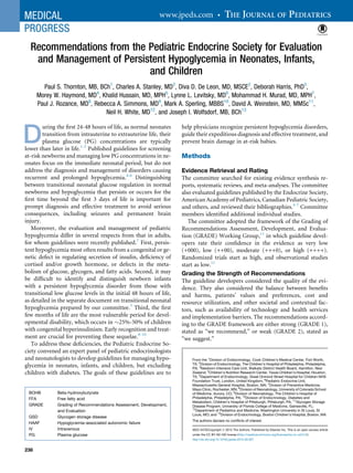
![Section 1: Which Neonates, Infants, and
Children to Evaluate for Hypoglycemia
1.1. For children who are able to communicate their symp-
toms, we recommend evaluation and management only of
those in whom Whipple’s triad (see below) is documented.
GRADE 1++++.
1.2. For infants and younger children who are unable to reli-
ably communicate symptoms, we suggest evaluation and
management only of those whose PG concentrations are
documented by laboratory quality assays to be below the
normal threshold for neurogenic responses (<60 mg/dL
[3.3 mmol/L]). GRADE 2+++0.
1.3. For those neonates who are suspected to be at high risk
of having a persistent hypoglycemia disorder, we suggest
evaluation when the infant is $48 hours of age so that the
period of transitional glucose regulation has passed and
persistent hypoglycemia may be excluded before discharge
home. GRADE 2++00.
Clinical Definition of Hypoglycemia
Clinical hypoglycemia is defined as a PG concentration low
enough to cause symptoms and/or signs of impaired brain
function.7
Hypoglycemia may be difficult to recognize
because the signs and symptoms are nonspecific, and a single
low PG concentration may be an artifact. For these reasons,
guidelines in adults emphasize the value of Whipple’s triad
for confirming hypoglycemia: symptoms and/or signs consis-
tent with hypoglycemia, a documented low PG concentra-
tion, and relief of signs/symptoms when PG concentration
is restored to normal. Young infants and children often
cannot dependably recognize and/or communicate their
symptoms, however; therefore, recognition of hypoglycemia
may require confirmation by repeated measurements of PG
concentration and formal testing. Nevertheless, suspected
hypoglycemia should be treated promptly to avoid potential
adverse consequences.
Hypoglycemia cannot be defined as a specific PG concen-
tration, because: (1) thresholds for specific brain responses to
hypoglycemia occur across a range of PG concentrations, and
these thresholds can be altered by the presence of alternative
fuels, such as ketones, and by recent antecedent hypoglyce-
mia; (2) it is not possible to identify a single PG value that
causes brain injury, and the extent of injury is influenced
by other factors, such as duration and degree of hypoglyce-
mia; and (3) potential artifacts and technical factors that
lead to inaccuracies in glucose determination may complicate
the interpretation of any single PG value.
Symptoms of Hypoglycemia
The symptoms of hypoglycemia reflect responses of the brain
to glucose deprivation and have been well delineated in
adults.12
Neurogenic (autonomic) symptoms result from
the perception of physiological changes caused by the sympa-
thetic nervous discharge triggered by hypoglycemia; these
include adrenergic responses (eg, palpitations, tremor,
anxiety) and cholinergic responses (eg, sweating, hunger,
paresthesias). Neuroglycopenic signs and symptoms,
including confusion, coma, and seizures, are caused by brain
dysfunction resulting from a deficient glucose supply to sus-
tain brain energy metabolism. Awareness of hypoglycemia
depends chiefly on perception of the central and peripheral
effects of neurogenic (as opposed to neuroglycopenic) re-
sponses to hypoglycemia. Brain glucose utilization becomes
limited at a PG concentration of approximately 55-65 mg/
dL (3.0-3.6 mmol/L).12
Neurogenic symptoms are perceived
at a PG concentration <55 mg/dL (<3.0 mmol/L), which in
older children and adults triggers a search for food or assis-
tance, an important defense against hypoglycemia. Cognitive
function is impaired (neuroglycopenia) at a PG concentra-
tion <50 mg/dL (<2.8 mmol/L).
Glucose Utilization
The adult brain accounts for more than one-half of total
glucose consumption. Because of their disproportionately
larger brain size relative to body mass, infants and young
children have a 2- to 3-fold higher glucose utilization rate
(4-6 mg/kg/min) per kilogram of body weight compared
with adults.13
Although the brain has an obligate requirement
for glucose, it also can use plasma ketones and lactate as
energy sources if the concentrations of these substances are
sufficiently elevated.14
However, in hypoketotic conditions,
such as hyperinsulinism or fatty acid oxidation disorders,
ketones and lactate are not available in sufficiently high
concentrations to substitute for glucose, and the risk of brain
energy failure is greater.
Neuroendocrine Defenses against Hypoglycemia
In normal individuals, the maintenance of normal PG
concentrations is highly protected. The first defense is sup-
pression of insulin secretion when PG concentration falls
below the normal postabsorptive mean of $85 mg/dL
(4.9 mmol/L).15
A further reduction of PG to 65-70 mg/dL
(3.6-3.9 mmol/L) elicits glucagon secretion and activation
of the sympathoadrenal system (reflected by increased
epinephrine concentration), which increases glucose release
from liver glycogen stores to raise the PG concentration. At
a PG concentration <65 mg/dL (3.6 mmol/L), levels of
plasma cortisol and growth hormone, important for mainte-
nance of glucose during prolonged fasting, increase as well.
Because the brain has only a few minutes worth of stored
fuel reserves in the form of glycogen,12
interruption of
glucose delivery can have devastating consequences. Whereas
recovery from brief periods of hypoglycemia is usually
complete, severe and prolonged hypoglycemia can cause
permanent brain injury.8-10,16
Metabolic Defenses against Hypoglycemia
In the postabsorptive phase, the liver supplies the brain
and other tissues with glucose by releasing glucose from
the breakdown of stored glycogen and by gluconeogenesis,
principally from gluconeogenic amino acids, such as
Vol. 167, No. 2 August 2015
239](data:image/gif;base64,R0lGODlhAQABAIAAAAAAAP///yH5BAEAAAAALAAAAAABAAEAAAIBRAA7)
Recommandé
Recommandé
Contenu connexe
Tendances
Tendances (20)
En vedette
En vedette (20)
Similaire à Recommendations for managing pediatric hypoglycemia
Similaire à Recommendations for managing pediatric hypoglycemia (20)
Plus de Hiperinsulinismo Congénito Argentina
Plus de Hiperinsulinismo Congénito Argentina (20)
Dernier
Dernier (20)
Recommendations for managing pediatric hypoglycemia
- 1. Recommendations from the Pediatric Endocrine Society for Evaluation and Management of Persistent Hypoglycemia in Neonates, Infants, and Children Paul S. Thornton, MB, BCh1 , Charles A. Stanley, MD2 , Diva D. De Leon, MD, MSCE2 , Deborah Harris, PhD3 , Morey W. Haymond, MD4 , Khalid Hussain, MD, MPH5 , Lynne L. Levitsky, MD6 , Mohammad H. Murad, MD, MPH7 , Paul J. Rozance, MD8 , Rebecca A. Simmons, MD9 , Mark A. Sperling, MBBS10 , David A. Weinstein, MD, MMSc11 , Neil H. White, MD12 , and Joseph I. Wolfsdorf, MB, BCh13 D uring the first 24-48 hours of life, as normal neonates transition from intrauterine to extrauterine life, their plasma glucose (PG) concentrations are typically lower than later in life.1-3 Published guidelines for screening at-risk newborns and managing low PG concentrations in ne- onates focus on the immediate neonatal period, but do not address the diagnosis and management of disorders causing recurrent and prolonged hypoglycemia.4-6 Distinguishing between transitional neonatal glucose regulation in normal newborns and hypoglycemia that persists or occurs for the first time beyond the first 3 days of life is important for prompt diagnosis and effective treatment to avoid serious consequences, including seizures and permanent brain injury. Moreover, the evaluation and management of pediatric hypoglycemia differ in several respects from that in adults, for whom guidelines were recently published.7 First, persis- tent hypoglycemia most often results from a congenital or ge- netic defect in regulating secretion of insulin, deficiency of cortisol and/or growth hormone, or defects in the meta- bolism of glucose, glycogen, and fatty acids. Second, it may be difficult to identify and distinguish newborn infants with a persistent hypoglycemia disorder from those with transitional low glucose levels in the initial 48 hours of life, as detailed in the separate document on transitional neonatal hypoglycemia prepared by our committee.3 Third, the first few months of life are the most vulnerable period for devel- opmental disability, which occurs in $25%-50% of children with congenital hyperinsulinism. Early recognition and treat- ment are crucial for preventing these sequelae.8-10 To address these deficiencies, the Pediatric Endocrine So- ciety convened an expert panel of pediatric endocrinologists and neonatologists to develop guidelines for managing hypo- glycemia in neonates, infants, and children, but excluding children with diabetes. The goals of these guidelines are to help physicians recognize persistent hypoglycemia disorders, guide their expeditious diagnosis and effective treatment, and prevent brain damage in at-risk babies. Methods Evidence Retrieval and Rating The committee searched for existing evidence synthesis re- ports, systematic reviews, and meta-analyses. The committee also evaluated guidelines published by the Endocrine Society, American Academy of Pediatrics, Canadian Pediatric Society, and others, and reviewed their bibliographies.4-7 Committee members identified additional individual studies. The committee adopted the framework of the Grading of Recommendations Assessment, Development, and Evalua- tion (GRADE) Working Group,11 in which guideline devel- opers rate their confidence in the evidence as very low (+000), low (++00), moderate (+++0), or high (++++). Randomized trials start as high, and observational studies start as low.11 Grading the Strength of Recommendations The guideline developers considered the quality of the evi- dence. They also considered the balance between benefits and harms, patients’ values and preferences, cost and resource utilization, and other societal and contextual fac- tors, such as availability of technology and health services and implementation barriers. The recommendations accord- ing to the GRADE framework are either strong (GRADE 1), stated as “we recommend,” or weak (GRADE 2), stated as “we suggest.” From the 1 Division of Endocrinology, Cook Children’s Medical Center, Fort Worth, TX; 2 Division of Endocrinology, The Children’s Hospital of Philadelphia, Philadelphia, PA; 3 Newborn Intensive Care Unit, Waikato District Health Board, Hamilton, New Zealand; 4 Children’s Nutrition Research Center, Texas Children’s Hospital, Houston, TX; 5 Department of Endocrinology, Great Ormond Street Hospital for Children NHS Foundation Trust, London, United Kingdom; 6 Pediatric Endocrine Unit, Massachusetts General Hospital, Boston, MA; 7 Division of Preventive Medicine, Mayo Clinic, Rochester, MN; 8 Division of Neonatology, University of Colorado School of Medicine, Aurora, CO; 9 Division of Neonatology, The Children’s Hospital of Philadelphia, Philadelphia, PA; 10 Division of Endocrinology, Diabetes and Metabolism, Children’s Hospital of Pittsburgh, Pittsburgh, PA; 11 Glycogen Storage Disease Program, University of Florida College of Medicine, Gainesville, FL; 12 Department of Pediatrics and Medicine, Washington University in St Louis, St Louis, MO; and 13 Division of Endocrinology, Boston Children’s Hospital, Boston, MA The authors declare no conflicts of interest. 0022-3476/Copyright ª 2015 The Authors. Published by Elsevier Inc. This is an open access article under the CC BY-NC-ND license (http://creativecommons.org/licenses/by-nc-nd/4.0/) http://dx.doi.org/10.1016/j.jpeds.2015.03.057 BOHB Beta-hydroxybutyrate FFA Free fatty acid GRADE Grading of Recommendations Assessment, Development, and Evaluation GSD Glycogen storage disease HAAF Hypoglycemia-associated autonomic failure IV Intravenous PG Plasma glucose 238
- 2. Section 1: Which Neonates, Infants, and Children to Evaluate for Hypoglycemia 1.1. For children who are able to communicate their symp- toms, we recommend evaluation and management only of those in whom Whipple’s triad (see below) is documented. GRADE 1++++. 1.2. For infants and younger children who are unable to reli- ably communicate symptoms, we suggest evaluation and management only of those whose PG concentrations are documented by laboratory quality assays to be below the normal threshold for neurogenic responses (<60 mg/dL [3.3 mmol/L]). GRADE 2+++0. 1.3. For those neonates who are suspected to be at high risk of having a persistent hypoglycemia disorder, we suggest evaluation when the infant is $48 hours of age so that the period of transitional glucose regulation has passed and persistent hypoglycemia may be excluded before discharge home. GRADE 2++00. Clinical Definition of Hypoglycemia Clinical hypoglycemia is defined as a PG concentration low enough to cause symptoms and/or signs of impaired brain function.7 Hypoglycemia may be difficult to recognize because the signs and symptoms are nonspecific, and a single low PG concentration may be an artifact. For these reasons, guidelines in adults emphasize the value of Whipple’s triad for confirming hypoglycemia: symptoms and/or signs consis- tent with hypoglycemia, a documented low PG concentra- tion, and relief of signs/symptoms when PG concentration is restored to normal. Young infants and children often cannot dependably recognize and/or communicate their symptoms, however; therefore, recognition of hypoglycemia may require confirmation by repeated measurements of PG concentration and formal testing. Nevertheless, suspected hypoglycemia should be treated promptly to avoid potential adverse consequences. Hypoglycemia cannot be defined as a specific PG concen- tration, because: (1) thresholds for specific brain responses to hypoglycemia occur across a range of PG concentrations, and these thresholds can be altered by the presence of alternative fuels, such as ketones, and by recent antecedent hypoglyce- mia; (2) it is not possible to identify a single PG value that causes brain injury, and the extent of injury is influenced by other factors, such as duration and degree of hypoglyce- mia; and (3) potential artifacts and technical factors that lead to inaccuracies in glucose determination may complicate the interpretation of any single PG value. Symptoms of Hypoglycemia The symptoms of hypoglycemia reflect responses of the brain to glucose deprivation and have been well delineated in adults.12 Neurogenic (autonomic) symptoms result from the perception of physiological changes caused by the sympa- thetic nervous discharge triggered by hypoglycemia; these include adrenergic responses (eg, palpitations, tremor, anxiety) and cholinergic responses (eg, sweating, hunger, paresthesias). Neuroglycopenic signs and symptoms, including confusion, coma, and seizures, are caused by brain dysfunction resulting from a deficient glucose supply to sus- tain brain energy metabolism. Awareness of hypoglycemia depends chiefly on perception of the central and peripheral effects of neurogenic (as opposed to neuroglycopenic) re- sponses to hypoglycemia. Brain glucose utilization becomes limited at a PG concentration of approximately 55-65 mg/ dL (3.0-3.6 mmol/L).12 Neurogenic symptoms are perceived at a PG concentration <55 mg/dL (<3.0 mmol/L), which in older children and adults triggers a search for food or assis- tance, an important defense against hypoglycemia. Cognitive function is impaired (neuroglycopenia) at a PG concentra- tion <50 mg/dL (<2.8 mmol/L). Glucose Utilization The adult brain accounts for more than one-half of total glucose consumption. Because of their disproportionately larger brain size relative to body mass, infants and young children have a 2- to 3-fold higher glucose utilization rate (4-6 mg/kg/min) per kilogram of body weight compared with adults.13 Although the brain has an obligate requirement for glucose, it also can use plasma ketones and lactate as energy sources if the concentrations of these substances are sufficiently elevated.14 However, in hypoketotic conditions, such as hyperinsulinism or fatty acid oxidation disorders, ketones and lactate are not available in sufficiently high concentrations to substitute for glucose, and the risk of brain energy failure is greater. Neuroendocrine Defenses against Hypoglycemia In normal individuals, the maintenance of normal PG concentrations is highly protected. The first defense is sup- pression of insulin secretion when PG concentration falls below the normal postabsorptive mean of $85 mg/dL (4.9 mmol/L).15 A further reduction of PG to 65-70 mg/dL (3.6-3.9 mmol/L) elicits glucagon secretion and activation of the sympathoadrenal system (reflected by increased epinephrine concentration), which increases glucose release from liver glycogen stores to raise the PG concentration. At a PG concentration <65 mg/dL (3.6 mmol/L), levels of plasma cortisol and growth hormone, important for mainte- nance of glucose during prolonged fasting, increase as well. Because the brain has only a few minutes worth of stored fuel reserves in the form of glycogen,12 interruption of glucose delivery can have devastating consequences. Whereas recovery from brief periods of hypoglycemia is usually complete, severe and prolonged hypoglycemia can cause permanent brain injury.8-10,16 Metabolic Defenses against Hypoglycemia In the postabsorptive phase, the liver supplies the brain and other tissues with glucose by releasing glucose from the breakdown of stored glycogen and by gluconeogenesis, principally from gluconeogenic amino acids, such as Vol. 167, No. 2 August 2015 239
- 3. alanine, and recycled lactate. With longer fasting and further suppression of insulin secretion, glucose utilization is restricted to the brain and a few glycolytic tissues, such as erythrocytes. Adipose tissue lipolysis releases glycerol, a gluconeogenic substrate, and free fatty acids (FFAs) that can replace glucose as an energy substrate in skeletal and heart muscle, but not in brain. FFAs are also converted by the liver to beta-hydroxybutyrate (BOHB) and acetoa- cetate for use by the brain. BOHB is the predominant ke- toacid, and its plasma level serves as a measure of ketogenesis. As ketoacid concentrations rise, they can partly support the brain’s energy needs. The changes in fuel metabolism during fasting in normal neonates after 2-3 days of age and in infants and children do not differ substantially from those in adults, except that PG concen- trations decrease more rapidly and hyperketonemia de- velops sooner, because of the energy needs of their relatively larger brains.17 Measurement of BOHB, FFA, and lactate at the time of hypoglycemia provides impor- tant information for diagnosing the cause of hypoglycemia (Figure). Altered Hypoglycemia Awareness Previous exposure to an episode of hypoglycemia can blunt, and repeated episodes can eliminate, neurogenic responses to subsequent hypoglycemic episodes.18 This leads to reduced or absent awareness of hypoglycemia and impairs hepatic glucose release, perpetuating hypoglycemia. This combination of events has been termed hypoglycemia- associated autonomic failure (HAAF).18 HAAF can persist for 24 hours after a single episode of hypoglycemia or even longer after repeated episodes of hypoglycemia. A similar impairment in neuroendocrine responses to hypo- glycemia also occurs during sleep and exercise.18 Thus, exposure to recurrent hypoglycemia can shift the usual glucose threshold for recognition of neurogenic symptoms of 55 mg/dL (3.0 mmol/L) to a lower level. Although previ- ous exposure to hypoglycemia lowers the glucose threshold for neurogenic responses, the threshold for neuroglycopenic symptoms is not altered acutely; whether adaptation occurs with repeated exposure to hypoglycemia is unknown. Fea- tures of HAAF have been demonstrated in infants as young as age 10-13 weeks.19 Potential Artifacts in Measurements of PG Concentration To diagnose hypoglycemia, PG concentration should be measured using a clinical laboratory method.12 Important considerations are that whole blood glucose values are $15% lower than PG concentrations, and that because of red cell glycolysis, delays in processing and assaying glucose can reduce the glucose concentration by up to 6 mg/dL/ hour (0.3 mmol/L/hour). Point-of-care meters provide a convenient screening method for detecting hypoglycemia, Metabolic Clues to Hypoglycemia Diagnosis Hypoglycemia HCO3 , BOHB , Lactate, FFA No Acidemia Acidemia BOHB↓ FFA ↑ OHB Acidem BOHB Gluconeogenesis Defects Fa y Acid Oxida on Defects Gene c Hyperinsulinism Hypopituitarism in newborns Transi onal Neonatal Hypoglycemia Perinatal Stress Hyperinsulinism BOHB↑BOHB↓ FFA ↓ Lactate ↑ Keto c Hypoglycemia Glycogenoses GH deficiency Cor sol deficiency Figure. Algorithm showing how the major categories of hypoglycemia can be determined with information from the critical sample. GH, growth hormone. THE JOURNAL OF PEDIATRICS www.jpeds.com Vol. 167, No. 2 240 Thornton et al
- 4. but their accuracy is limited to approximately Æ10-15 mg/dL (0.6-0.8 mmol/L) in the range of hypoglycemia. Therefore, before establishing a diagnosis of hypoglycemia in neonates, infants, and children, it is essential to confirm low PG con- centration using a clinical laboratory method. Normal PG Concentrations in Neonates Aged 48 Hours, Infants, and Children After the first 48 hours of life, PG concentration and the physiology of glucose homeostasis do not differ to any great extent with age. Mean PG concentration in the postabsorp- tive state in normal neonates after $2 days of age and in infants and children does not differ from that in adults (70-100 mg/dL [3.9-5.5 mmol/L])17,20,21 ; however, children under age 4 years may have a PG concentration 70 mg/dL (3.9 mmol/L) and hyperketonemia after overnight fasting because of limited fasting tolerance.17,20,21 Other evidence that the normal PG concentration in children does not differ from that in adults include the following: (1) fasting hyper- ketonemia develops at a similar PG concentration in infants, children, and adults21,22 (hyperketonemia does not occur during transitional neonatal hypoglycemia in normal new- borns in the first 1-2 days of life3 ); (2) plasma lactate rises as PG falls below $70 mg/dL ($3.9 mmol/L) in children with glucose-6-phosphatase deficiency23 ; and (3) lethargy and tachycardia are observed in patients with defects of fatty acid oxidation when PG decreases to 60-70 mg/dL (3.3- 3.9 mmol/L). PG Concentrations in Neonates Aged 48 Hours In normal newborn infants, PG concentration commonly de- creases immediately after birth to levels below those in older infants and children. The interpretation and response to PG concentration during the first 2 days of life have been contro- versial.24 Whether the brain of newborn infants has greater or lesser susceptibility to hypoglycemic injury is controversial as well.25,26 As discussed previously,3 the committee’s review of available data on transitional neonatal hypoglycemia in normal newborns (ie, hypoketonemic hypoglycemia with inappropriately large glycemic responses to glucagon or epinephrine stimulation) suggests that it is a mild and tran- sient form of hyperinsulinism in which the mean PG threshold for suppression of insulin secretion is $55- 65 mg/dL (3.0-3.6 mmol/L) shortly after birth, compared with $80-85 mg/dL (4.4-4.7 mmol/L) in older infants, chil- dren, and adults. As the glucose stimulated-insulin secretion mechanism matures, mean PG concentration in normal new- borns increases and by 72 hours of age is similar to those in older infants and children1,2 ; therefore, the standards for normal neonates must never be extrapolated beyond 2-3 days after birth. Because of the difficulty in distinguishing a suspected persistent hypoglycemia disorder from transi- tional neonatal glucose concentrations during the first 48 hours of life, we suggest delaying diagnostic evaluations until 2-3 days after birth. Neonates Aged 48 Hours Who May Be at Risk for Persistent Hypoglycemia Disorders Some neonates can be identified by various clinical features as being at high risk for severe hypoglycemia during the first 48 hours after delivery,16,26 and a subset of those neonates are also at increased risk for persistent hypoglycemia beyond 48 hours of life (Table).27-30 These include not only the rare infants with genetic hypoglycemia disorders, such as congenital hyperinsulinism or hypopituitarism,31 but also those with relatively more common prolonged neonatal hyperinsulinism (also referred to as perinatal stress hyperinsulinism) associated with birth asphyxia, intrauterine growth restriction, or toxemia.27,28,30 To provide a practical guide and in the light of scarce ev- idence in this area, the committee used a consensus process to make the following technical suggestions: For neonates with a known risk of a genetic or other persis- tent form of hypoglycemia (eg, congenital hyperinsulinism, glucose-6-phosphatase deficiency, fatty acid oxidation disor- der), consultation with a specialist should be considered before planning discharge from the nursery. Diagnostic tests may be available to exclude the possibility of a specific disor- der within a few weeks (eg, genetic mutation analysis, plasma acyl-carnitine profile). In these cases, a “safety” fasting test Table. Recognizing and managing neonates at increased risk for a persistent hypoglycemia disorder Neonates at increased risk of hypoglycemia and require glucose screening: 1. Symptoms of hypoglycemia 2. Large for gestational age (even without maternal diabetes) 3. Perinatal stress a. Birth asphyxia/ischemia; cesarean delivery for fetal distress b. Maternal preeclampsia/eclampsia or hypertension c. Intrauterine growth restriction (small for gestational age) d. Meconium aspiration syndrome, erythroblastosis fetalis, polycythemia, hypothermia 4. Premature or postmature delivery 5. Infant of diabetic mother 6. Family history of a genetic form of hypoglycemia 7. Congenital syndromes (eg, Beckwith-Wiedemann), abnormal physical features (eg, midline facial malformations, microphallus) Neonates in whom to exclude persistent hypoglycemia before discharge: 1. Severe hypoglycemia (eg, episode of symptomatic hypoglycemia or need for IV dextrose to treat hypoglycemia) 2. Inability to consistently maintain preprandial PG concentration 50 mg/dL up to 48 hours of age and 60 mg/dL after 48 hours of age 3. Family history of a genetic form of hypoglycemia 4. Congenital syndromes (eg, Beckwith-Wiedemann), abnormal physical features (eg, midline facial malformations, microphallus) August 2015 MEDICAL PROGRESS Recommendations from the Pediatric Endocrine Society for Evaluation and Management of Persistent Hypoglycemia in Neonates, Infants, and Children 241
- 5. should be done before discharge from the nursery to ensure that PG concentration can be maintained above 70 mg/dL if a feeding is missed (ie, a minimum of 6-8 hours). If no definitive test is available (which is often the case with hyper- insulinism), then the fasting test should be extended to exclude diagnostic features of the suspected disorder following the paradigm shown in the Figure. For at-risk neonates without a suspected persistent hypo- glycemia disorder and in whom hypoglycemia is considered likely to resolve within a short time, a “safety” fast of 6-8 hours should be considered before discharge, to determine whether a PG concentration 60 mg/dL can be maintained, or whether additional management or investiga- tions may be required. Values Hypoglycemia disorders in older infants and children are un- common. Thus, the recommendation to require the presence of Whipple’s triad before undertaking an evaluation avoids exposing patients without a specific disorder to unnecessary procedures, potential risks, and expense without any likely benefit. For neonates, infants, and younger children in whom Whipple’s triad cannot be applied, unnecessary inves- tigation can be avoided by confirming a PG concentration at or below the range at which neurogenic symptoms usually occur. The recommendations regarding which neonates to evaluate for hypoglycemia focus on those high-risk neonates who require close monitoring and/or intervention, such as intravenous (IV) dextrose, for treatment of hypoglycemia (Table). They take into account that transitional hypoglycemia in normal newborns should be completely resolved before the usual time for discharge from the nursery at 2-3 days. The committee emphasizes the necessity of carefully excluding persistent hypoglycemia disorders before discharge to home in the small subset of high-risk neonates (Table) to ensure recognition and facilitate treatment. Section 2. Workup/Investigation of Persistent Hypoglycemia 2.1. We recommend that investigations be carried out to di- agnose the underlying mechanism of persistent hypoglyce- mia disorders to provide specific management. GRADE 1++++. In patients with hypoglycemia, a thorough history should include the episode’s timing and its relationship to food, birth weight, gestational age, and family history. Physical ex- amination should include looking for evidence of hypopitu- itarism (eg, micropenis or cleft lip or palate, short stature), glycogenosis (eg, hepatomegaly), adrenal insufficiency (eg, recurrent abdominal pain, hyperpigmentation, anorexia, weight loss), or Beckwith-Wiedemann syndrome (eg, om- phalocele, hemihypertrophy, macroglossia). Whenever possible, specimens (a “critical sample”) for identifying the etiology of hypoglycemia should be obtained at the time of spontaneous presentation and before treatment that may rapidly alter the levels of BOHB, FFA, and insulin. Assays for PG, bicarbonate, BOHB, and lactate are readily available and useful for distinguishing categories of hypogly- cemia disorders (Figure). Extra plasma can be held in reserve for specific tests (eg, plasma insulin, FFA, and C-peptide for suspected hyperinsulinism; plasma total and free carnitine and acyl-carnitine profile for a suspected disorder of fatty acid oxidation). Laboratory reference ranges for some of these tests (eg, insulin, BOHB) may be inappropriate for interpreting values obtained during childhood hypoglycemia. In the absence of a “critical sample,” a provocative fasting test is the most informative method for identifying the etiol- ogy of hypoglycemia disorders.20,32 The duration of fasting and the specimens obtained should be tailored to the sus- pected diagnosis. With few exceptions, blood tests performed while glucose concentrations are normal are not informative. For safety, a fasting test should be done with frequent moni- toring of vital signs, PG, and BOHB concentrations. A PG concentration of 50 mg/dL (2.8 mmol/L) is sufficiently low to elicit the metabolic and neuroendocrine responses required for diagnosis. After appropriate specimens have been obtained, the fast may be terminated. The fast also can be stopped earlier for signs or symptoms of distress or a plasma BOHB measurement of 2.5 mmol/L. For suspected hyperinsulinism, the fasting test can be terminated when the PG concentration is 50 mg/dL (2.8 mmol/L) with administration of glucagon (1 mg IV, intramuscularly, or subcutaneously) to evaluate the glycemic response. An exaggerated glycemic response (30 mg/dL [1.7 mmol/L]) is nearly pathognomonic of hyperinsulin- ism.33 Because plasma insulin concentration is sometimes not above the lower limit of detection,34 it is important to include the following tests when assessing the possibility of hypoglycemia due to hyperinsulinism: plasma BOHB and FFA (both inappropriately low; BOHB 1.5 mmol/L [15 mg/dL] and FFA 1.0-1.5 mmol/L [28-42 mg/dL]), and an increased glycemic response to glucagon. Based on the suspected diagnosis, additional tests performed on spec- imens obtained at the time that fasting is terminated should be considered, such as growth hormone, cortisol, total and free carnitine, acyl-carnitine profile, C-peptide, proinsulin, and drug screening. Adrenal insufficiency, congenital or acquired, may mani- fest with hypoglycemia. Simultaneous measurement of plasma adrenocorticotropic hormone and cortisol is recom- mended if primary adrenal insufficiency is suspected. An ad- renocorticotropic hormone stimulation test may be needed to confirm suspected secondary or tertiary adrenal insuffi- ciency. The possibility of drug administration (accidental or sur- reptitious) or a toxin may need to be considered. Specimens of plasma and urine obtained at the time of an acute episode may be informative. High plasma insulin level but low C-peptide level during hypoglycemia suggest exogenous in- sulin administration. The drugs most commonly associated with hypoglycemia include insulin, sulphonylureas, alcohol, THE JOURNAL OF PEDIATRICS www.jpeds.com Vol. 167, No. 2 242 Thornton et al
- 6. beta-blockers, and salicylates. Not all commercially available insulin assays measure the insulin analogues; thus, the finding of low insulin and C-peptide levels at time of hypo- glycemia might not rule out surreptitious insulin administra- tion. (Consultation with the testing laboratory should be considered if clinical suspicion is high.) Postprandial hypoglycemia in infants and children is usu- ally a complication of fundoplication for gastroesophageal reflux (incidence 25%-30%) or gastric bypass bariatric sur- gery for morbid obesity. In these patients, a meal may trigger hypoglycemia 1-3 hours later. Postprandial hypoglycemia is often preceded by exaggerated glucose, glucagon-like pep- tide-1, and insulin responses, which can be detected on an oral glucose or mixed-meal tolerance test.35 Values Failure to investigate a neonate, infant, or child with sus- pected hypoglycemia increases the risk of delaying a defini- tive diagnosis and instituting effective treatment. Expeditiously identifying the specific cause of hypoglycemia, as outlined above, will enable prompt institution of appro- priate treatment and decrease the risk of permanent brain injury from persistent and recurrent severe hypoglycemia. The decision to subject an infant or child to a diagnostic investigation for suspected hypoglycemia, which includes a monitored assessment of fasting adaptation that may expose the patient to the risk of another episode of hypoglycemia, places a higher value on achieving diagnostic certainty and a lower value on avoiding the discomfort, inconvenience, and cost of such procedures. Section 3. Management of Neonates, Infants, and Children with a Persistent Hypoglycemia Disorder 3.1. For neonates with a suspected congenital hypoglycemia disorder and older infants and children with a confirmed hy- poglycemia disorder, we recommend that the goal of treat- ment be to maintain a PG concentration 70 mg/dL (3.9 mmol/L). GRADE 1++00. 3.2. For high-risk neonates without a suspected congenital hypoglycemia disorder, we suggest the goal of treatment be to maintain a PG concentration 50 mg/dL (2.8 mmol/L) for those aged 48 hours and 60 mg/dL (3.3 mmol/L) for those aged 48 hours. GRADE 2+000. 3.3. We recommend an individualized approach to man- agement with treatment tailored to the specific disorder, tak- ing into account patient safety and family preferences. Ungraded best practice statement. Neonates, Infants, and Children with Hypoglycemia Disorders Neurogenic and neuroglycopenic symptoms usually occur when the PG concentration decreases to 50-70 mg/dL (2.8-3.9 mmol/L). Recurrent PG levels in this range may result in the development of HAAF, which increases the risk of subsequent hypoglycemia and attenuates its recogni- tion.7,36 Therefore, treatment targets are generally aimed at avoiding activation of neuroendocrine responses and HAAF by maintaining PG concentration within the normal range of 70-100 mg/dL (3.9-5.6 mmol/L).7 For this reason, we recommend that the same goal for treatment of adults and children with diabetes (ie, PG 70 mg/dL [3.9 mmol/ L]) be used for neonates with a confirmed hypoglycemia dis- order and for infants and children with a persistent hypogly- cemia disorder. For disorders such as hyperinsulinism, the aim is to pre- vent recurrent hypoglycemia that increases the risk of subse- quent, possibly unrecognized, hypoglycemic episodes. For disorders such as defects in glycogen metabolism and gluco- neogenesis, maintenance of PG concentration in the normal range prevents metabolic acidosis and growth failure, and possibly the development of long-term complications. Any episode of severe symptomatic hypoglycemia should be rapidly corrected with IV dextrose infusion. The initial dose is 200 mg/kg, followed by infusion of 10% dextrose at a maintenance rate for age. In cases of hyperinsulinism, glucagon can be expected to raise PG concentration to normal or above within 10-15 minutes and to maintain that concentration for at least 1 hour. Doses of 0.5-1.0 mg (independent of weight), given IV, intramuscularly, or sub- cutaneously, are usually effective; lower doses (0.03 mg/kg) may carry less risk of transient nausea and vomiting, but may be ineffective unless given IV. Long-term therapy for hypoglycemia disorders should be based on the specific etiology of the disorder, in consultation with a physician experienced in the diagnosis and manage- ment of hypoglycemia in infants and children and with care- ful consideration of patient and family preferences. Medications are available for some disorders, such as hyper- insulinism, and for cortisol and growth hormone deficiency. Surgery may be necessary in some children with hyperinsu- linism who are unable to maintain PG concentration in a safe range through medical therapy. Nutritional therapy is the cornerstone of treatment for some disorders, such as dis- orders of glycogen metabolism or hereditary fructose intoler- ance. Some milder disorders may be treated adequately by avoidance of prolonged fasting. Patients diagnosed with ketotic hypoglycemia who have recurrent episodes should be reevaluated for other specific disorders, such as glycogen synthase deficiency (GSD 0), GSD type VI, GSD type IX (especially in males), and MCT1 gene deficiency.37,38 Nonspecific treatment with glucocorticoids for neonates with hypoglycemia is discouraged. High-Risk Neonates without a Suspected Congenital Hypoglycemia Disorder The mean PG concentration in normal newborns during the first hours after birth ($55-60 mg/dL [$3 mmol/L]) is similar to the threshold for neurogenic symptoms of hypo- glycemia in older children and adults, but above the threshold for neuroglycopenic symptoms. This appears to August 2015 MEDICAL PROGRESS Recommendations from the Pediatric Endocrine Society for Evaluation and Management of Persistent Hypoglycemia in Neonates, Infants, and Children 243
- 7. be tolerated during the first few hours after birth. In normal newborns, the mean PG concentration rises to 70 mg/dL (3.9 mmol/L) after 48 hours of age.1 This is similar to the mean glucose concentration in older children and adults. There is no evidence that the concentration at which PG be- comes limiting for brain function is different in neonates than in older children and adults. Of concern, as noted pre- viously,3 plasma ketone levels are suppressed during hypogly- cemia in high-risk neonates, making them particularly vulnerable to hypoglycemia-induced brain damage, and there is no assurance the plasma lactate level will be suffi- ciently elevated to compensate for low glucose.27,28,30,39 Given the absence of evidence on the short-term or long- term consequences of different treatment targets, the com- mittee focused on evidence related to physiology, etiology, and mechanism, and balanced the risks and benefits of in- terventions. The committee also focused on postnatal age, because ongoing hypoglycemia beyond the first 48 hours of life (and particularly beyond the first week) increases the concern for an underlying hypoglycemia disorder, such as prolonged neonatal hyperinsulinism,30 or a genetic disorder. The committee’s consensus was that during the first 48 hours of life, for a high-risk neonate without a sus- pected congenital hypoglycemia disorder and on normal feedings, a safe target PG concentration should be close to the mean for healthy newborns on the first day of life and above the threshold for neuroglycopenic symptoms (50 mg/dL [2.8 mmol/L]). The committee recommended raising the glucose target after 48 hours of age (60 mg/ dL [3.3 mmol/L]) to above the threshold for neurogenic symptoms and close to the target for older infants and chil- dren, because of the increased concern for an underlying hypoglycemia disorder. The suggested target glucose con- centrations were considered adequate while assessing whether hypoglycemia will resolve over time. Because at- risk neonates who require interventions beyond normal feedings, such as IV dextrose, to treat hypoglycemia in the first 48 hours of life are likely to have more severe and pro- longed hypoglycemia, the initial treatment target should be above the thresholds for both neurogenic and neuroglyco- penic symptoms (60 mg/dL [3.3 mmol/L]). For neonates who require dextrose infusion, transitioning to normal feedings alone can be attempted once PG concentration is stabilized at 60 mg/dL (3.3 mmol/L). The committee’s consensus to accept a lower PG target in high-risk neonates without a suspected congenital hypogly- cemia disorder (Section 3.2) than in those with a suspected congenital hypoglycemia disorder (Section 3.1) was based on balancing the risks of intervention compared with a brief period of undertreatment in this group. Higher treatment targets (Section 3.1) should be considered for those with a suspected genetic hypoglycemia disorder and for symptom- atic neonates, because the risks of undertreatment outweigh those of overtreatment in these patients. n Submitted for publication Aug 8, 2014; last revision received Mar 3, 2015; accepted Mar 31, 2015. Reprint requests: Paul S. Thornton, MD, Cook Children’s Medical Center, 1500 Cooper St, Second Floor, Fort Worth, TX 76104. E-mail: Paul.thornton@ cookchildrens.org References 1. Cornblath M, Reisner SH. Blood glucose in the neonate and its clinical significance. N Engl J Med 1965;273:378-81. 2. Srinivasan G, Pildes RS, Cattamanchi G, Voora S, Lilien LD. Plasma glucose values in normal neonates: a new look. J Pediatr 1986;109:114-7. 3. Stanley CA, Rozance PJ, Thornton PS, De Leon DD, Harris D, Haymond MW, et al. Re-evaluating “transitional neonatal hypoglycemia”: mechanism and implications for management. J Pediatr 2015;166:1520-5. 4. Canadian Paediatric Society. Screening guidelines for newborns at risk for low blood glucose. Paediatr Child Health 2004;9:723-40. 5. Adamkin DH. Postnatal glucose homeostasis in late-preterm and term infants. Pediatrics 2011;127:575-9. 6. Wight N, Marinelli KA. ABM clinical protocol 1: guidelines for glucose monitoring and treatment of hypoglycemia in breastfed neonates. Breastfeed Med 2006;1:178-84. 7. Cryer PE, Axelrod L, Grossman AB, Heller SR, Montori VM, Seaquist ER, et al. Evaluation and management of adult hypoglycemic disorders: an Endocrine Society clinical practice guideline. J Clin Endo- crinol Metab 2009;94:709-28. 8. Menni F, de Lonlay P, Sevin C, Touati G, Peigne C, Barbier V, et al. Neurologic outcomes of 90 neonates and infants with persistent hyper- insulinemic hypoglycemia. Pediatrics 2001;107:476-9. 9. Steinkrauss L, Lipman TH, Hendell CD, Gerdes M, Thornton PS, Stanley CA. Effects of hypoglycemia on developmental outcome in chil- dren with congenital hyperinsulinism. J Pediatr Nurs 2005;20:109-18. 10. Meissner T, Wendel U, Burgard P, Schaetzle S, Mayatepek E. Long-term follow-up of 114 patients with congenital hyperinsulinism. Eur J Endo- crinol 2003;149:43-51. 11. Atkins D, Best D, Briss PA, Eccles M, Falck-Ytter Y, Flottorp S, et al. Grading quality of evidence and strength of recommendations. BMJ 2004;328:1490. 12. Cryer PE. Hypoglycemia in diabetes:Pathophysiology,prevalence,and pre- vention. 2nd ed. Alexandria (VA): American Diabetes Association; 2013. 13. Bier DM, Leake RD, Haymond MW, Arnold KJ, Gruenke LD, Sperling MA, et al. Measurement of “true” glucose production rates in infancy and childhood with 6,6-dideuteroglucose. Diabetes 1977;26: 1016-23. 14. Veneman T, Mitrakou A, Mokan M, Cryer P, Gerich J. Effect of hyper- ketonemia and hyperlacticacidemia on symptoms, cognitive dysfunc- tion, and counterregulatory hormone responses during hypoglycemia in normal humans. Diabetes 1994;43:1311-7. 15. Cryer PE. Hypoglycemia, functional brain failure, and brain death. J Clin Invest 2007;117:868-70. 16. Koivisto M, Blanco-Sequeiros M, Krause U. Neonatal symptomatic and asymptomatic hypoglycaemia: a follow-up study of 151 children. Dev Med Child Neurol 1972;14:603-14. 17. Chaussain JL, Georges P, Calzada L, Job JC. Glycemic response to 24- hour fast in normal children, III: influence of age. J Pediatr 1977;91: 711-4. 18. Cryer PE. Diverse causes of hypoglycemia-associated autonomic failure in diabetes. N Engl J Med 2004;350:2272-9. 19. Hussain K, Bryan J, Christesen HT, Brusgaard K, Aguilar-Bryan L. Serum glucagon counterregulatory hormonal response to hypoglycemia is blunted in congenital hyperinsulinism. Diabetes 2005;54:2946-51. 20. Bonnefont JP, Specola NB, Vassault A, Lombes A, Ogier H, de Klerk JB, et al. The fasting test in paediatrics: application to the diagnosis of path- ological hypo- and hyperketotic states. Eur J Pediatr 1990;150:80-5. 21. van Veen MR, van Hasselt PM, de Sain-van der Velden MG, Verhoeven N, Hofstede FC, de Koning TJ, et al. Metabolic profiles in children during fasting. Pediatrics 2011;127:e1021-7. 22. Saudubray JM, Marsac C, Limal JM, Dumurgier E, Charpentier C, Ogier H, et al. Variation in plasma ketone bodies during a 24-hour THE JOURNAL OF PEDIATRICS www.jpeds.com Vol. 167, No. 2 244 Thornton et al
- 8. fast in normal and in hypoglycemic children: relationship to age. J Pe- diatr 1981;98:904-8. 23. Wolfsdorf JI, Plotkin RA, Laffel LM, Crigler JF Jr. Continuous glucose for treatment of patients with type 1 glycogen-storage disease: compar- ison of the effects of dextrose and uncooked cornstarch on biochemical variables. Am J Clin Nutr 1990;52:1043-50. 24. Hay WW Jr, Raju TN, Higgins RD, Kalhan SC, Devaskar SU. Knowledge gaps and research needs for understanding and treating neonatal hypo- glycemia: workshop report from the Eunice Kennedy Shriver National Institute of Child Health and Human Development. J Pediatr 2009; 155:612-7. 25. Kim M, Yu ZX, Fredholm BB, Rivkees SA. Susceptibility of the devel- oping brain to acute hypoglycemia involving A1 adenosine receptor activation. Am J Physiol Endocrinol Metab 2005;289:E562-9. 26. Vannucci RC, Vannucci SJ. Hypoglycemic brain injury. Semin Neonatol 2001;6:147-55. 27. Collins JE, Leonard JV. Hyperinsulinsim in asphyxiated and small- for-dates infants with hypoglycaemia. Lancet 1984;2:311-3. 28. Collins JE, Leonard JV, Teale D, Marks V, Williams DM, Kennedy CR, et al. Hyperinsulinaemic hypoglycaemia in small for dates babies. Arch Dis Child 1990;65:1118-20. 29. Hume R, McGeechan A, Burchell A. Failure to detect preterm infants at risk of hypoglycemia before discharge. J Pediatr 1999;134:499-502. 30. Hoe FM, Thornton PS, Wanner LA, Steinkrauss L, Simmons RA, Stanley CA. Clinical features and insulin regulation in infants with a syndrome of prolonged neonatal hyperinsulinism. J Pediatr 2006;148: 207-12. 31. Mohamed Z, Arya VB, Hussain K. Hyperinsulinaemic hypoglycaemia: genetic mechanisms, diagnosis and management. J Clin Res Pediatr Endocrinol 2012;4:169-81. 32. Morris AA, Thekekara A, Wilks Z, Clayton PT, Leonard JV, Aynsley- Green A. Evaluation of fasts for investigating hypoglycaemia or suspected metabolic disease. Arch Dis Child 1996;75:115-9. 33. Finegold DN, Stanley CA, Baker L. Glycemic response to glucagon dur- ing fasting hypoglycemia: an aid in the diagnosis of hyperinsulinism. J Pediatr 1980;96:257-9. 34. De Leon DD, Stanley CA. Determination of insulin for the diagnosis of hyperinsulinemic hypoglycemia. Best Pract Res Clin Endocrinol Metab 2013;27:763-9. 35. Calabria AC, Gallagher PR, Simmons R, Blinman T, De Leon DD. Post- operative surveillance and detection of postprandial hypoglycemia after fundoplasty in children. J Pediatr 2011;159:597-601.e1. 36. Clarke W, Jones T, Rewers A, Dunger D, Klingensmith GJ. Assessment and management of hypoglycemia in children and adolescents with dia- betes. Pediatr Diabetes 2009;10(Suppl 12):134-45. 37. Wolfsdorf JI, Weinstein DA. Glycogen storage diseases. Rev Endocr Metab Disord 2003;4:95-102. 38. van Hasselt PM, Ferdinandusse S, Monroe GR, Ruiter JP, Turkenburg M, Geerlings MJ, et al. Monocarboxylate transporter 1 defi- ciency and ketone utilization. N Engl J Med 2014;371:1900-7. 39. Harris DL, Weston PJ, Williams CE, Pleasants AB, Battin MR, Spooner CG, et al. Cot-side electroencephalography monitoring is not clinically useful in the detection of mild neonatal hypoglycemia. J Pediatr 2011;159:755-60.e1. August 2015 MEDICAL PROGRESS Recommendations from the Pediatric Endocrine Society for Evaluation and Management of Persistent Hypoglycemia in Neonates, Infants, and Children 245