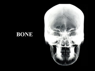
Bones (2)
•
5 j'aime•1,225 vues

The Indian Dental Academy is the Leader in continuing dental education , training dentists in all aspects of dentistry and offering a wide range of dental certified courses in different formats.for more details please visit www.indiandentalacademy.com

Recommandé
Contenu connexe
Tendances
Tendances (20)
Similaire à Bones (2)
Similaire à Bones (2) (20)
Plus de Indian dental academy
Plus de Indian dental academy (20)
Dernier
Dernier (20)
Bones (2)
- 2. Bones are the units of the skeletal system. www.indiandentalacademy.com
- 3. Classification of bone tissues 1. Shape of bones • Long,short,flat,irregular,pneumatic, sesamoid,supernumerary 2. Regions of long bone • Epiphysis • Diaphysis • Metaphysis 3. Macroscopic appearance • Compact bone • Trabecular/cancellous/spongy bone 4. Developmental origin • Intramembranous/dermal bone • Intracartilaginous/endochondral bone www.indiandentalacademy.com
- 5. Histology of bone • Periosteum White fibrous layer Elastic osteogenic layer www.indiandentalacademy.com
- 6. Histology of bone • Haversian system Haversian canals Volkmann’s canals Lamellae Lacunae Canaliculli www.indiandentalacademy.com
- 7. Histology of bone • Types of lamellae: Osteonic Interstitial Circumferential www.indiandentalacademy.com
- 8. High power view of part of an osteon in transverse section seen with transmitted light. www.indiandentalacademy.com
- 9. Single osteon viewed with polarization optics to illustrate lamellar structurewww.indiandentalacademy.com
- 10. Histological sections of bone illustrating the various kinds of lamellaewww.indiandentalacademy.com
- 11. Osteocyte in lacuna.Arrow shows canalliculi.www.indiandentalacademy.com
- 12. Histogenesis of bone • Intramembranous & Endochondral ossification. • Histologically both bones are identical. • Both are formed from mesenchymal tissue. www.indiandentalacademy.com
- 13. Intramembranous bone formation A. Condensation of mesenchyme around capillary network at specific points-Centres of ossification B. Cells converted into osteoblasts • Open faced • nucleus,prominent nucleolus • RER & Golgi bodies • Lysosomes containing alk.phos. C. Matrix(osteoid) production:collagen,proteo glycans etc. Osteoblasts,blood vessels line the growing bone spicule which is full of osteocytes. matrix www.indiandentalacademy.com
- 14. D. Calcification & woven bone formation www.indiandentalacademy.com
- 16. Endochondral bone formation A. Mesenchymal condensation B. Cartilaginous model formed in hyaline cartilage. www.indiandentalacademy.com
- 17. • Cartilage cells enlarge(beginning from ossification centre) • Surrounding cartilage matrix calcifies • Cartilage cells die • Calcified matrix left behind(primary areolae) • Perichondrium forms. www.indiandentalacademy.com
- 18. • Blood vessels invade from perichondrium • Perivascular cells (OSTEOBLAST) and OSTEOCLAST come along with blood vessels. • Osteoclast erode the primary alveolae (secondary alveolae) • Osteoblast form new bone over secondary alveolae • Woven bone formed. www.indiandentalacademy.com
- 22. An alizarin stained & cleared fetus of about 14 weeks in utero.Note the degree of progression of ossification from primary centres.Sternum, carpals, tarsals & secondary ossification centres are yet to ossify. www.indiandentalacademy.com
- 23. Further development, growth & remodelling All bone initially formed is woven bone(seen in infants, alveolar bone in which orthodontic tooth movement has recently occurred & fracture repair sites) www.indiandentalacademy.com
- 24. Conversion of woven bone to mature bone www.indiandentalacademy.com
- 25. Bone remodelling = resorption + deposition Old bone is being removed in segment A by osteoclasts,forming the cutting cone.In segment B, osteoblasts begin to synthesize the osteoid(filling cone), the osteoid mineralizes, becoming new bone.www.indiandentalacademy.com
- 26. Origin of the cells of the bone •Osteoblasts are derived from paravascular connective tissue as an independent process. •Circulating osteoclast precursor cells derived from marrow,are the origin of the osteoclasts. www.indiandentalacademy.com
- 27. Bone resorption Classical features of a osteoclast:multinucleation, numerous mitochondria(M), ruffled border(RF), golgi saccules(G) www.indiandentalacademy.com
- 28. Re(modelling). A schematic cross section of cortical bone shows surface modelling (M), which is the process of uncoupled resorption and formation. Remodelling(R) is the turn over of existing bone. www.indiandentalacademy.com
- 29. Re(modelling). Surface modelling is noted the PDL and the periosteal surfaces. Bone remodelling assists with cortical bone resorption on the right and replaces immature bone on the left side the alveolar process www.indiandentalacademy.com
- 30. Bone growth & modelling www.indiandentalacademy.com
- 32. Secondary ossification centres appear at a later stage of growth. www.indiandentalacademy.com
- 33. Bone growth-the epiphysial growth plate Hyaline cartilage Zone of cell multiplication Zone of cell hypertrophy Matrix calcification Chondrolysis & ossification www.indiandentalacademy.com
- 34. Bone growth & modelling Scheme of the patterns of remodelling occuring in the growth of a long bone Periosteal resorption Endosteal resorption Endochondral bone formation Periosteal deposition Endosteal deposition www.indiandentalacademy.com
- 35. Bones & their response to applied stresses • 1867 Meyer(anatomist) & Culman(mathematician): propounded the Trajectorial theory of bone formation.Trabeculae develop along lines of stresses calculated mathematically that enable it to best resist stresses to which it is subjected during function. • 1870’s Julius Wollf: trabecular arrangement can change with a change in intensity & direction of forces. www.indiandentalacademy.com
- 37. • 1925 Beninghoff:Studied architecture of cranial & facial skeleton & so called stress trajectories. The trajectories obeyed no bone limits but rather the demands of the functional forces. • 1955 Sicher: Pillars of trajectories. Bones & their response to applied stresses www.indiandentalacademy.com
- 39. Frontal section of the maxilla and the mandible in the plane of the first molars.Because it transmits masticatory load to the entire cranium the maxilla has thin cortices connected by fine trabeculae.the mandible is however loaded in bending and torsion;it therefore is composed of thick cortical bone connected by coarse oriented trabeculae. Bones & their response to applied stresses www.indiandentalacademy.com
- 40. 2D vectorial analysis of stress in the frontal section of the human skull.relative to a bilateral bitting force 100 arbitary units , the load is distributed to the vertical components of the mid face as compressive stress(-ve).the horizontal structures are loaded in tension (+ve) Bones & their response to applied stresses www.indiandentalacademy.com
- 41. • 1990 Frost : Mechanostat theory-Mechanical loading is essential to skeletal health.Control of most bone modelling & some remodelling process is related to strain history. The mechanostat provides a useful reference for the hierarchy of biomechanical responses to applied loads. Bones & their response to applied stresses www.indiandentalacademy.com
- 42. Mechanostat theory Schematic drawing of the Mechanostat concept of Frost. Bone formation(F) & resorption(R) are the remodelling phenomena that change the shape or form (or both) of a bone.The peak strain history determines whether atrophy, maintenance, hypertrophy or fatigue failure occurs. uE= microstrain= percent of deformation x 1/10000 www.indiandentalacademy.com
- 43. Joints of the cranium • Temporomandibular joints • Sutures • Synchondroses • Gomphoses • Symphysis ? www.indiandentalacademy.com
- 44. Sutures: Limited to the skull. Bones are separated by sutural ligament.growth occurs by apposition by cambial layer. Finally obliterated 20’s Joints of the cranium www.indiandentalacademy.com
- 45. Types of sutures: • Plane • Serrate • Denticulate • Squamous Joints of the cranium www.indiandentalacademy.com
- 46. Synchondrosis: temporary cartilaginous junctions between the skull components developing in chondrocranium. These are : Spheno occipital: 13-15 years. Spheno ethmoidal: 6 years . Intrasphenoidal & intraerthmoidal: at birth. Histologically:a two sided epiphysis. Joints of the cranium www.indiandentalacademy.com
- 48. Symphysis bone bone hyaline cartilage hyaline cartilage fibrous cartilagewww.indiandentalacademy.com
- 49. Is ‘symphysis menti’ a symphysis really? The articulation between the two halves of the mandible is usually described as a symphysis & the symphysis menti does bear some resemblance histologically.It is rapidly abolished during the 1st postnatal year 7 since it is improbable that it is the site of any functionally significant movement while present,it is of doubtful propriety to assign it to this group of joints. www.indiandentalacademy.com
- 50. Chemical composition of bone www.indiandentalacademy.com
- 51. Calcium metabolism ICF 11000 mg ECF 900 mg Glomerular Filtrate 10,000 mg Excretion in urine 100 mg Bone exchangeable 400 mg Stable 1000 mg Diet 1000 mg GIT 1000 mg Excretion in faeces 900 mg Absorption 600mg Rapid exchange Resorption 300mg Deposition 300mg www.indiandentalacademy.com
- 52. Decreased blood Ca level parathyroid kidney parathormone Vitamin D Bone Increase in resorption & release of Ca Intestine Increase in absorption of Ca Increased blood Ca level thyroid calcitonin Bone Inhibition of bone resorption and deposition of Ca Normal calcium levelwww.indiandentalacademy.com
- 53. Factors affecting bone metabolism Stimulation Inhibition Bone formation GH Calcitonin Insulin Testosterone Estrogen Cortisol Mineralization Calcitonin Insulin Vit D Cortisol Bone resorption Parathormone Thyroxine Cortisol Testosterone www.indiandentalacademy.com
- 54. Cholecalciferol LIVER (inactive form of Vit D3) 25 Hydroxycholecalciferol KIDNEY 1,25 Dihydroxycholecalciferol (active form Vit D) INTESTINE Absorption of CALCIUM Vitamin D metabolism www.indiandentalacademy.com
- 55. Orthodontics is a bone manipulative therapy & favourable calcium metabolism is an important consideration. Because of the interaction of structure & metabolism,a through understanding of the osseous structure & function is fundamental to patient selection,risk assessment,treatment planning & retention of desired denofacial relationsips. Orthodontics:current principles & technique T.M. Graber & R.L.Vanarsdall www.indiandentalacademy.com
- 56. However if the metabolic problems (particularly –ve calcium balance) are resolved with medical treatment, these patients can be treated orthodontically assuming sufficient skeletal structure remains. No age limit is specified for orthodontic treatment; however the clinician must carefully assess the probability of metabolic bone disease.In addition to osteoporosis orthodontist must be particularly vigilant for osteomalacia and renal osteodystrophy. Orthodontics:Current principles & techniques T.M. Graber & R.L. Vanarsdall;225,3rdEd Orthodontics is contraindicated in patients with active metabolic bone disease because of excessive resorption and poor bone formation. www.indiandentalacademy.com
- 57. Osteoporosis: Generic term for low bone mass. Risk factors: • Age (after 3rd decade). • Long term glucocorticoid therapy. • Slight stature. • Menopause. • Excessive smoking,alcohol. • Low physical activity. • Low calcium,vit. D diet. • Kidney failure,liver disease. Features: Low bone mass,low radiographic density of jaws, thin cortices, excessive bone resorption www.indiandentalacademy.com
- 58. Vitamin D deficient rickets Any disorder in the vit.D-Calcium phosphorus axis which results in hypomineralized bone matrix. Cessation of calcification of epiphysial growth plate cartilage.However the cartilage continues to grow.Since unmineralized bone canot bear weight they bow. www.indiandentalacademy.com
- 59. Adult rickets Seen in postmenopausal women with low calcium intake and little exposure to UV light. Features:softening,distortion,increased tendency towards fracture. Stress bearing bones have asymmetric deformities & long bones have hairline fracture. www.indiandentalacademy.com
- 60. Vitamin D resistant rickets (familial hypophosphatemia,refractory rickets,phosphate diabetes) • Hypophosphatemia & hyperphosphaturia associated with decreased renal tubular reabsorption of inorganic phosphates. • Familial occurrence,X-linked dominant trait. • Do not respond to the usual doses of vit. D. • Normocalcaemia with high normal PTH levels. • Diminished intestinal calcium & phosphate absorption. • Decreased growth & short stature. • Normal vit. D metabolism. www.indiandentalacademy.com
- 61. Renal rickets(renal osteodystropy) Common finding in patients with chronic renal disease.Results from the inability of diseased kidneys to convert 25-hydroxy cholecalciferol to the active form of vit.D. Hypophosphatasia Low alkaline phosphatase levels.Results in rachitic deformity. www.indiandentalacademy.com
- 62. Vitamin C It has a role in the hydroxylation of proline in collagen synthesis. Vit C furthers the normal development of intercellular ground substance in bone,dentin & connective tissue. Effects on bone: Osteoblasts fail to form osteoid.Calcified cartilage (scorbutic lattice) is formed but no bone develops.The calcified cartilage is liable to fracture (trummerfeld zone or zone of complete destruction). www.indiandentalacademy.com
- 63. Hyperparathyroidism Bone pain,pathologic fractures,gerneralized osteoporosis,giant cell tumours of the jaw. Histologically: The most characteristic change in bone is osteoclastic resorption of the trabeculae of the spongiosa & along the blood vessels in the haversian system of the cortex. Fibroblasts are found as a mass in some areas. www.indiandentalacademy.com
- 64. Clinical considerations Is it easier to move teeth in younger patients? The alveolar bone in young persons who comprise the majority of our patients is not very dense. It contains large marrow spaces. Since tooth movement is facilitated by the formation of resorptive cells,whose number again increases according to the number of marrow spaces, the anatomic condition can hardly be more favourable than in the supporting structures of majority of the young orthodontic patients. www.indiandentalacademy.com
- 65. Clinical Considerations Is it easier to move teeth in younger patients? www.indiandentalacademy.com
- 66. Clinical considerations Why does cortical bone offer good anchorage? The marrow space contains a large surface area for cellular activity which is indispensable for tooth movement.On the other hand if bone involved in tooth movement is of a compact character the surface area where the cellular reaction can take place is greatly reduced. When one is planning orthodontic treatment ,the tooth should remain in spongy bone during movement.On the other hand when teeth are pitted against cortical bone they can be used to our advantage to provide more anchorage. www.indiandentalacademy.com
- 67. Clinical considerations How soon after extraction can we start treatment ? Extraction spaces contain tissue undergoing reconstruction which is rich in cells & vascular supply.Such an area is ideally suitable for tooth movement & due advantage should be taken of this by commencing treatment as soon as possible following extraction. Thereby one avoids atrophy & narrowing of the alveolar process,resulting in bone loss & cortical bone formation at the extraction site. www.indiandentalacademy.com
- 68. Clinical considerations Why do teeth move faster in the upper arch? A common example is space closure in a Class I four premolar extraction case.It is often necessary to use headgear on the maxillary 1st molars to maintain the Class I relationship.The relative resistance of the mandibular molars to mesial movements is well known. www.indiandentalacademy.com
- 69. Clinical considerations Why do teeth move faster in the upper arch? Why are mandibular molars more difficult to move mesially than maxillary molars ? 2 physiologic factors hold the answer: • Thin cortices & trabecular bone of the maxilla offer less resistance to resorption than thick cortices & more coarse trabeculae of the mandible. • The leading root of mandibular molars being translated mesially forms bone that is far more dense than the bone formed by translating maxillary molars mesially.Why this occurs is not known. www.indiandentalacademy.com
- 70. Clinical considerations Why teeth move faster in the upper arch … www.indiandentalacademy.com
- 71. Clinical considerations Why do I have to be careful with cases suffering from periodontitis? Moving teeth when progressive periodontal disease is present invites disaster. Osteoclasts thrive in the diseased tissue environment. On the other hand osteoblast histogenesis is suppressed by inflammatory disease. When teeth are moved in the presence of active periodontal disease resorption is normal or even enhanced & bone formation inhibited.this may exacerbate the disease process,resulting in a rapid loss of supporting bone. www.indiandentalacademy.com
- 72. Clinical considerations We need healthy bone for adequate retention.. Teeth that have been moved recently are surrounded by lightly calcified woven bone.Thus the teeth are not adequately stabilized & have a tendency to move to their original position.During retention phase the bone matures. www.indiandentalacademy.com