Cleft lip & palate management in orthodontics
•
35 j'aime•10,459 vues

The Indian Dental Academy is the Leader in continuing dental education , training dentists in all aspects of dentistry and offering a wide range of dental certified courses in different formats.for more details please visit www.indiandentalacademy.com

Recommandé
Contenu connexe
Tendances
Tendances (20)
En vedette
En vedette (20)
Similaire à Cleft lip & palate management in orthodontics
Similaire à Cleft lip & palate management in orthodontics (20)
Plus de Indian dental academy
Plus de Indian dental academy (20)
Dernier
Dernier (20)
Cleft lip & palate management in orthodontics
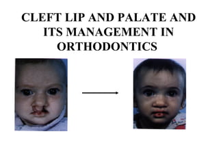
- 1. CLEFT LIP AND PALATE AND ITS MANAGEMENT IN ORTHODONTICS
- 2. Introduction • Clefts of the lip and or palate unfortunately, are by far the most common major facial malformations in mankind. Fortunately, as a result of technical advancements in the fields of medicine and their families for treatment, much can be done and achieved for them. • The orthodontist, by virtue of having gained in depth knowledge of the craniofacial complex, it’s growth and development, and expertise in tooth movement, has to play a role of prime importance in making critical decisions, planning treatment and rendering care to these patients.
- 3. INCIDENCE Incidence of cleft lip and Incidence of different palate in different races: types of cleft Geographic Section/Race Incidence per 1000 llve birth Negro 0.5 Caucasians 1.0 Japanese 2.34 American 2.91 Type of cleft Incidence (Percent of all cleft cases) Cleft lip alone 25% Cleft plate alone 25% Cleft lip and palate both 50%
- 4. • Prevalence of different types of cleft in India • Predicted incidence of the defect with affected relatives • CLP males > female • CP females > males • ULCLP is very common that to left side • Consanguis Marriages Palace Type of cleft with percentage CLP CP CL Dharwad 44.3 12.8 42.9 Delhi 68 18 14 Chennai 84.7 1.9 13.3 Affected relatives Predicted incidence (%) of cleft lip and palate One sibling 4.4 Two sibling 9.0 One sibling one parent 3.2 Two sibling one parent 15.8
- 5. ASSOCIATED SYNROMES 1. Down’s Syndrome (Trisomy 21) 2. Wardenburg’s Syndrome (Abnormalities of pigmentation of hair, iris and skin deafness). 3. Vander woude’s Syndrome (lip pits) 4. Orofacial digital syndrome: median cleft lip associated with bilateral accessory toes is found only in indians. 5. Treacher Collins Syndrome (hypoplastic zygoma, micrognathia, external and middle ear defects.) 6. Pierre Robin syndrome (micrognathia, glossoptosis with respiratory obstruction). 7. Klippel Feil Syndrome (short neck with an abnormal or missing cervical vertebra)
- 6. Development Of Palate Four week of IUL 5 brachial arches develop at the site of the future neck. • The first arch (mandibular arch) Development of naso- maxillary complex. • From first arch, maxillary arch is formed and give rise to upper lip, upper port of cheek. • Palate developed from 1- Frontonasal process give - primitive palate – premaxilla. 2- Maxillary processes give - Palatal process - Hard palate and soft palate
- 7. Development of CLP A. Complete BCLP B. Unilateral complete CLP C. Midline cleft extending in to hard palate D. Bifid Uvulae E. Cleft of soft palate
- 9. CLASSIFICATION OF CLP Morphological Embryological Morphological – Dow & Ritchie (1922) Veau (1931) have given morphological classification Group I - Cleft soft palate only Group II - Cleft of hard & soft plate Group III - Complete unilateral Group IV - Complete Bilateral
- 10. EMBRYOLOGICAL CLASSIFICATION I Fogit & Andersen 1942 - Hair lip (includes alveolus as far bat as incisive foramen) - Hair lip & Cleft palate - Isolated clefts of palate as far back as incisive foramen II. KERNAHAN & STARKS 1985 Group I – Cleft of the primary palate only • Unilateral • Bilateral • Total • Subtotal Group III – Cleft of the secondary palate only • Total • Subtotal • Submenus
- 11. Group IV – Cleft both primary & secondary palate • Unilateral - Total, Subtotal • Medium - Total, Subtotal • Bilateral - Total, Subtotal III SPINA CLASSIFICATION Group I - Pre- incisive foramen clefts • Unilateral • Bilateral • Median (cleft of the lip with /without an alveolar cleft) total, partial. Group II - Transincisive foramen clefts (cleft of the lip, alveolus & palate). • Unilateral • Bilateral Group III – Post incisive foramen clefts • Total • Partial
- 12. Dais & Ritchie classification (1922) Based on the location of the cleft relative to the alveolar proven ‘3’groups Group I Pre alveolar clefts – involving lip are sub classified as - Unilateral - Bilateral - Median Group II Post alveolar clefts Different hard & soft palate clefts that extend up to the alveolar ridge Group III Alveolar clefts Complete clefts involving the palate , alveolar ridge and the lip. Subdivided into Unilateral Bilateral Median
- 13. SCHUCHARDT & PFEIFER’S SYMBOLIC CLASSI FICATION
- 14. KERNAHAN’S stripped ‘Y’ classification
- 15. LAHSHAL CLASSIFICATION Simple classification presented by OK ring in 1987. LAHSHA in a paraphrase of the anatomic areas affected by the cleft. L- Lip A- Alveolus H- Hard palate S- Soft palate H- Hard palate A – Alveolus L- Lip. E.g L----S - Cleft fright lip & soft palate t-----S – Cleft right lip Alveolus, soft palate with combination of left cleft lip
- 16. Internationally approved classification Internal ion confederation for plastic & constructive surgery 1967 Rome. Classification of cleft of the lip, alveolus & palate this classification is bared on embryonic principle. Group I – Clefts of anterior ( primary palate) Lip: Right and / or left b) Alveolus : Right & / or left Group II – Cleft of anterior posterior (Primary and secondary ) palate a. Lip: Right / or left b. Alveolus Right / or left c. Hard plate : Right / or left Group III – Clefts of posterior (secondary) palate a. Hard palate : Right / or left b. Soft palate : Medial
- 17. FACIAL GROWTH IN CLP CASES Prenatal Growth • Over maxillary segment on non cleft side - Pull of lip & cheek muscles - Tongue pressure - Relatively unstrained nasal septum growth • Over maxillary segment on the cleft side. - Intrinsic deficiency - Presence from alas base due to stretching of the nostril
- 18. In compel unilat CLP cases: - Large maxillary segment on non deft side thorns a scare deviation of the midline away from the cleft - Smaller maxillary segment retro positioned / collapse / prohibition - Nose toward normal side - Ala grow in deformed matrix In Bilat CLP cases: - Primarily tilts forward / shifts one side due to tongue procure - Smaller maxillary segments cither side similar growth pattern as in • Due to above forces the defects produced in CLP basics are
- 19. FACIAL GROWTH IN UNREPAIRED CLP - A functionally normal facial skeleton develops in unpaired cases - Presence pf local bony defect due to intrinsic deficiency in the humiliate area of the cleft - Absence normal lip pressure abnormal development of dento alveolar process can occur - Mestre et al 1960 Ortiz monarteri et al 1961 & Birhona 1976 fond no significant difference in overall size with normal adults in un-operated clefts - Biohara 1973 found retro poritional maxilla & mandible than normal but no significant differences in any parameters b/co operated & unoperated cases. - Localized growth defects in un-operated subjects, region immediate to cleft o alveolar arch defects occurred particularly in lateral & vertical growth at the region of the alveolar cleft. - STARK 1968 – A deficiency of mesodermal tissue rather than a more failure of fusion of embryonic elements pathogenic back ground - FUSTER & LAUELLE 1971 – the teeth in cleft subjects significantly smaller AMETON 1967 reduction in subject of the elements of maxilla at all ages, didn’t progressively greater with age.
- 20. FACIAL GROWTH AFTER SURGICAL REPAIR • Effect of lip repair - Increased tighten in the upper part of the upper lip inhibits growth banal bone of maxilla increased tighten in the lower pout of the upper lip ratiocination of dento alveolar an structures • HAYASHI et al 1976 The growth of the maxilla repaired cleft lip and palate tends reduced in all planes of space Effect of palate repair- Inhibit the growth of the maxilla scar contractor, reduction in the maxillary size may occurs
- 21. PROBLEMS ASSOCIATE WITH CLEFTS 1. Dental • Congenital missing teeth (usually ) • Neonatal teeth • Super numorary teeth • Ectopically erupting teeth • Peg shaped teeth • Delayed eruption if teeth • Enamel hypoplasia • Microdontia / macrodontia • Fused teeth • Spacing / crowding • Posterior & anterior cross bite
- 22. 3. Hearing and speech • Associated with disorders with of the middle ear affect hearing Differ cuties in language uptake speech. Speech defects CP patients • When sound M.N & N.G • Speech defects when sounds UL / BL CL patients are not approximating • When upper teeth maloccluded sounds th, F,V,S, ZE, SH. • Surgical operations • Speech therapy 4. Psychological Problems Psychological stress due to their abnormal facial appearance they have to put up with staring, curiosity, pity et •
- 23. 2. Esthetic problems The clefts involving the lip can result in lot of facial disfigurement. The oro-facial structures may be malformed and congenitally missing. Deformities of nose can also occur thus esthetes greatly affected
- 24. MANAGEMENT OF CLP • Multi-disciplinary team for cleft lip and palate patients S.No. Experts involved Role 1. Obstetrician - Refers to plastic surgeon and pediatrician counseling the parents 2. Pediatrician or Neonatologist - Refers to the plastic surgeon 3. Plastic surgeon - Leads the team of CLP cases discusses the case with members of the team phayngoplasty reversionary lip 4. Oromaxillofacial surgeon - Bone grafting 5. Neurosurgeon - Craniogacial syndrome is associated 6. Pedodontist - Presurgical orthopedic treatment for the baby monitors the growth and development to maintain oral health to guide the occlusion and facial growth 7. Orthodontist - Provides presurgical dental orthopedic consultation definitive orthodontic treatment once the full permanent dentition is erupted. 8. Speech pathologist Monitors the speech development to normal 9. Audiologist - Hearing in the young child 10. Otolaryngologist - Nasopharyngeal tissues, including tonsils, adenoids and middle ear blockage 11. Psychologist - Plays an important role under stress.
- 25. • CLP difficult management problem. • Esthetics, speech, respiration and mastication that most be dealt with in the rehabilitation of cleft child • The orthodontist has to give maximum attention to the child bs of the longitudinal nature of his cancer
- 26. MANAGEMENT IN GENERAL 1. Parent counseling at birth 2. Feeding advise 3. Pre-dental orthodontics 4. Surgical approach 5. Post-dental orthodontics - Primary dentition period - Secondary dentition period - Permanent dentition period
- 27. Orthodontic management by fish man a) Predental treatment – prior to eruption of primary molars Pre-surgical post-surgical b) Deciduous dentition (3-6yrs) – After eruption of primary duration. c) Early mixed dentition treatment (7-9yrs) –after during eruption of maxillary permanent incisors d) Late mixed permanent dentition treatment (9 12 yrs on words)
- 28. PRE- DENTAL • Initiated by Mc NEIL 1954 and developed by Burstone 1958 • Advantages of this appliance - To facilitate feeding - To help in psychological boost to the parents - To assist the surgeon in his initial repairs - To stimulate palatal growth - To restore the oro-facial ‘functional matrix’ - To help in reducing ear infections - To expand or prevent collapsed segment - To reduce the need for later orthodontic treatment - To guide tooth eruption - To improve esthetics - To re-establish proper sutural growth patterns early.
- 29. IMPRESSION • Extend into the nasal chamber undercut • Custom trays are preferred • Posterior limit – up to maxillary puberosity • Fabrication of a combination soft and hard acrylic passive maxillary orthopedic appliance OBTURATOR • Placement of the appliance and surgical closure of the lip. This is usually at 10 days to 2 weeks autogenous alveolar bone graft. • Stabilization of the graft 2 months or so, using the orthopedic appliance as a splint. OBTURATOR -USES • It facilitates lip repair • It facilitates the feeding • It prevents the collapse of the palatal segments
- 31. METHODS OF PRE SURGICAL ALIGNMENT • Discussed in two aspects I. Control of maxillary elements • Achieve upper arch form which confirms to that of lower arch • Stabilization of the arch can be achieved by simple upper appliance • Expansion of the upper arch can be achieved by screw appliance or pre-expanded appliance • Retention of above appliances by the force of the turn and the lower jaw, the extra orally ‘whiskers’ are facilitate handling of appliance
- 32. A. In bilateral clefts the displaced premaxilla is readapted to confirm to the arch B. In unilateral clefts the displaced greater segment is readapted to conform to the arch
- 33. II. Control of Pre-maxillary elements Both unilateral and bilateral clefts extra-oral strapping across the pre maxilla, attached either directly to the face Retrusion of the pre-maxilla can only be achieved at the extends of distortion of the nasal septum. All these procedure should be done with in 3 months Huddart 1974 found that use of an intra – oral appliance restricted further widening of the maxillary arch allowed normal growth to proceed at margins of cleft.
- 34. Lantham appliance – Retraction, Incomplete BCLP
- 35. Lanthem Appliance with retention Pin
- 36. PRIMARY BONE GRAFTING • Objective – It is to make good the hard tissue deficiency • Good surgical grafting improve the oral contours improved function Advantages claimed a) Prevention of collapse of anterior portion of arch b) Allowing eruption of teeth into the graft site c) Allow mid line adjustment that is so necessary to minimize the asymmetry of treated cleft lip and palate.
- 37. PRIMARY DENTITION PHASE • Common problems encountered a) Cross bite of incisor or buccal teeth b) A functional shift of the mandible c) An oronasal fistula d) Premaxillary protrusion in bilateral clefts • Expansion fixed maxillary lingual arch appliance either W-arch or an Arnold expander • Anterior cross bites a removable inclined plane on lower incisors or maxillary Hewley retainer with finger spring. Supernumerary teeth should be extracted
- 38. MIXED DENTION PHASE • Problems are more severe a short period & effective of orthodontic treatment a) Sagittal deficiency of the maxilla, b) Anterior cross bite c) Downward rotation of the maxillary plane d) Function deviation of the Mandible e) Missing teeth f) Dental crowding
- 40. Protraction headgear – 5 – 8 yrs, acrylic maxillary splint with occlusal coverage, cemented on maxillary teeth with heavy elastics. 500g forces, 10o to occlusal plane, for 14-16hrs day for 6 to 12 months. Leads remodeling changes in the anterior maxilla and a backward rotation of the mandible. Contra indicated in mandibular prognatism and high mandibular plane cases.
- 41. OTHER AUXILIARY APPLIANCES Cross bite central incisor • Negative overjet and maxillary incisor rotations are corrected simultaneously by lower bite planes, removable appliance with Z-spring are used. Maxillary contraction (Transverse / buccal cross bite) • Expansion of the maxillary arch by : – Quad Helix appliance 036 blue engiloy provides controlled force appliance – NiTi palatal expanders effect maxillary expansion more dentoalveolar than skeletal effects.
- 43. LINGUAL RETAINERS Open end of U shape retainer facing posteriorly- located canine and premolar area. Uses of semi fixed and lingual sheaths type designs have 4 advantages – 1. Adjustments / replacement easy 2. Transition from expansion to retention is immediate 3. Avoid interference during bone grafting 4. Allows rapid replacement with easy secondary design.
- 44. Retainers
- 45. Functional deviation of the Mandible Occurs due to interference of incisors Correction of incisors cross bite and expansion of the maxillary arch invariably permit to the mandible to achieve a normal path of closure
- 46. Missing Teeth - Most common in 2 2 next to the cleft followed by second bicuspids I both the arches and maxillary third molars - Retainers replacing missing teeth should be worn until permanent dentition treatment
- 47. Dental crowding Because of the incisors retruction arch length is often reduced I both the arches A class III tendency man require an oven greater retruction of the mandibular incisors to establish normal overjet. Serial extraction is the mode of treatment If should be also noted that the class III elastics are usually worn in permanent dentition so that if first biocuspids have been removed space closure in the mandibular arch may be difficult to accomplish without excessive lingual tipping of the mandibular incisors.
- 48. Alveolar Bone Grafting Indicated- To stabilize the premaxilla, to close oronasal fistula and anterior palatal clefts To ensure better periodontal support for the erupting teeth and the teeth burdening the cleft, to build up the piriform rim and alar base symmetric with the normal side, To lend sup[port to the depressed lip[ over the cleft] To provide bony continuity in the alveolous for eruption and orthodontic movement of the teeth adjacent to the cleft in their optimal positions.
- 49. -Cancellous iliac crest grafts done between 9 and 11 years of age allow quite favorable eruption of the maxillary canine through the graft either by itself or by prthodontic mechantotherapy, with the best possible periodontal support to be expected. -Secondary bone grafts or greater acceptance than the primary bone grafting
- 50. Permanent dentition - Most difficult case of treatment - In case analysis three considerations must be stressed a. Stability of teeth and maxillary segment b. Facial esthetics c. Preparation for a prosthetics Surgical procedures which are particularly traumatic which must be assessed on an individual basis and treatment must be planned accordingly Problems includes, a. Vertical jaw relations b. Jaw relation c. Lateral jaw relations and dental irregularities
- 51. Vertical jaw relations Freeway space is atmost important treating this problems this excess free way space can be corrected by a. Vertical elastics b. Vertical development plate – used for both open bite closure which is usually in cuspid region and forced eruption of all maxillary teeth c. Mandibular acrylic bite opening plate in conjunction with vertical elastics – Best method, because the plate covers mandibular incisors and provides lingual rest for maxillary incisors
- 52. Anterio –Posterior jaw relations - This directly related to the freeway space, so that the A-P must be made with the mandible in rest position - A-P relations in females is known to stabilize at 13-14 years where as in males it is 17-18 years. So stable results may not be achieved until this age Surgery for severe A-P discrepancy cases - May be advised if large excess freeway and mandibular over closure -Maxillary advancement – preferred in case of mid face deficiency -Surgery to be delayed until puberatal growth spurt. 14 years for females and 17 years for males -Beginning of orthodontic treatment will be after surgery
- 53. Lateral Jaw relationships Expansion should be overdone in order to maintain for several years with a palatal retainers Dental irregularities Teeth frequently crupt in the palate (because of surgery). Lateral cuspids are commonly involved in which case retention of these teeth after may be alignment may be difficult particularly, if the root apex is palatably placed. Serial extraction often it is indicated it is satisfactory to remove one or more mandibular incisors to relieve crowding, this makes treatment much simpler and co-ordination to mandibular incisors is less critical in cleft cases
- 54. Surgery Time of surgery is quite a contraoversy Different according to different school of thought generally, Cleft lip- 3-6months Cleft palate – 1 1/2 – 2years Most surgeons follows the rule of 10- -10 weeks of age -Weight 10pounds -Hb 10% -WBC should be lesser than 10,000
- 55. Cleft lip surgical repair -Various methods of CL repair aimed to lengthening the philithral ridge on the cleft side to correct the nose deformaties have much as possible -The lip is closed in three layers – mucosa, muscles,skin - Most favorable surgical operations are Millerd’s rotations advancement flap, Tennison Randal repair, Veau repair
- 56. Millard’s Repair of CL Millard’s rotation flap (a) and Columella flap(c) or planned medial side of the cleft . After full thickness of the lip cut along the markings a rotation gap is produced on the medial side which is filled by an advancement flap(b) planned on the lateral side of the cleft
- 57. Tennison Randall repairs • A triangular flap is created on the lateral side of the cleft to fit in to the triangular defect produced on the medial side of the cleft. This procedure can be planned exactly after initially measurements. The results cannot be modified once the lip is cut. The scar is more prominent than in other procedures.
- 58. Veau repair It is the simplest one stage straight line closure and produces satisfactory results u a bilateral cleft lip. In this method vermilion flap from either lateral side of the cleft is brought down over the prolabium to augment the vermilion I the centre of the upper lip
- 59. Cleft palate surgical Repairs 1. Von Langen beck’s simple palato plasty of CBCLP
- 60. 2.Von Langen beck’s palatoplasty for CUCLP
- 61. 3. Veu Wardill Kilner V-Y push back palatoplasty Cleft palate repair by a double opposing Z- plasty of Furlow is relatively new procedure aimed are reducing the midfacial growth retradation problem. However more experience is required with this procedure before reaching any conclusion
- 62. Distraction Osteogenesis - This technique is Increasing popular and has a good potential for severe maxillofacial deformities, where in a bone segments are lengthened by using intra oral, extra oral devices - Maxillary distraction for CLP cases has been shown to be particularly useful when the maxilla to moved forward enlarged distance, which by orthognathic surgery alone may not be technique feasible or stable. Since this technique involves fixing of considerable hardware on the cranium for a few months, the choice is restricted to select cases having a large maxillary retraction
- 63. Relapse and retention CLP cases requires long term if not permanent retention for the following reasons: • In adequate bone support • Absence of some teeth • Presence of stretched scar tissue - For this reason, retention, the orthodontist’s final frontier must be planned in detail Retainers of choice i. Soldered lingual retainers ii. Hawley’s retainers with Pontiac to replace missing tooth iii. Bonded spiral wire retainers for derotated incisors
- 64. CONCLUSION The course of the individual CLP patients ’s rehabilization depends not only on the quality of the individual components of the treatment, but also on organization and co-ordination to ensure the right timing, sequence and balance during the often protruded course of treatment. For this reason, a well adapted CLP protocol, with a collectively operating inter disciplinary rather than multidisciplinary approach to provide integrated cleft care seems to be absolutely imperative.
- 65. The myriad manifestation of the malocclusion typical in Cleft lip and palate necessities a harmonious medley of a multitude of mechanotherapy procedures that emancipates the orthodontist from the monotonous momentum of mundane tooth moving procedures. CLP rehabiltative treatment is the privilege and real reward. In that endeavourer, our responsibilities go beyond orthodontics. We can always go further; we can always work harder; we can always find newer possibilities; but for that we must keep going and doing