1. Appendix G
Characterization of chemical warfare G-agent hydrolysis products
by surface-enhanced Raman spectroscopy
Frank Inscore, Alan Gift, Paul Maksymiuk, and Stuart Farquharson*
Real-Time Analyzers, 87 Church Street, East Hartford, CT 06108
ABSTRACT
The United States and its allies have been increasingly challenged by terrorism, and since the September 11, 2001 attacks
and the war in Afghanistan and Iraq, homeland security has become a national priority. The simplicity in manufacturing
chemical warfare agents, the relatively low cost, and previous deployment raises public concern that they may also be
used by terrorists or rogue nations. We have been investigating the ability of surface-enhanced Raman spectroscopy
(SERS) to detect extremely low concentrations (e.g. part-per-billion) of chemical agents, as might be found in poisoned
water. Since trace quantities of nerve agents can be hydrolyzed in the presence of water, we have expanded our studies
to include such degradation products. Our SERS-active medium consists of silver nanoparticles incorporated into a sol-
gel matrix, which is immobilized in a glass capillary. The choice of sol-gel precursor allows controlling hydrophobicity,
while the porous silica network offers a unique environment for stabilizing the SERS-active silver particles. Here we
present the use of these silver-doped sol-gels to selectively enhance the Raman signal of the hydrolyzed products of the
G-series nerve agents.
Keywords: chemical warfare agent detection, CWA, hydrolysis, SERS, Raman spectroscopy
1. INTRODUCTION
The potential use of chemical and biological warfare agents by terrorist organizations directed against U.S. military and
Coalition forces in the Middle East, and civilians at home, is an issue that has generated considerable concern in the post
9/11 era. The ability to counter such attacks, requires recognizing likely deployment scenarios, among which includes
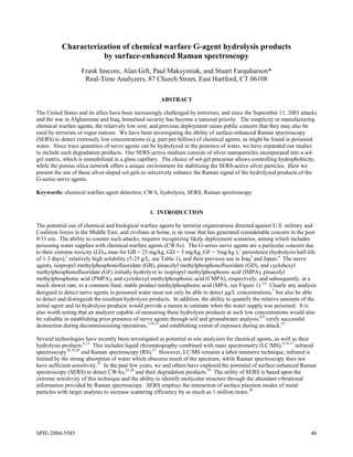
poisoning water supplies with chemical warfare agents (CWAs). The G-series nerve agents are a particular concern due
to their extreme toxicity (LD50 man for GB = 25 mg/kg, GD = 5 mg/kg, GF = 5mg/kg ),1 persistence (hydrolysis half-life
of 1-3 days),2 relatively high solubility (5-25 g/L, see Table 1), and their previous use in Iraq3 and Japan.4 The nerve
agents, isopropyl methylphosphonofluoridate (GB), pinacolyl methylphosphonofluoridate (GD), and cyclohexyl
methylphosphonofluoridate (GF) initially hydrolyze to isopropyl methylphosphonic acid (IMPA), pinacolyl
methylphosphonic acid (PMPA), and cyclohexyl methylphosphonic acid (CMPA), respectively, and subsequently, at a
much slower rate, to a common final, stable product methylphosphonic acid (MPA, see Figure 1).5,6 Clearly any analysis
designed to detect nerve agents in poisoned water must not only be able to detect µg/L concentrations,7 but also be able
to detect and distinguish the resultant hydrolysis products. In addition, the ability to quantify the relative amounts of the
initial agent and its hydrolysis products would provide a means to estimate when the water supply was poisoned. It is
also worth noting that an analyzer capable of measuring these hydrolysis products at such low concentrations would also
be valuable in establishing prior presence of nerve agents through soil and groundwater analysis,8,9 verify successful
destruction during decommissioning operations,5,10,11 and establishing extent of exposure during an attack.12
Several technologies have recently been investigated as potential at-site analyzers for chemical agents, as well as their
hydrolysis products.6,13 This includes liquid chromatography combined with mass spectrometry (LC/MS),9,14-17 infrared
spectroscopy18,19,20 and Raman spectroscopy (RS).21 However, LC/MS remains a labor intensive technique, infrared is
limited by the strong absorption of water which obscures much of the spectrum, while Raman spectroscopy does not
have sufficient sensitivity.21 In the past few years, we and others have explored the potential of surface-enhanced Raman
spectroscopy (SERS) to detect CWAs,22-28 and their degradation products.29 The utility of SERS is based upon the
extreme sensitivity of this technique and the ability to identify molecular structure through the abundant vibrational
information provided by Raman spectroscopy. SERS employs the interaction of surface plasmon modes of metal
particles with target analytes to increase scattering efficiency by as much as 1 million times.30
SPIE-2004-5585 46
2. In our studies, we have employed metal-doped sol-gels to promote the SERS effect. The porous silica network of the
alkoxide sol-gel matrix offers a unique environment for immobilizing and stabilizing SERS-active metal particles of both
silver and gold.31-34 The choice of metal and Si-alkoxide composition provides a means for chemically selecting the
target analyte to be enhanced based on charge and polarity. Electropositive silver or electronegative gold particles can
selectively enhance the Raman signals of negative or positive chemical species, respectively, while different alkoxides
(or combinations of) can be used to select for polar or non-polar molecules. Previously, we used glass vials internally
coated with the SERS-active sol-gel to measure cyanide, HD, VX, and MPA.28 More recently, we have developed glass
capillaries filled with the SERS-active sol-gel that can be attached to a syringe to perform simple and rapid sample
extraction and SERS analysis.35 This paper employs these extractive and SERS-active capillaries to examine the ability
of SERS to measure and distinguish the hydrolysis products of GB, GD, and GF. Both Raman and surface-enhanced
Raman spectra are presented along with preliminary vibrational mode assignments.
Table 1. Properties of chemical agents and their primary hydrolysis products investigated in the present study.2
Chemical Agent Hydrolysis ½ life Water Solubility at 25°C
Sarin (GB) 39 hr (pH 7) completely miscible
IMPA stable (can hydrolyze to MPA) 4.8 g/L
MPA very stable (resistant to further degradation) >1000 g/L
Soman (GD) 45 hr (pH 6.6) 21 g/L (@20°C)
PMPA stable (can hydrolyze to MPA) no data
Cyclosarin (GF) slower than GB 3.7 g/L
CMPA no data (can hydrolyze to MPA) no data
H2O
O H2O O
IMPA 2-propanol + MPA
GB HF +
P P
O F O OH
H2O H2 O
GD HF + PMPA 2-pinacolyl + MPA
O O
P P
O F O OH
H2O
H2O
GF O + O CMPA cyclohexanol + MPA
HF
P P
O F O OH
Figure 1. Hydrolysis pathways for G-Series nerve agents.
2. EXPERIMENTAL
The hydrolysis degradation chemicals measured in this study (IMPA, PMPA, CMPA) were obtained as analytical
reference materials from Cerilliant (Round Rock, TX) and used without further purification. MPA and all chemicals
used to prepare the silver-doped sol-gel coated capillaries were acquired from Sigma-Aldrich (St. Louis, MO) and used
as received. For the purpose of safety, samples were prepared in a chemical hood, transferred to a sampling device and
sealed prior to being measured. All samples were measured initially by Raman in their pure state at room temperature;
MPA as a solid powder, with IMPA, and PMPA as neat liquids. CMPA was obtained in forensic quantities (1 mg/mL in
MeOH), and was not amenable to RS studies at these concentration levels.
Methanol or water (HPLC grade) was used to prepare solutions of the target chemicals for SERS measurements at a
SPIE-2004-5585 47
3. concentration of 1 mg/mL from solid powders or 0.1% v/v from neat liquids unless noted otherwise. Lower
concentrations were prepared from these solutions by serial dilution, and all solutions were stored at 10°C until needed.
The Raman and SERS spectra of the target chemicals presented here were all measured in capillaries.
SERS-active capillaries were prepared using the following general methodology. A silver-doped sol-gel solution,
prepared according to previous published procedures from a mixture of two precursor solutions,31 was drawn via a
syringe into pre-cleaned 1-mm diameter capillaries. This procedure was modified for the SERS-active capillaries, in
particular by replacing TMOS with an alkoxide mixture composed of tetramethyl orthosilicate (TMOS),
octadecyltrimethoxysilane (ODS), and methyltrimethoxysilane (MTMS) at a v/v/v ratio of 1/1/5.
A 50 µL sample from each of the prepared analyte solutions was drawn into a SERS-active capillary for measurement.
The capillaries were mounted horizontally on an XY positioning stage (Conix Research, Springfield, OR), such that the
focal point of an f/0.7 aspheric lens was positioned just inside the glass wall. The probe optics and fiber optic interface
have been described previously.35 A Fourier transform Raman spectrometer (Real-Time Analyzers, model IRA-785,
East Hartford, CT) equipped with a 785 nm diode laser (Process Instruments Inc. model 785-600, Salt Lake City, UT)
and a silicon photo-avalanche detector (Perkin Elmer model C30902S, Stamford, CT) was used to deliver 100 mW of
power to the SERS and RS samples and generate spectra with 8 cm-1 resolution.
3. RESULTS AND DISCUSSION
The SERS spectra of chemicals are often different than their Raman spectral counterparts due to the surface interactions
that can enhance various vibrational modes to different extents. Therefore the Raman spectra were measured and
included in this study to aid interpretation of the corresponding SERS spectra. The simplest chemical specific to the G
series nerve agents is methylphosphonic acid, which has been well characterized by IR and Raman spectroscopy,36,37 and
subsequent normal coordinate analysis for assigning the vibrational modes.38 The Raman spectrum of MPA contains 10
discernable peaks between 350 and 1650 cm-1 (Figure 2B). Four PO3 bending modes are observed at 408, 462, 491
(shoulder) and 504 cm-1. The PC symmetric stretch is the most intense peak observed at 774 cm-1. A CH3 rocking mode
occurs at 892 cm-1 with little intensity, while the PO3 stretching mode produces a peak to 956 cm-1. Two additional CH3
and PO3 modes produce peaks at 1004 and 1054 cm-1, also with moderate intensity. The 10th mode in this region is a
CH3 bending mode which occurs at 1424 cm-1.
A A
B B
Figure 2. A) SERS and B) Raman spectra of MPA. Figure 3. A) SERS and B) Raman spectra of IMPA.
Conditions: A) 0.1 mg/ml in water, TMOS/ODS/MTMS Conditions as in Fig. 2, but: A) 0.1 % v/v in MeOH, B)
sol-gel in capillary, 1-min acquisition time. B) solid, 5- neat liquid.
min acquisition time.
The SERS spectrum of MPA (Figure 2A) is considerably simpler than that of the solid powder Raman spectrum, with
weak peaks observed at 469, 521, 958, 1003, 1038, and 1420 cm-1. These SERS spectral peaks can all be assigned to the
modes observed at similar frequencies in the Raman spectrum, albeit the 521 and 1038 cm-1 peaks have shifted
significantly from the 504 and 1054 cm-1 Raman spectral peaks. The most characteristic SERS spectral peaks are the
SPIE-2004-5585 48
4. intense 756 cm-1 peak and the unique peak at 1300 cm-1. The former peak clearly corresponds to a nearly pure PC
symmetric stretch, while the latter is likely a CH3 twist.
The next hydrolysis product studied was isopropyl methylphosphonic acid. Like MPA, both the Raman and SERS
spectra of IMPA are dominated by a peak in the 700 cm-1 region, specifically at 728 and 716 cm-1, respectively (Figure
3). However, these peaks are not simply a PC stretch, but include a considerable amount of the backbone CPOCC mode
created by the addition of the isopropyl group. Both spectra also contain moderate peaks at 782 and 772 cm-1 that may
also be PC containing backbone modes, as has been suggested by a theoretical treatment for sarin.39 It is also worth
noting that the Raman spectrum of IMPA is very similar to that of a published spectrum of sarin.21 A number of the
peaks assigned to PO3 modes for MPA have shifted moderately from the Raman to the SERS spectra for IMPA, and
includes the following respective peaks; 510 and 508 cm-1, 938 and 931 cm-1, and 1006 and 1004 cm-1. The latter peak
likely contains significant methyl character. Similarly, the methyl rocking and bending modes observed for MPA are
now at 880 and 874 cm-1, and 1420 and 1416 cm-1 in the respective Raman and SERS spectra of IMPA. Not
surprisingly, the isopropyl group not only increased the intensity of these bands, but also gives rise to a CH deformation,
and additional CH3 and CH2 wagging modes, at 1359 and 1349 cm-1, 1390 and 1388 cm-1 and 1453 and 1451 cm-1, in the
respective Raman and SERS spectra. The isopropyl group also gives rise to a CC bend at 421 and 424 cm-1, and a CC
stretch at 1179 and 1173 cm-1 in the respective Raman and SERS spectra. In the Raman spectrum of IMPA a peak also
appears at 1104 cm-1 that is characteristic of CO or CC stretches, while in the SERS spectrum a peak appears at 1055
cm-1 and is assigned to a PO3 stretch, as was the 1038 cm-1 peak in the MPA SERS spectrum.
The Raman spectrum of pinacolyl methylphosphonic acid, like IMPA, contains an increasing amount of CC and CHn
character (Figure 4B). This includes new peaks at 541, 934, 977, 1212 and 1264 cm-1 that are assigned to a CC3 wag, a
CC3 bend, a CCC bend, and two CC stretching modes based on a theoretical treatment for soman.39 The 1300 to 1500
cm-1 region again contains a number of CHn bending modes, and the peaks are assigned accordingly. The most obvious
change in the spectrum is that the PC plus backbone mode in the IMPA spectrum has split into two distinct peaks at 732
and 761 cm-1. The SERS spectrum for PMPA is dominated by these latter peaks, except that they overlap considerably
producing a peak centered at 750 cm-1 with a shoulder at 729 cm-1 (Figure 4A). The remaining SERS peaks are evident,
but have little intensity, except for the CC3 wag at 543 cm-1, the PO3 stretch at 1037 cm-1, and the CH2 bend at 1444 cm-1.
Cyclohexyl methylphosphonic acid was only available as 1 mg/mL in methanol and a Raman spectrum at this
concentration could not be obtained. The SERS spectrum in many ways is like IMPA with the addition of cyclohexane
modes (Figure 5). This includes peaks at 622, 1023, and 1262 cm-1, that are attributed to ring CC stretching modes, and
a peak at 811 cm-1 that is assigned to a ring CH2 bending mode. The most intense peak observed at 747 cm-1 is again
assigned to a PC stretch plus backbone mode.
A
B
Wavenumber (cm-1)
Figure 4. A) SERS and B) Raman spectra of PMPA. Figure 5. SERS spectrum of CMPA. Conditions as in
Conditions as in Fig. 3. Fig. 3, but A) 1 mg/mL in MeOH.
In general, the SERS spectra for these alkyl methylphosphonic acids have two common features, the PC stretch produces
the most intense peak, more so than the Raman spectra when compared to the intensity of the other peaks, and the most
SPIE-2004-5585 49
5. substantial shift in peak frequencies occurs for PO3 modes when compared to the Raman spectra. The increased
intensity of the PC mode suggests that it is perpendicular to the surface, based on previous research that has shown that
modes couple to the plasmon field more effectively in this orientation.40 The shift in the PO3 frequencies suggests strong
surface interactions through this group. Taken together, the SERS data suggests that these molecules are oriented with
the PO3 group interacting with the silver surface and the methyl group away from the surface. In the case of MPA,
especially for the doubly deprotonated anion, the three oxygens could form the base of a tripod on the surface. This
orientation may become less likely for the other molecules as the alkoxide groups replace the hydroxide group with
surface interaction through the other two oxygens. This change in orientation along with increasing amounts of
backbone character to the PC stretch could explain the shift and splitting of this mode.
Table 2. Tentative vibrational mode assignments for Raman and SERS peaks for VX and its hydrolysis products.
MPA IMPA PMPA CMPA Tentative Assignmentsa
RS SERS RS SERS RS SERS SERSb
408 421 424 PO3 bend
462c,d 469 441 442 441 PO3 bend
491c 475 PO3 bend
504c 521 510 508 514 495 C-PO3 bend
541e 543 549 C-C3 bend
622 Ring breathing
728 716 732 729sh PC stretch and backbone
774 756 782 772 761 750 747 PC stretch and backbone
799 792 CH bend
811 Ring CH2
880e 874 869e 863 857 CCC bend
892c,d 902 888 896 CH3 rock
934e 929 C-C3 bend
c,d
956 958 938 931 PO3 stretch
977e CCC stretch
1004 1003 1006 1004 1015 1000 PO3 or CH3 bend
1023 Ring breathing sym
1054 1038d 1055 1052 1037 1050 PO3 stretch
1079 1073 CCC bend
1104 1116 OC or CC stretch
1143 1132 1150 CC stretch
1179 1173 1212e 1206 CC stretch
1224 1236 1243 CH2 bend or above
1264e 1257 1262 CC stretch
1300 1291 CH3 bend
1359 1349 1355 1347 CH deformation
1374 CHn bend
1390 1388 1390 1394 1393 CH3 rock
1424c,d 1420 1420 1416 1420 1415 1416 CH3 bend (bound to P)
1453 1451 1447 1444 1443 CH2 rock
a - Assignment terminology is simplified since assignments refer to multiple molecules. b - no Raman spectrum
measured, c = Ref. 36, d = Ref. 37, e = Ref. 39.
4. CONCLUSION
The ability to measure and identify the various hydrolysis degradation products with our SERS-active silver-doped sol-
gel coated capillaries has been demonstrated. The SERS spectra of these chemicals were somewhat different than their
Raman spectral counterparts, which is attributed to the interaction of these chemicals with the silver. In general, the
Raman and SERS spectra for the alkyl methylphosphonic acid hydrolysis products were dominated by one or two peaks
between 715 and 765 cm-1, which have been assigned to PC stretching modes with varying amounts of backbone mode
SPIE-2004-5585 50
6. contributions. The spectral intensity of this mode and the shift in frequency of the PO3 modes in the SERS spectra
suggest a strong surface interaction for these molecules. It is clear from the present study that the hydrolysis products
can easily be identified as a class by these 700 cm-1 peaks, but quantifying each in a mixture is likely to require chemical
separations or chemometric approaches. These approaches, as well as measurements to determine the detection limits
and pH dependence of these hydrolysis products are in progress.
5. ACKNOWLEDGMENTS
The authors are grateful for the support of the U.S. Army (DAAD13-02-C-0015, Joint Service Agent Water Monitor
program), and the National Science Foundation (DMI-0215819), and would like to thank Dr. Steve Christesen for
helpful discussions, and Mr. Chetan Shende for sol-gel chemistry development.
6. REFERENCES
1. Committee on Toxicology. Review of Acute Human-Toxicity Estimates for Selected Chemical-Warfare Agents,
Nat. Acad. Press (Washington, D.C.) 1997.
2. Munro, N.B., Talmage, S.S., Griffin, G.D., Waters, L.C., Watson, A.P., King, J.F., and Hauschild V. “The Sources,
Fate, and Toxicity of Chemical Warfare Agent Degradation Products”, Environ. Health Perspect., 107, 933-974
(1999).
3. Hoenig, S.L. Handbook of Chemical Warfare and Terrorism, Greenwood Press (Westport, CT) 2002.
4. Nozaki, H. and Aikawa, N. “Sarin poisoning in Tokyo subway”, Lancet, 345 1446-1447 (1995).
5. Wagner, G. and Yang, Y. “Rapid nucleophilic/oxidative decontamination of chemical warfare agents”, Ind. Eng.
Chem. Res., 41, 1925-1928, (2002).
6. Creasy, W., Brickhouse, M., Morrissey, K., Stuff, J., Cheicante, R., Ruth, J., Mays, J., Williams, B., O’Connor, R.,
and Durst, H. “Analysis of chemical weapons decontamination waste from old ton containers from Johnston atoll
using multiple analytical methods”, Environ. Sci. Technol., 33, 2157-2162, (1999).
7. McKone, T.E., Huey, B.M., Downing, E., and Duffy, L.M., Editors. Strategies to Protect the Health of Deployed
U.S. Forces: Detecting, Characterizing, and Documenting Exposures, Nat. Acad. Press (Washington, D.C.) p.207,
1999.
8. Johnston, R.L., Hoefler, C.M., Fargo, J.C., and Moberley, B. “The Defense Nuclear Agency’s Chemical/
Biochemical Weapons Agreements Verification Technology Research, Development, Test and Evaluation Program
and its Requirements for On-Site Analysis”, AT-ONSITE, 5-8 (1994).
9. D’Agustino, P.A, Hancock, J.R., and Provost, L.R. “Determination of sarin, soman and their hydrolysis products in
soil by packed capillary liquid chromatography-electrospray mass spectrometry”, J. Chromatography A, 912, 291-
299 (2001).
10. Yang, Y., Baker, J., and Ward, J. “Decontamination of chemical warfare agents”, Chem. Rev., 92, 1729-1743
(1992).
11. Christesen, S., MacIver, B., Procell, L., Sorrick, D., Carrabba, M., and Bello, J. “Nonintrusive analysis of chemical
agent identification sets using a portable fiber-optic Raman spectrometer”, Appl. Spec., 53, 850-855 (1999).
12. Hui, D.-M. and Minami, M. “Monitoring of fluorine in urine samples of patients involved in the Tokyo sarin
disaster, in connection with the detection of other decomposition products of sarin and the by-products generated
during sarin synthesis”, Clin. Chim. Acta, 302, 171-188 (2000).
13. “The Chemical Weapons Convention Redefines Analytical Challenge”, Analytical Chemistry News & Features,
June 1, 397A (1998).
14. Sega, G.A., Tomkins, B.A., and Griest, W.H. “Analysis of methylphosphonic acid, ethyl methylphosphonic acid
and isopropyl methylphosphonic acid at low microgram per liter levels in groundwater” J. Chromatography A, 790,
143-152 (1997).
15. Creasy, W.R. “Postcolumn Derivatization Liquid Chromatography/Mass Spectrometry for Detection of Chemical-
Weapons-Related Compounds” Am. Soc. Mass Spectrom., 10, 440-447 (1999).
16. Katagi…, J. Chromatography A, 833, 169-179 (1999).
17. Liu, Q., Hu, X., and Xie, J. “Determination of nerve agent degradation products in environmental samples by liquid
chromatography–time-of-flight mass spectrometry with electrospray ionization”, Analytica Chimica Acta, 512, 93-
101 (2004).
SPIE-2004-5585 51
7. 18. Hoffland, L.D., Piffath, R.J., and Bouck, J.B. “Spectral signatures of chemical agents and simulants”, Optical
Engineering, 24, 982-984, (1985).
19. Braue, E.H.J., and Pannella, M.G. “CIRCLE CELL FT-IR Analysis of Chemical Warfare Agents in Aqueous
Solutions”, Applied Spectroscopy, 44, 1513-1520, (1990).
20. Kanan, S. and Tripp, C. “An infrared study of adsorbed organophosphonates on silica: a prefiltering strategy for the
detection of nerve agents on metal oxide sensors”, Langmuir, 17, 2213-2218, (2001).
21. Christesen, S.D. “Raman cross sections of chemical agents and simulants”, Appl. Spec., 42, 318-321 (1988).
22. Lee, Y. and Farquharson, S. “Rapid chemical agent identification by SERS”, SPIE, 4378, 21-26 (2001).
23. Farquharson, S., Maksymiuk, P., Ong, K., and Christesen, S. “Chemical agent identification by surface-enhanced
Raman spectroscopy”, SPIE, 4577, 166-173 (2001).
24. Spencer, K.M., Sylvia, J., Clauson, S. and Janni, J. “Surface Enhanced Raman as a Water Monitor for Warfare
Agents in Water”, SPIE, 4577, 158-165 (2001).
25. Premasiri, W., Clarke, R., Londhe, S., and Womble, M. “Determination of cyanide in waste water by low-resolution
surface enhanced Raman spectroscopy on sol-gel substrates”, J. Ram. Spec., 32, 919-922 (2001).
26. Tessier, P., Christesen, S., Ong, K., Clemente, E., Lenhoff, A., Kaler, E., and Velev, O. “On-line spectroscopic
characterization of sodium cyanide with nanostructured Gold surface-enhanced Raman spectroscopy substrates”,
App. Spectrosc., 56, 1524-1530 (2002).
27. Christesen, S.D., Lochner, M.J., Ellzy, M., Spencer, K.M., Sylvia, J., and Clauson, S. “Surface Enhanced Raman
Detection and Identification of Chemical Agents in Water”, 23rd Army Science Conf., Orlando, 2002.
28. Farquharson, S., Gift, A., Maksymiuk, P., Inscore, F., Smith, W., Morrisey, K., and Christesen, S. “Chemical agent
detection by surface-enhanced Raman spectroscopy”, SPIE, 5269, 16-22 (2004).
29. Farquharson, S., Gift, A., Maksymiuk, P., Inscore, F., and Smith, W. “pH dependence of methyl phosphonic acid,
dipicolinic acid, and cyanide by surface-enhanced Raman spectroscopy”, SPIE, 5269, 117-125 (2004).
30. Weaver, M.J., Farquharson, S., and Tadayyoni, M.A. “Surface-enhancement factors for Raman scattering at silver
electrodes”, J. Chem. Phys., 82, 4867-4874 (1985).
31. Lee, Y. and Farquharson, S. “SERS Sample Vials Based on Sol-Gel Process for Trace Pesticide Analysis”, SPIE,
4206, 140-146 (2000).
32. Farquharson, S. and Lee, Y. “Trace Drug Analysis by Surface-Enhanced Raman Spectroscopy”, SPIE, 4200-16
(2000).
33. Lee, Y., Farquharson, S., and Rainey, P.M. “Surface-Enhanced Raman Sensor for Trace Chemical Detection in
Water”, SPIE, 3857, 76-84 (1999).
34. Lee, Y, Farquharson, S., Kwong, H., and Shahriari, M. “Surface-Enhanced Raman Sensor for Surface-Enhanced
Raman Spectroscopy”, SPIE, 3537, 252-260 (1998).
35. Farquharson, S., Gift, A., Maksymiuk, P., and Inscore, F. “Rapid dipicolinic acid extraction from Bacillus spores
detected by surface-enhanced Raman spectroscopy”, Appl. Spec. 58, 351-354 (2004).
36. Nyquist, R. “Vibrational spectroscopic study of (R-PO3)2¯”, J. Mol. Struct., 2, 123-135, (1968).
37. Van der Veken, B.J. and Herman, M.A. “Vibrational analysis of methylphosphonic acid and its anions: I.
Vibrational spectra”, J. Molec. Struct., 15, 225-236 (1973).
38. Van der Veken, B.J. and Herman, M.A. “Vibrational analysis of methylphosphonic acid and its anions: II. Normal
coordinate analysis”, J. Molec. Struct., 15, 237-248 (1973).
39. Hameka, H. and Jensen, J. “Theoretical prediction of the infrared spectra of nerve agents”, CRDEC-TR-326, 1992.
40. Suh, J.S. and Moskovitz, M. “SERS of amino acids and nucleotide bases adsorbed on silver” J. Am. Chem. Soc.
108, 4711-4718 (1986).
SPIE-2004-5585 52
8. Appendix H
Surface-enhanced Raman spectra of VX and its hydrolysis products
STUART FARQUHARSON,∗ ALAN GIFT, PAUL MAKSYMIUK, AND FRANK INSCORE
Real-Time Analyzers, East Hartford, CT 06108
Detection of chemical agents as poisons in water supplies, Table I. Hydrolysis half-lifea and water solubilityb for VX
not only requires µg/L sensitivity, but also requires the and its primary hydrolysis products.
ability to distinguish their hydrolysis products. We have Chemical Agent Hydrolysis Half-life Water Solubility
been investigating the ability of surface-enhanced Raman VX >3 days (pH 7) 150 g/L
spectroscopy (SERS) to detect chemical agents at these EA2192 > 10 x VX ∞ sol.
concentrations. Here we expand these studies and present DIASH stable ca. 1000 g/L
the SERS spectra of the nerve agent VX (ethyl S-2- EMPA >8 days 180 g/L
diisopropylamino ethyl methylphosphonothioate) and its MPA very stable >1000 g/L
hydrolysis products; ethyl S-2-diisopropylamino a = Ref. 1, b = Ref. 2, c at 25°C
methylphosphonothioate, 2-(diisopropylamino) ethanethiol,
ethyl methylphosphonic acid, and methylphosphonic acid. molecule interacts with the surface plasmon modes of metal
Vibrational mode assignments for the observed SERS peaks nanoparticles, such as gold or silver,12 which will provide the
are also provided. Overall, each of these chemicals necessary sensitivity. Typical enhancements on the order of 1
produces a series of peaks between 450 and 900 cm-1 that million times have been reported for MPA,6 and calculated
are sufficiently unique to allow identification. SERS limits of detection (LOD) at 50 to 100 µg/L,8,9 are close to the
measurements were performed in silver-doped sol-gel filled required 10 µg/L LOD for nerve agents in water.13 The
capillaries that are being developed as part of an extractive expected success of SERS is also based on the unique set of
point sensor. Raman spectral peaks due to the specific molecular vibrations
of each chemical that will allow unequivocal identification of
INTRODUCTION the nerve agents and their hydrolysis products. Towards
fulfilling this second expectation, we have measured the SERS
In the post 9/11 era the use of chemical and biological spectra of VX and its hydrolysis products; EA2192, DIASH,
warfare agents by terrorist organizations directed against U.S. EMPA, and MPA, and provide preliminary vibrational mode
and Coalition forces in Afghanistan and Iraq, as well as assignments. In this study, a silver-doped sol-gel has been
civilians at home is an undeniable possibility. Countering incorporated into a glass capillary to both chemically extract
future attacks requires recognizing likely deployment scenarios, the target analytes and promote the SERS effect.14
among which includes poisoning of water supplies. In this
instance, the nerve agent ethyl S-2-diisopropylamino ethyl EXPERIMENTAL
methylphosphonothioate (VX) is of particular concern, because
in addition to an oral LD50 of 0.012 mg/kg in humans, it is DIASH and EMPA were obtained as analytical reference
reasonably soluble (150g/L), and somewhat persistent with a materials from Cerilliant (Round Rock, TX) and used without
hydrolysis half-life greater than 3 days.1 Furthermore, one of further purification. MPA and all chemicals used to prepare
its hydrolysis products, ethyl S-2-diisopropylamino the silver-doped sol-gel coated capillaries were acquired from
methylphosphonothioate (EA2192), is considered just as Sigma-Aldrich (St. Louis, MO) and also used as received.
deadly, more soluble and more persistent (Table I).2 In fact, For the purpose of safety, all samples were prepared in a
VX can hydrolyze according to two different pathways (Fig. 1, chemical hood, transferred to a capillary and sealed prior to
Reaction Pathways 1 and 2).3,4 In one case, 80% of VX is being measured. The Raman spectra of VX and EA2192 were
converted to 2-(diisopropylamino) ethanethiol (DIASH), which measured as a pure liquid and a pure solid, respectively at the
is stable in water, and ethyl methylphosphonic acid (EMPA), U.S. Army’s Edgewood Chemical Biological Center. The
which further hydrolyzes to form methylphosphonic acid Raman spectra of EMPA was measured as a pure liquid, while
(MPA) and ethanol. In the other case, 20% of VX is converted both DIASH and MPA were measured near the point of
to EA2192 and ethanol, and as previously indicated, EA2192 saturation as 1 g/mL in HPLC grade water samples. In the
eventually hydrolyzes and forms DIASH and MPA. case of surface-enhanced Raman spectral measurements,
Previously, we5-8and others 9-11 reported the surface- EMPA was prepared as 0.1% v/v in methanol, DIASH as 1
enhanced Raman spectra of VX, EA2192, and MPA as mg/mL in methanol, VX as 1% v/v in water, MPA as 0.1
preliminary data to demonstrate the potential of developing a mg/mL in water, and EA2192 as 1 mg/mL in water. VX and
portable analyzer capable of measuring µg/L concentrations of EA2192 were measured in 2-ml glass vials internally coated
chemical agents in less than 10 minutes. The expected success with a layer of silver-doped sol-gel (Real-Time Analyzers,
of surface-enhanced Raman spectroscopy (SERS) is based on Simple SERS Sample Vials, East Hartford, CT), while MPA,
the enormous increase in Raman scattering efficiency when a EMPA, and DIASH were measured in 1-mm diameter glass
∗
Author to whom correspondence should be sent.
Applied Spectroscopy, 59, 2005 654
9. HO
O H2O
DIASH P
Pathway 1 N
+ EMPA EtOH + O
VX OH
HS P
O OH MPA
O
H2O
P N
O S
HO H2O
EtOH + EA2192
Pathway 2 P N DIASH + MPA
O S
FIG. 1. Hydrolysis pathways for VX.3,4
capillaries filled with silver-doped sol-gel. The latter were RESULTS AND DISCUSSION
prepared according to previously published methods,15 except
for the following modification: the alkoxide, tetramethyl The assignment of SERS peaks to vibrational modes is less
orthosilicate (TMOS), was replaced by an alkoxide mixture straightforward than for Raman spectral peaks due to the
composed of TMOS, methyltrimethoxysilane (MTMS), and metal-to-molecule surface interactions that shift and enhance
octadecyltrimethoxysilane (ODS) in a v/v/v ratio of 1/1/5. This various modes to different extents. For this reason, the Raman
latter alkoxide combination produced a more non-polar sol-gel spectra for all of the chemicals investigated were also
that better extracted the MPA, EMPA, and DIASH from the measured and included in the spectral analysis. The analysis
solvent. begins with methyl phosphonic acid, the final hydrolysis
Both SERS-active sampling devices were mounted product, since it is the simplest molecule, and the vibrational
horizontally on an XY positioning stage (Conix Research, modes have been assigned.17-19 This approach provides
Springfield, OR), such that the focal point of an f/0.7 aspheric greater confidence in the assignments of the more complex
lens was positioned just inside the glass wall. The probe optics molecules, in particular VX. It should be realized that ethanol
and fiber optic interface have previously been described.15 In is also a hydrolysis product, but is SERS-inactive and
all cases a 785 nm diode laser (Process Instruments Inc. model consequently not included in this study. Table II summarizes
785-600, Salt Lake City, UT) was used to deliver ~100 mW of the assignments of the measured spectral peaks to vibrational
power to the SERS samples and 100 to 300 mW to the Raman modes for a 1 g/mL aqueous MPA solution. Six of the
spectroscopy samples. A Fourier transform Raman possible 24 vibrational modes for this molecule with Cs
spectrometer (Real-Time Analyzers, model IRA-785) equipped symmetry occur in the solution Raman spectrum between 350
with a silicon photo-avalanche detector (Perkin Elmer model and 1650 cm-1 (Fig. 2A). The dominant spectral feature at 763
C30902S, Stamford, CT) was used to collect both the Raman cm-1 is assigned to the symmetric PC stretch, which in essence
and SERS spectra at 8 cm-1 resolution and at 5-min and 1-min bonds methyl and phosphate tetrahedral-like structures.
acquisition times, respectively, except in the case of the Raman Moderately intense peaks at 444 and 954 cm-1 are assigned to
spectra of VX and EA2192. These two measurements, a symmetric PO3 bend and a symmetric PO3 stretch,
performed at Aberdeen, used a 785 nm diode laser to deliver respectively. The other three peaks of moderate intensity at
100 to 150 mW to the sample. A dispersive spectrometer and a 488, 883, and 1423 cm-1 are assigned to a PO3 bend, a CH3
silicon-based CCD detector were used to acquire 1 cm-1 rock, and a CH3 bend, respectively.
resolution spectra in 1-min acquisitions (InPhotonics, The SERS spectrum of 0.1 mg/mL MPA is very similar to
Norwood, MA).16 the Raman spectrum in general appearance (Fig. 2B),
All samples were measured within 1 hour of preparation to dominated by the peak at 756 cm-1, which is again assigned to
ensure minimum hydrolysis. Only in the case of VX, with the the symmetric PC stretch. This peak has gained intensity
shortest hydrolysis half-life, would any significant product relative to all of the other peaks, suggesting that this mode is
form in this time frame (< 1%). Furthermore, once the samples perpendicular to the surface, based on previous research that
were introduced into the vials or capillaries they were measured has shown that modes couple to the plasmon field more
within 10 minutes. For the vials, this appears to be sufficient effectively in this orientation.20 While shifts in the peaks at
time for the sample to diffuse through the sol-gel to the silver 954 and 1423 cm-1 to 958 and 1420 cm-1, respectively, are
surface, as no time dependence was observed for the spectra. minor, shifts in the peaks at 444 and 488 cm-1 to 469 and 521
For the capillaries, the sample is drawn through the sol-gel cm-1, respectively, are more substantial. Nevertheless, these
minimizing the amount of diffusion required to reach latter peaks are consistent with Raman spectra of monobasic
equilibrium, and again no time dependence was observed for anion of methylphosphonic acid (MPA-), which have been
the spectra. reported at 462 and 507 cm-1, respectively.18 This is further
Applied Spectroscopy, 59, 2005 655
10. supported by recent pH dependent SERS studies of MPA, that modes are no longer pure PC and can not be oriented
show that MPA- is the predominant species at neutral pH and completely perpendicular to the surface. Nevertheless,
very low concentrations.8 Two additional peaks appear at 1038 interaction with the silver is still most favored through the
and 1300 cm-1. The former has also been reported for the oxygen atoms, which not only shifts the PO2 stretch from 1047
Raman spectrum of MPA- at 1040 cm-1 and has been assigned to 1059 cm-1, but also produces significant enhancement. The
to a symmetric PO2 stretch, while the latter peak has been remaining POn and CHn modes shift by less than 10 cm-1 and
observed in infrared spectra at 1310 cm-1, and assigned to a are less enhanced by interaction with silver.
symmetric CH3 bend.18 Taken together, the shift in the
frequency of these PO3 peaks and the increased intensity of the
PC mode, the SERS data suggests that MPA is oriented with
Raman Intensity (relative)
the PO3 group interacting with the silver surface and the methyl B
group away from the surface.
Raman Intensity (relative)
B
A
450 650 850 1050 1250 1450 1650
Wavenumber (∆cm-1)
FIG. 3. A) Raman and B) SERS spectra of EMPA. Conditions as in Fig. 2,
but A) neat liquid, 100 mW of 785 nm, 5-min, B) 0.1 % v/v in MeOH.
A
The other major hydrolysis product of VX according to
Pathway 1 is 2-(diisopropylamino) ethanethiol. The normal
450 650 850 1050 1250 1450 1650 Raman spectrum can be analyzed in terms of an alkanethiol
Wavenumber (∆cm-1) and an alkyl substituted tertiary amine. For example, the
FIG. 2. A) Raman and B) SERS spectra of MPA. Conditions: A) 1g/mL MPA former chemical type produces a CSH bending mode and two
in water, 300 mW of 785 nm, 5-min acquisition time, B) 0.1 mg/ml in water,
MTMS/ODS/TMOS sol-gel in glass capillary, 100 mW of 785 nm, 1-min
CS stretching modes between 650 and 750 cm-1, and an SH
acquisition time. stretching mode at 2570 cm-1.21,22 DIASH contains peaks at
667, 721, 738, and 2569 cm-1 (Fig. 4A), which are assigned to
The next simplest hydrolysis product of VX is ethyl these respective modes. The latter chemical type produces
methylphosphonic acid, formed according to Pathway 1. The one NC3 breathing mode in the 400-500 cm-1 region and a
replacement of a hydroxy with an ethoxy group quickly second breathing mode near 950 cm-1, an NCC bending mode
increases the number of predicted vibrational modes to 42, near 570 cm-1, an NC stretching mode near 1200 cm-1, and in
decreases the symmetry of the molecule as well as the purity of concert CH bending modes near 740 and 1450 cm-1.23,24
the modes, and adds a CPOCC backbone. In addition to the DIASH contains peaks at 481, 945, 585, 1184, 738, and 1441
appearance of several new peaks, the dominant PC symmetric cm-1, which are assigned to these respective modes. Note that
stretch at 763 cm-1 is replaced by a peak at 730 cm-1 in the the assignment of the peak at 738 cm-1 has been assigned to
Raman spectrum (Fig. 3A), which is now assigned as a both a CS stretch and a CH bend. Also the most intense peak
backbone stretch containing PC and OCC character. The in the spectrum appears at 814 cm-1 and is attributed to a
asymmetry of this peak suggests an additional, underlying backbone mode consisting of SC stretching and NC3 breathing
peak, which may also be due to a backbone mode. The CH3 modes. The Raman spectrum also contains two low frequency
rock and bending modes that occurred for MPA at 883, 1300 peaks at 416 and 435 cm-1 that are attributed to CC or CN
(SERS) and 1423 cm-1, are still apparent at 893, 1293 and 1420 bending modes, while more than 12 moderately intense peaks
cm-1, while additional CH2 rock, and CH3 and CH2 bending appear between 1000 and 1400 cm-1, which are variously
modes occur at 792, 1454 and 1480 cm-1. The MPA PO3 assigned to CC or CN stretches, or CHn bending modes.
bending modes at 444 and 488 cm-1 are replaced by PO2 The SERS spectrum of DIASH is dominated by the
bending modes at 475 and 503 cm-1, while a new peak at 1047 nitrogen and sulfur containing modes (Fig. 4B), specifically
cm-1 is assigned to a PO2 stretch, as was the 1038 cm-1 peak in peaks at 482, 587, 811, and 938 cm-1 can be attributed to
the MPA SERS spectrum. The second most intense peak in the modes at similar frequencies in the Raman spectrum. This is
Raman spectrum at 1098 cm-1 is characteristic of CO or CC expected for the sulfur modes, since DIASH can couple
stretches, and is assigned as such without differentiation. strongly to the silver surface through a deprotonated sulfur.
Changes, similar to MPA, occur in the SERS spectrum of Deprotonation is supported by the absence of the 667 and
EMPA (Fig. 3B). Again, the PC stretch, or at least the PC 2569 cm-1 peaks assigned to the CSH and SH modes,
containing backbone modes, which are now resolved at 727 and respectively, in the SERS spectrum. It is also believed that
746 cm-1, are enhanced the most. However, this enhancement this interaction shifts the CS mode from 738 to 698 cm-1. A
relative to the other peaks, is less than for MPA, since the similar shift of 26 cm-1 has been observed for simple
Applied Spectroscopy, 59, 2005 656
11. alkanethiols in the Raman and SERS spectra.25-27 It is also PO2S bend, the OPC stretch, and a PO2 stretch. The
believed that the 738 cm-1 peak of moderate intensity in the appearance of the SC stretching mode at 693 cm-1 indicates
SERS spectrum of DIASH is the CH bend component of the that sulfur still interacts with silver significantly. But then, the
Raman peak. An additional peak occurs in the SERS spectrum absence of the PO2S stretching mode at 1054 cm-1 is difficult
at 1032 cm-1 that likely contains some S character. The to explain, and the Raman assignment is therefore, in doubt.
enhancement of the two NC3 modes at 482 and 938 cm-1 is
somewhat surprising since these modes are sterically excluded
by the isopropyl groups from interacting with the surface.
Consequently, the enhancement is attributed to a molecular
Raman Intensity (relative)
orientation with these modes perpendicular to the surface,
which is easily attained. B
Raman Intensity (relative)
B
A
450 650 850 1050 1250 1450 1650
Wavenumber (∆cm-1)
A
FIG. 5. A) Raman and B) SERS spectra of EA2192. Conditions: A) pure
solid, 150 mW of 785 nm, 1-min, 1 cm-1, B) 1 mg/mL in water, 100 mW of
785 nm, 1-min in standard SERS vial.
450 650 850 1050 1250 1450 1650 The Raman spectra of VX and EA2192 are surprisingly
Wavenumber (∆cm-1) different. This may be attributed, at least to some degree, to
FIG. 4. A) Raman and B) SERS spectra of DIASH. Conditions as in Fig. 3, but
A) 1g/mL in water, B) 1 mg/mL in MeOH. the fact that VX was measured as a pure liquid, while EA2192
was measured as a solid, the natural states for these two
The last hydrolysis product studied in this series is EA2192, chemicals at room temperature. The change in state can
and most of the observed Raman peaks can be assigned to the certainly account for the peaks in the VX spectrum to be
same modes assigned for the Raman peaks of MPA, EMPA and broader, overlap, and change relative intensity (Fig. 6A).
DIASH. Specifically, the Raman peaks at 418, 484, 587, 814, Nevertheless, the following peaks are found at near the same
1132, 1183, 1219, 1306, 1343, 1399, and 1460 cm-1 (Fig. 5A), frequency as the EA2192 peaks; 372, 461, 484, 528, 696, 744,
can be assigned to the following DIASH modes; a CC or CN 836, 856, 891, 931, 1015, 1101, 1170, 1214, 1300, 1366,
bending mode, an NC3 breathing mode, an NCC bending mode, 1394, 1443, and 1462 cm-1, and are assigned accordingly (see
the SCNC3 backbone mode, three NC stretching modes, and Table II). The addition of the ethyl group produces two new
four CHn bending modes. Similarly, the peaks at 732 and 1418 peaks at 1101 and 1228 cm-1, which are assigned to an OC
cm-1 can be assigned to MPA or EMPA modes; an OPC stretching mode (see EMPA) and a CH2 bending mode. The
backbone mode and the CH3 wagging mode of the isolated reappearance of the PC stretching mode at 769 cm-1 suggests
methyl group bound to phosphorous. The PS bond connecting that this peak and the 731 cm-1 peak contain significant OPC
the MPA and DIASH moieties also produces several new
peaks. For example, the peaks at 386, 513, and 1054 cm-1 (the
Raman Intensity (relative)
latter being the most intense peak in the spectrum) are assigned
to SPO bending, PO2S bending and PO2S stretching modes,
respectively. The peak at 947 cm-1 is assigned to an NC3 B
stretch based on the DIASH spectrum, while a less intense peak
at 966 cm-1 is assigned to a PO2 stretch based on the MPA
spectrum. It is also worth noting that the peaks at 667 and 2569
cm-1 that were observed for DIASH due to SH modes are
absent, as expected.
Just as the Raman spectrum of EA2192 is dominated by
DIASH peaks, so is the SERS spectrum (Fig. 5B). This A
includes peaks at 481, 584, 693, 811, 939, and 1125 cm-1,
assigned to an NC3 breathing mode, an NCC bending mode, the
shifted CS stretching mode, the SCNC3 backbone mode, 450 650 850 1050 1250 1450 1650
another NC3 stretching mode, and a NCC stretching mode. Wavenumber (∆cm-1)
Three additional peaks of significant intensity occur at 526, FIG. 6. A) Raman and B) SERS spectra of VX. Conditions as in Fig. 5, but
735, and 971 cm-1, and are all attributed to phosphate modes, a A) pure liquid, and B) 1% v/v in methanol.
Applied Spectroscopy, 59, 2005 657
12. Table II. Tentative vibrational mode assignments for Raman and SERS peaks for VX and its hydrolysis products
MPA EMPA DIASH EA2192 VX Tentative Assignmentsa
NR SER NR SER NR SER NR SER NR SER
386 372 376 SPO bend
423 416 418 CC or CN bend
435 CC or CN bend
444b,c 469c 453 456 461 458 POn bend
481d 482 484 481 484 484 NC3 breathing
488b 475 482 499 POn bend
521c 503 505 513 526 528 539 POn(S) bend
585d 587 587 584 NCn bend
645 667 622 PSC bend
667e CSH bend
697f 693 696 CS stretch
730 727 721e 732 735 744 731 PC stretch + backbone (CPOCC)
738d,e 738 CH bend and/or CS stretch
763 756 741sh 746 769 769 PC stretch and/or backbone
792 779 790 CH bend
817 811 814 811 805 SC stretch + NC3 breathing
827 830 831 830 836 820
883b,c 893 891 889 863 856 CH3 bend
904 903 905 891 891 885 OPC stretch / CCN stretch
929 925
946d 938 947 939 931 939 NC3 stretch
954b,c 958 945 966 971 965 POn stretch
1003 1010 1006 1015 1006 POn or CH3 bend
1043 1032 1040 1029 SCCN bend
1038c 1047 1059 1054 PO2(S) stretch
1070
1098 1094 1095 1101 1096 OC or CC stretch
1129 1120 1132 1125 1121 NC stretch
1162
1184d 1205 1183 1170 NC stretch
1224 1219 1214 1220 NC stretch
1228 1237 CH2 bend
1253
1300 1293 1287 1299 1306 1300 1301 CH3 bend
1329 1327
1365 1355 1343 CN bend + CC bend
1366 1365 1366
1397 1399 1394 1400 CH3 bend / NC3 stretch
1423b,c 1420 1420 1416 1418 CH3 bend
1454 1441 1449d 1427 1443 1439 CH2 bend
1451 CHn bend
1480 1461 1459 1460 1464 1462 1462 CHn bend
1493
1547 CH3 bend
a Assignment terminology is simplified since assignments refer to multiple molecules.
b = Ref. 17, c = Ref. 18, d = Refs. 22 and 23, e = Refs. 20 and 21, f = Refs. 24-26
character. Most of these assignments are consistent with those isopropyl groups.
of a computer predicted Raman spectrum,28 especially since the The SERS spectrum of VX is reasonably similar to the
VX modes are significantly delocalized and only the primary Raman spectrum, with corresponding peaks at 376, 458, 539,
contributions are listed. The most intense peaks were predicted 731, 939, 1096, 1301, 1439, and 1462 cm-1 readily observed
at 455, 546, 713, 759, 762, 880, 1093, 1216, 1414, 1441, and (Fig. 6B). In fact the greatest difference is that the CC and
1463 cm-1, and assigned to a PS stretch or CPO bend, PO2SC CHn modes are not enhanced, as expected, and little can be
wag, SC stretch, PC stretch, OCC stretch, CC stretch or CH3 said about the orientation of the molecule to the surface, other
rock, OC stretch or CH3 rock, NC stretch, the CH3 bend of the than the PO2S group interacts sufficiently to be enhanced
phosphorous methyl group, and two CH bends of the producing the peak at 539 cm-1. It is worth noting that the
Applied Spectroscopy, 59, 2005 658
13. SERS spectra of VX and EA2192 are not that similar. In like to thank Dr. Steve Christesen for helpful discussions, and Mr. Chetan
Shende for sol-gel chemistry development.
particular, the NC3 modes have little intensity in the VX
spectrum. More interestingly, perhaps, is the similarity ____________________________
between the EA2192 and DIASH SERS spectra. The principle 1. Y. Yang., Acc. Chem. Res. 32, 109 (1999).
difference being the addition of the PC stretching mode at 735 2. Y. Yang, J. Baker and J. Ward, Chem. Rev. 92, 1729 (1992).
cm-1. This may simply be due to the fact that both molecules 3. W. Creasy, M. Brickhouse, K. Morrissey, J. Stuff, R. Cheicante, J. Ruth,
J. Mays, B. Williams, R. O’Connor, and H. Durst, Environ. Sci. Technol.
interact through the sulfur with the metal surface to similar 33, 2157 (1999).
extents resulting in similar orientations. However, it is also 4. Q. Liu, X. Hu, and J. Xie, Anal. Chim. Acta 512, 93 (2004).
possible that the EA2192 spectrum is of DIASH. This is 5. Y. Lee and S. Farquharson, SPIE-Int. Soc. Opt. Eng. 4378, 21 (2001).
possible if EA2192 either hydrolyzed or photodegraded. Since 6. S. Farquharson, P. Maksymiuk, K. Ong, and S. Christesen, SPIE-Int. Soc.
Opt. Eng. 4577, 166 (2001).
the sample was prepared and measured within 1 hour, and the 7. S. Farquharson, A. Gift, P. Maksymiuk, F. Inscore, W. Smith, K.
hydrolysis half-life is on the order of weeks,1 the former Morrisey, and S. Christesen, SPIE-Int. Soc. Opt. Eng. 5269, 16 (2004).
explanation seems unlikely. Since the peak intensities did not 8. S. Farquharson, A. Gift, P. Maksymiuk, F. Inscore, W. Smith, SPIE-Int.
change during these measurements, photodegradation catalyzed Soc. Opt. Eng. 5269, 117 (2004).
9. K. M. Spencer, J. Sylvia, S. Clauson, and J. Janni, SPIE-Int. Soc. Opt.
by silver also seems unlikely. Further experiments are Eng. 4577, 158 (2001).
required to clarify this point. 10. P. Tessier, S. Christesen, K. Ong, E. Clemente, A. Lenhoff, E. Kaler, and
O. Velev, Appl. Spectrosc. 56, 1524 (2002).
CONCLUSION 11. S. D. Christesen, M. J. Lochner, M. Ellzy, K. M. Spencer, J. Sylvia, and
S. Clauson, 23rd Army Science Conference, Orlando (2002).
12. D. L. Jeanmaire and R. P. Van Duyne, J. Electroanal. Chem. 84, 1 (1977).
We have reported the SERS spectra of VX and its hydrolysis 13. T. E. McKone, B. M. Huey, E. Downing, and L. M. Duffy, Strategies to
products, EA2192, DIASH, EMPA, and MPA. Tentative Protect the Health of Deployed U.S. Forces: Detecting, Characterizing,
vibrational mode assignments for the observed SERS peaks and Documenting Exposures (National Academy Press, Washington,
D.C., 2000) p.207.
have also been provided. This was accomplished with the aid 14. S. Farquharson and P. Maksymiuk, Appl. Spectrosc. 57, 479 (2003).
of the corresponding Raman spectra for these chemicals. 15. S. Farquharson, A. Gift, P. Maksymiuk, and F. Inscore, Appl. Spectrosc.
Overall the SERS spectra consisted of unique peaks at 58, 351 (2004).
approximately 460, 530, 730, 760, and 890 cm-1, assigned to 16. S. Christesen, B. MacIver, L. Procell, D. Sorrick, M. Carrabba, and J.
Bello, Appl. Spectrosc. 53, 850 (1999).
POnX (X= O or S) and PC and PS backbone modes. The 17. R. A. Nyquist, J. Mol. Struct. 2, 123 (1968).
contribution of these modes had sufficient variability that each 18. B. J. Van Der Veken and M. A. Herman, J. Mol. Struct. 15, 225 (1973).
chemical could be uniquely identified by its SERS spectrum in 19. B. J. Van Der Veken and M. A. Herman, J. Mol. Struct. 15, 237 (1973).
this low frequency region. However, quantifying each of these 20. J. S. Suh and M. Moskovitz, J. Am. Chem. Soc. 108, 4711 (1986).
21. M. Hayashi, Y. Shiro, H. Murata, Bull. Chem. Soc. Jpn. 39, 112 (1966).
chemicals in an aqueous mixture may require chemical 22. T. Torgrimsen and P. Kleboe, Acta Chem. Scand. 24, 1139 (1970).
separations or chemometric approaches. Such approaches, 23. C. Crocker and P. L. Goggin, J. Chem. Soc. Dalton Trans. 5, 388 (1978).
along with establishing detection limits and pH dependence for 24. C. Gobin, P. Marteau, and J.-P. Petitet, Spectrochim. Acta 60, 329 (2004).
these chemicals are currently being pursued. 25. T. H. Joo, K. Kim, and M. S. Kim, J. Phys. Chem. 90, 5816 (1986).
26. C. H. Kwon, D. W. Boo, H. J. Hwang, and M. S. Kim, J. Phys. Chem. B
103, 9610 (1999).
ACKNOWLEDGMENTS
27. A. Kudelski, Langmuir 19, 3805 (2003).
28. H. Hameka and J. Jensen, ERDEC-TR-065 (1993).
The authors are grateful for the support of the U.S. Army (DAAD13-02-C-
0015, Joint Service Agent Water Monitor program). The authors would also
Applied Spectroscopy, 59, 2005 659
14. Appendix I
Detect-to-treat:
development of analysis of Bacilli spores in nasal mucus by
surfaced-enhanced Raman spectroscopy
Frank E. Inscore, Alan D. Gift, and Stuart Farquharson*
Real-Time Analyzers, Inc., East Hartford, Connecticut 06108
ABSTRACT
As the war on terrorism in Afghanistan and Iraq continue, future attacks both abroad and in the U.S.A. are expected. In
an effort to aid civilian and military personnel, we have been investigating the potential of using a surface-enhanced
Raman spectroscopy (SERS) sampling device to detect Bacillus anthracis spores in nasal swab samples. Such a device
would be extremely beneficial to medical responders and management in assessing the extent of a bioterrorist attack and
making detect-to-treat decisions. The disposable sample device consists of a glass capillary filled with a silver-doped
sol-gel that is capable of extracting dipicolinic acid (DPA), a chemical signature of Bacilli, and generating SERS spectra.
The sampling device and preliminary measurements of DPA extracted from spores and nasal mucus will be presented.
Keywords: Dipicolinic acid; Bacillus spores; Anthrax; Surface-enhanced Raman spectroscopy.
1. INTRODUCTION
In the autumn of 2001 the threat of conventional suicide-bombing terrorism and bioterrorism within the United States
became a grave reality. Consequently, future terrorist attacks both at home and abroad against civilian and military
personnel alike are undeniable possibilities. In the case of using anthrax causing spores as a terrorist weapon, much was
learned from the distribution of endospores through the U.S. postal system.1-6 For example, it was established that
detection of exposure within the first few days allowed successful treatment of victims using Ciproflaxin, deoxycycline
and/or penicillin G procaine.5 However, the National Naval Medical Center who processed 3,936 nasal swab samples
from the Capitol Hill, DC and Brentwood, NJ postal facility employees, required 2-3 days of growing microorganisms in
culture media to establish that all but six employees were uninfected.6 The remaining six employees were also
uninfected, but the samples required further analysis. This process was reported as “extremely time-consuming and
labor-intensive”. This re-emphasizes the much stated need for methods to rapidly detect Bacillus anthracis spores so
that emergency responders and management can assess the extent of the event and make detect-to-treat decisions.
Nevertheless, the challenges are formidable considering that the Center for Disease Control (CDC) estimates that
inhalation of 10,000 anthracis endospores or 100 nanograms will be lethal to 50% of an exposed population (LD50).7
Although polymerase chain reactions (PCR)8,9 and immunoassays5,10,11 have been developed to augment or replace the
standard laboratory method of culture growth, they still have significant limitations. PCR still requires hours to perform
and each analyzer is limited to the number of samples that can be measured, while the latest immunoassays designed to
detect the response of immunoglobulin G to the protective antigen of B. anthracis are only 80% specific and require at
least 10 days after infection to be detected.5
As an alternative to these methods, several researchers have been investigating the analysis of calcium dipicolinate
(CaDPA) as a B. anthracis signature.12-14 This approach is viable because only spore forming bacteria contain CaDPA,
and the most common, potentially interfering spores, such as pollen and mold spores, do not. It has been long known
that Raman spectra of Bacilli spores are dominated by bands associated with CaDPA15 and that these spectra may
provide a suitable anthrax signature at the genus level.16 With this in mind, we have been investigating the potential of
using a surface-enhanced Raman spectroscopy (SERS) sampling device to detect spores in nasal swab samples. The
design, intended for medical responders, employs disposable SERS-active capillaries (one per analysis) that can be
easily analyzed using a portable Raman analyzer.17 This approach is based on our previous SERS measurements of
dipicolinic acid (DPA), the acid of CaDPA, both in water18,19 and extracted from B. cereus spores.20
SPIE-5585 2004 53
15. 2. EXPERIMENTAL
Lyophilized B. cereus spores, prepared according to literature,16 were supplied by the University of Rhode Island and
used as received. Dipicolinic acid (2,6-pyridinedicarboxylic acid), dodecylamine (DDA), and all chemicals used to
prepare the silver-doped sol-gel coated capillaries were obtained from Sigma-Aldrich (Milwaukee, WI) and used without
further purification. The SERS-active capillaries were prepared according to previous published procedures for the
Simple SERS Sample Vials using a silver amine precursor and an alkoxide precursor with the following modifications.17
The alkoxide precursor employed a combination of methyltrimethoxysilane (MTMS) and tetramethyl orthosilicate
(TMOS) in a v/v ratio of 6/1, which was mixed with the amine precursor in a v/v ratio of 1/1. Approximately 15 microL
of the mixed precursors were then drawn into a 1-mm diameter glass capillary coating a 15-mm length. After sol-gel
formation, the incorporated silver ions were reduced with dilute sodium borohydride.
The serial diluted samples of DPA were prepared in HPLC grade water. B. cereus samples were prepared using ~0.1
mm3 particles with a typical mass of 0.1 mg. The sample masses were consistent with a previous determination of spore
density at 0.081 g/mL that indicated a high degree of entrained air. These particles were carefully divided into 3 or 10
equal specks prior to the addition of DDA or nasal mucus (see RESULTS AND DISCUSSION). DPA or B. cereus
spores were artificially added to nasal mucus samples that were collected in 20 mL glass vials by expulsion. The DPA
in mucus samples were prepared by mixing equal volumes of 1mg/mL DPA in water and mucus. The B. cereus in
mucus samples were prepared by adding a finely diced 0.1 mg spore sample to 100 microL of mucus.
For each of the spore samples, either specks or 100 microL of spore containing mucus, 100 µL drop of a 50 mM DDA
solution in ethanol, pre-heated to 78 oC, was added and allowed to digest the spore coat for 1 minute. The resultant
solutions, as were the DPA in water samples, were drawn into SERS-active capillaries for analysis. This was
accomplished by mounting the capillaries horizontally to an XY positioning stage (Conix Research, Springfield, OR)
just inside the focal point of an f/0.7 aspheric lens. The probe optics and fiber optic interface have been described
previously.20 A Fourier transform Raman spectrometer (Real-Time Analyzers, model IRA-785, East Hartford, CT)
equipped with a 785 nm diode laser (Process Instruments Inc. model 785-600, Salt Lake City, UT) and a silicon photo-
avalanche detector (Perkin Elmer model C30902S, Stamford, CT) was used to deliver 100 mW of power to the SERS
samples and generate spectra with 8 cm-1 resolution.
3. RESULTS AND DISCUSSION
Previously we reported SERS spectra of dipicolinic acid at a series of concentrations obtained in 2-mL glass vials
internally coated with a silver-doped sol-gel as the SERS-active media.19 This included samples as low as 1 mg/L using
100 mW of 785 nm and 1-min acquisition time. For this concentration the signal was barely discernable above the noise
for the 1008 cm-1 peak (signal-to-noise, S/N =5.6), and a
limit of detection (LOD, defined as a S/N of 3) was
estimated just below the measured value at 540 microg/L.
One limitation of these vials is that the sample must
A
diffuse through the porous sol-gel to the silver surface for
SERS to occur. Since this might limit sensitivity or
require allowance for diffusion, we have developed sol-gel
filled capillaries. A syringe allows drawing the sample
through the sol-gel in a couple of seconds forcing analyte-
B
to-surface interactions. In an effort to establish that these
SERS-active capillaries provide better sensitivity, a set of
serially diluted solutions of DPA in HPLC grade water
were prepared and measured.
Figure 1 shows that, as desired, a significantly better DPA
SERS spectrum was obtained for 1 mg/L using the Figure 1. SERS spectra of DPA in water at A) 1 mg/L
capillaries rather than the vials. In fact 10 microg/L and B) 10 microg/L (100 pg in 10 microL sample) using
samples repeatedly produced spectra (Figure 1B). Intense the SERS-active capillaries, 100 mW of 785 nm and 1-
peaks are observed at 815, 1008, and 1382 cm-1, moderate min acquisition time.
SPIE-5585 2004 54
16. peaks are observed 657, 758, 1049, 1182, 1428 cm-1, and 1567 cm-1. Several of these peaks have been previously
assigned based on the Raman spectrum of DPA as follows:15,16,20 the 1008 cm-1 peak to the symmetric ring stretch, the
1382 cm-1 peak to the O-C-O symmetric stretch, the 1428 cm-1 peak to the symmetric ring C-H bend, and the 1567 cm-1
peak to the asymmetric O-C-O stretch. The 10 microg/L sample was used to estimate an LOD of 1 microg/L (S/N
equaled 33 for the 1008 cm-1 peak). This was consistent with the fact that attempted measurements of 1 microg/L
samples did yield spectra, but not in every case. It is also worth noting that only 10 microL samples were used to
generate the spectra, or in the case of the 10 microg/L sample, 100 pg of DPA.
Previously, the SERS-active capillaries were used to measure DPA extracted from ~10 microg of Bacillus cereus spores,
and preliminary spectra were reported.20 The procedure is described here (Figure 2). Three 0.1 mg samples of B.
cereuswere weighed and then each diced into ~ 10 equal parts (~10 microg or 10 million spores), which allowed
performing 30 measurements. To each particle 100 microL of 50 mM DDA in ethanol at 78 oC was added. After 1
minute the solution was drawn into a SER-active capillary, which was then mounted above a laser excitation beam such
that the surface-enhanced Raman spectrum could be acquired. Figure 2E shows a representative spectrum for one of
these capillaries using a 1-min acquisition time. The primary DPA peaks at 657 cm-1, 815 cm-1, 1008 cm-1, 1382 cm-1,
and 1428 cm-1 are easily seen. Again, the S/N of the 1008 cm-1 peak, which was measured as 120, was used to estimate
an LOD of 250 ng or 25,000 B. cereus spores in 100 microL DDA. Since it is known that B. cereus spores contain 10-
15% DPA (as calcium dipicolinate),21 and that the majority of the DPA is extracted by hot DDA,14 this LOD can be
compared to DPA in water. Accordingly, the 10 microg of spores per 100 microL DDA is approximately equivalent to
10 mg of DPA per L water, and consequently the LOD is equivalent to 250 microg/L, which is considerably less
sensitive than the 10 microg/L measured for DPA in water.
A B
E
C
F
Figure 2. Sample preparation includes A) three initial 0.1
mg B. cereus spore samples, B) addition of 100 microL 78
o
C 50 mM DDA to ~10 microg portion, C) drawing 10
microL into SERS-active capillary, and D) mounting
D capillary in Raman sample compartment. E) SERS
spectrum of representative 10 microg sample using 150
mW of 785 nm and 1-min acquisition time. F) SERS
spectrum of representative 2 microg sample using 100 mW
of 785 nm and 1-min acquisition time.
In an effort to measure fewer spores, anhydrous ether was used to disperse spores on a surface to the point of being
invisible to the unaided eye. In this series of experiments a 0.1 mg B. cereus sample was divided into three near
equivalent specks. To each speck 600 microL of ether was added and allowed to dry. The dispersed spores and ether
produced a solvent ring ~5 cm in diameter with a significant portion of the spores at the edge. A non-cotton swab was
used to collect the residual spores in the center 1/3rd of this area. The swab was added to a vial containing 100 microL of
50 mM DDA in ethanol heated to 78 oC. After 1-min, ~ 10 microL of this solution was extracted into a SERS-active
capillary and measured as before. The peaks in the SERS spectrum, acquired in 1-min, are ~ 1/5th the intensity of those
in the previous experiment, suggesting a collected sample of ~2 microg (Figure 2F). The measured S/N of 25 for the
SPIE-5585 2004 55
17. 1008 cm-1 peak suggests an LOD of 250 ng. Although this LOD is equivalent to the previous experiment, this
experiment has at least lowered the measured amount of spores by a factor of 5. In either case, comparison to the
measurement of 10 microg/L DPA, suggests that these procedures include considerable losses in extracting the DPA
from the spores and transferring it to the silver surface. Conversely, if the efficiency of these procedures can be
improved then 1 ng or 100 spores should be able to be detected.
In an effort to establish baseline sensitivity for spores
contained in nasal mucus, several samples were prepared
and measured. Although nasal mucus is mostly water, it
contains sulfate, sugars, proteins (including albumin), A
protective enzymes and phagocytes, as well as mucin, a
glycoprotein. Consequently, the first samples consisted
only of DPA added to nasal mucus to evaluate the
potential chemical and spectral interferences that could
result from this matrix. Approximately 10 microL of a 0.5 B
mg/mL DPA in a 50/50 mucus/water mixture was drawn
into a SERS-active capillary without any pretreatment and
measured. Although the matrix produced a significant
offset of the baseline, the primary, characteristic spectral
peaks of DPA were easily observed (Figure 3).
Figure 3. SERS spectra of A) 0.5 mg/mL DPA in a 50/50
Next finely divided specks of B. cereus were added to nasal mucus/water mixture and B) 1 mg/ml DPA in HPLC
nasal mucus, thoroughly mixed, and treated with hot DDA. water for comparison. Conditions as in Fig. 1, but A) 5-
Again 10 microL samples were drawn into the SERS- min.
active capillaries and measured. Unfortunately, no peaks
were observed, even when the sample was kept at 78 oC for 10 minutes. Several possibilities may explain this result. It
is possible that chemicals within mucus 1) react with or coat the spores protecting them from digestion by the DDA, 2)
react with DDA making it ineffective in digesting the spores, 3) effectively clog the sol-gels preventing released DPA
from reaching the silver particles, 4) react with the silver particles and deactivate their Raman signal enhancing
properties, 5) react with DPA making it unavailable for measurement, or 6) any combination of these possibilities. The
successful measurement of DPA in nasal mucus suggests that possibilities 3 and 4 are not the major reason for being
unable to detect DPA extracted from spores contained in mucus. Experiments are currently being designed and tested to
determine which of these possibilities is hindering the measurement.
4. CONCLUSION
Towards the goal of developing a simple SERS-active sample device to measure Bacillus anthracis spores in nasal
mucus, we have measured 100 pg dipicolinic acid in a 10 microL water sample, suggesting that as few as 100 spores
could be measured. However, only 0.2 microg of B. cereus spores in a 10 microL sample were measured lowering
expectations to 20,000 spores. Furthermore, SERS spectra were not obtained for B. cereus spores artificially added to
nasal mucus. Current research is aimed at determining the factors that hindered this last measurement, and at developing
the appropriate separation methods to overcome this limitation. However, it is worth noting that the presented method
can be used to detect spores on surfaces, and may have value in determining the extent of facility contamination.
ACKNOWLEDGEMENTS
The authors are grateful for the support of the National Science Foundation (DMI-0296116 and DMI-0215819) and the
U.S. Army (DAAD13-02-C-0015, Joint Service Agent Water Monitor program). The authors are indebted to Chetan
Shende for preparing the SERS-active capillaries. The authors would also like to thank James Gillespie, Nicholas Fell,
and Augustus Fountain for providing important background information, and Professor Jay Sperry of the University of
Rhode Island for supplying B. cereus spores.
SPIE-5585 2004 56
18. REFERENCES
1 Jernigan, JA et al. “Bioterrorism-related inhalation anthrax: The first 10 cases reported in the United States”,
Emerg. Infect. Dis. 6, 933-944 (2001).
2 Klietmann, WF, and KL Ruoff “Bioterrorism: implications for the clinical microbiologist,” Clin. Microbiol. Rev. 14,
364-381 (2001).
3 Rotz, LD, AS Khan, SR Lillibridge, SM Ostroff, and JM Hughes, “Public health assessment of potential biological
terrorism agents,” Emerg. Infect. Dis. 8, 225-230 (2002).
4 Dewan, PK et al. “Inhalational Anthrax Outbreak among Postal Workers, Washington, D.C., 2001,” Emerg. Infect.
Dis. 8, 1066-1072 (2002).
5 Bell DM, PE Kozarsky, D. Stephens, “Clinical issues in the prophylaxis, diagnosis, and treatment of anthrax,”
Emerg. Infect. Dis. 8, 222-225 (2002);
6 Kiratisin, P et al. “Large-scale screening of nasal swabs for Bacillus anthracis: Descriptive summary and discussion
of the National Institute of Health’s experience”, J. Clin. Microbio., 3012-3016 (2002)
7 Ingelsby TV, et al. “Anthrax as a biological weapon, 2002: Updated recommendations for management,” J. Amer.
Med. Ass. 287, 2236-52 (2002)
8 Glick, BR, and JJ Pasternak, Molecular biology: Principles and Applications of Recombinant DNA, ASM Press,
Wash. D.C. (1994).
9 Bell CA, Uhl JR, Hadfield TL, David JC, Meyer RF, Smith TF, Cockerill III FR, ”Detection of Bacillus Anthracis
DNA by LightCycler PCR” J. Clin. Microbiol. 40, 2897 (2002).
10 Gatto-Menking DL, Yu H, Bruno JG, Goode MT, Miller M, Zulich AW “Sensitive detection of biotoxoids and
bacterial spores using an immunomagnetic electrochemiluminescence sensor” Biosens. Bioelectron. 10, 501-507
(1995).
11 Quinlan JJ and Foegeding PM, J. Rapid Methods Automation Microbiol. 6: 1(1998)
12 Nudelman R, Bronk BV, Efrima S “Fluorescence Emission Derived from Dipicolinate Acid, its Sodium, and its
Calcium Salts” App. Spectrosc. 54, 445-449 (2000)
13 Rosen DL, Sharpless C, and McBrown LB “Bacterial spore detection and determination by use of terbium
dipicolinate photoluminescence,” Anal. Chem. 69, 1082-1085 (1997)
14 Pellegrino PM, Fell Jr NF, and Gillespie JB “Enhanced spore detection using dipicolinate extraction techniques,”
Anal. Chim. Acta 455, 167-177 (2002)
15 Woodruff WH, Spiro TG, and Gilvarg C “Raman Spectroscopy In Vivo: Evidence on the Structure of Dipicolinate
in Intact Spores of Bacillus Megaterium,” Biochem. Biophys. Res. Commun. 58, 197 (1974)
16 Ghiamati E, Manoharan R, Nelson WH, and Sperry JF “UV Resonance Raman spectra of Bacillus spores” Appl.
Spectrosc. 46, 357- 364 (1992)
17 Farquharson, S and P Maksymiuk, “Simultaneous chemical separation and surface-enhancement Raman spectral
detection using silver-doped sol-gels,” Appl. Spectrosc., 57, 479-482 (2003)
18 Farquharson S, Smith WW, Elliott S and Sperry JF “Rapid biological agent identification by surface-enhanced
Raman spectroscopy,” SPIE 3855: 110-116 (1999)
19 Farquharson, S, A Gift, P Maksymiuk, F Inscore, and W Smith, “pH dependence of methyl phosphonic acid,
dipicolinic acid, and cyanide by surface-enhanced Raman spectroscopy”, SPIE 5269, 117-125 (2004)
20 Farquharson, S., A. Gift, P. Maksymiuk, and F. Inscore, “Rapid dipicolinic acid extraction from Bacillus spores
detected by surface-enhanced Raman spectroscopy”, Appl. Spectrosc., 58, 351- 354 (2004).
21 F.W. Janssen, A.J. Lund, and L.E. Anderson, Science, 127, 26, (1958).
SPIE-5585 2004 57
19. Detecting Bacillus Spores by Raman Exosporium
Outer core wall
and Surface-Enhanced Raman DNA
Ribosomes
Spectroscopy (SERS)
Inner core wall
Intensity (arbitrary units)
Cortex Core
(a) Spore coat
Raman spectroscopy has been employed to detect Bacillus cereus
spores, an anthrax surrogate, collected from a letter as it passed
2+
through a mail sorting system. Raman spectroscopy also has the (b)
Ca
capability to identify many common substances used as hoaxes. A
Calcium dipicolinate
three-step method also is decribed for the detection of dipicolinic
acid extracted from surface spores by SERS.
1000 1500 2000 2500 3000
∆cm-1
Stuart Farquharson, Wayne Smith, Carl Brouillette, and Frank Inscore
Appendix J
I
Figure 1. Raman spectra of (a) Bacillus cereus spores and (b) calcium dipicolinate.
mmediately following the September other bacteria and from each other (1,2). Conditions: 500 mW of 1064 nm at the sample, 5-min acquisition time.
11, 2001 terrorist attacks, four letters From this bioterrorist attack, it became
containing anthrax causing spores clear that considerably faster methods of
were mailed through the U.S. postal sys- analysis were required. This would expe- in the form of hoax letters (5–7). Literally very small samples can be measured with-
tem infecting 22 individuals, five fatally. dite assessment of the scale of an attack as tens of thousands of letters containing out preparation. The sample need only be
The anxiety caused by this bioterrorist well as the extent of facility contamina- harmless powders have been mailed to placed at the focal spot of the excitation
attack was exacerbated by the extensive tion. This information, in turn, could be create additional fear (8). Consequently, laser and measured. Moreover, the rich
time required for positive identification used to minimize fatalities, because it was an analyzer must not only be able to dif- molecular information provided by
of the Bacillus anthracis spores and the learned that if exposure is detected with- ferentiate B. anthracis spores from other Raman spectroscopy usually allows
unknown extent of their distribution in the first few days, the majority of vic- biological materials, but must be able also unequivocal identification of chemicals
along the east coast. The delay in identi- tims can be treated successfully using to identify these harmless powders to and biochemicals. As early as 1974, the
fication was due to the fact that spores ciprofloxacin, doxycycline, and penicillin eliminate fear and potentially costly Raman spectrum of Bacillus megaterium
had to be germinated and grown in cul- G procaine (3). However, the challenges shutdowns (9). was measured and shown to be domi-
ture media to sufficient cell numbers so of developing such an analyzer are formi- In the case of postal-targeted terror- nated by calcium dipicolinate (CaDPA,
that the 16S rRNA gene unique to B. dable considering that the CDC estimates ism, we have been investigating the 14). This chemical can be used as a signa-
anthracis could be measured. Conse- that inhalation of 10,000 anthracis utility of Raman and surface-enhanced ture since only spore forming bacteria
quently, the Center for Disease Control endospores or 100 nanograms will be Raman spectroscopy (SERS) to meet contain CaDPA, at ~10% by weight
and Prevention (CDC) employed a com- lethal to 50% of an exposed population the analytical challenges of speed, sen- (15–17), and the most common spores,
bination of biological analyses of culture (4). An additional challenge has emerged sitivity, and selectivity by identifying such as pollen and mold spores, do not.
grown colonies and polymerase chain since the 2001 attacks, in that a secondary visible and invisible particles on sur- The ability of Raman spectroscopy to
reactions to differentiate bacilli from type of postal-terrorism has proliferated faces, respectively (10–13). measure and identify spores is exempli-
fied in Figure 1. Here an ~1-mm3 spec
Stuart Farquharson is president and CEO, Wayne Smith is vice-president of Raman Spectroscopy — Bacilli (~100 mg) of Bacillus cereus spores, a
Raman products, Carl Brouillette is a senior instrument design engineer, and Spores and Hoax Materials nontoxic surrogate for B. anthracis spores,
Frank Inscore is a senior Raman applications specialist, all with Real-Time Analyzers, Raman spectroscopy is attractive because was placed on a glass surface, positioned
Inc. (East Hartford, CT). E-mail: stu@rta.biz.