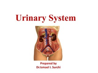
4. The kidneys remove waste products of
metabolism, excess water and salts from blood
and maintain the pH .
Ureters convey urine from the kidneys to the
urinary bladder.
The urinary bladder is the muscular reservoir of
urine.
Urethra is the channel to the exterior.
5. THE URETERS
• Definition
• The Ureters are a pair of
narrow , thick walled muscular
tubes which convey urine from
the kidneys to urinary bladder.
• Each Ureters is about 25cm (10
inch)long
• The upper half lies in the
abdomen and the lower half in
the pelvis.
• The urine is propelled from the
kidney to the urinary bladder
by the peristaltic contractions
of the smooth muscle of the
wall of the ureter.
6. Dimensions
• It measures 3mm diameter, but it slightly constricted
at three places.
– At the pelviureteric junction
– At the brim of lesser pelvis
– At its passage through the bladder wall
• For the purpose of description, ureter is divided into 2
parts
– From the site of origin to pelvic brim- abdominal part
– From pelvic brim to entry into urinary bladder- pelvic
part
7. COURSE IN ABDOMINAL
PART
• The ureter begins as a
downward continuation of a
funnel shaped renal pelvis at
the medial margin of the lower
end of the kidney.
• The ureter passes downward and
slight medially on the psoas
major, which separates it from
the transverse processes of the
lumbar vertebrae.
• Enters the pelvic cavity by
crossing in front of the
bifurcation of the common iliac
artery at the pelvic brim in front
of the sacroiliac joint.
8. COURSE IN PELVIS
• In the pelvis, the ureter first
runs downward, backward,
and laterally along the anterior
margin of the greatersciatic
notch.
• Opposite to the ischial
spine, it turns forward and
medially to reach the base of
the urinary bladder.
• Where it enters the bladder
wall obliquely.
11. Relations of RightUreter
Anteriorly:
• Theduodenum, the
terminal part ofthe ileum,
the right colic andileocolic
vessels,the right testicular
or ovarian vessels,andthe
root of the mesentery of
the smallintestine.
Posteriorly:
• Theright psoasmuscle,
which separates it fromthe
lumbar transverse
processes, and the
bifurcation of the right
common iliac artery
12. Relations of LeftUreter
Anteriorly:
• Thesigmoid colon and
sigmoid mesocolon, the left
colic vessels,and the left
testicular or ovarianvessels.
• The inferior mesenteric vein
lies along the medial side of
the leftureter
Posteriorly:
• Theleft psoasmuscle,
which separates it fromthe
lumbar transverse
processes, and the
bifurcation of the left
common iliac artery.
13. Blood supply ofUreter
• Upperendreceives blood supply from the renal
arteries,
• Themiddlepart may receive branches from the
abdominal aorta, the testicular or ovarian
arteries, and the common iliacarteries,
• In the pelviccavity, the ureters are supplied by
one or more arteries from branches of the
internal iliac arteries and inferior vesicalarteries.
14. Blood Supply
• Ureter is supplied by
branches of
» Renal artery
» Gonadal artery
» Abdominal aorta
» Common iliac artery
» Internal iliac artery
» Uterine artery (F)
» Middle rectal artery
» Vaginal artery (F)
» Inferior vesical artery(M)
» Superior vesical artery(M)
15. LymphaticDrainageof TheUreters
Lymphatic drainage of the
ureters follows apattern
similar to that of the arterial
supply:
• Theupper part of eachureter
drains to the lumbarnodes;
• Themiddle part of eachureter
drains to lymph nodes
associated with the common
iliac vessels;
• Theinferior part of each
ureter drains to lymphnodes
associated with the external
and internal iliacvessels.
18. Urinary Bladder
• The urinary
bladder is a hollow
, muscular organ ,
which functions as
the reservoir for
the urine received
from the kidneys
and to discharge it
out periodically
19. Urinary Bladder
• The bladder is the most
anterior element of the pelvic
viscera.
• It is entirely situated in the
pelviccavitywhen empty,it
expands superiorly into the
abdomen whenfull.
• Theempty bladderis shaped
likea three- sidedpyramid.
• It has anapex,a base,a
superior surface,and two
inferolateral surfaces.
20. UrinaryBladder
• Theapexof the bladderis
directedtoward the top of
the pubic symphysis; a
structure known as the
median umbilical ligament
(aremnant of the
embryologic urachus that
contributes to the
formation of the bladder)
continues from it
superiorly up theanterior
abdominal wall to the
umbilicus.
21. UrinaryBladder
• Thebaseof the bladder is
shaped likean inverted
triangle and faces
posteroinferiorly.The two
ureters enter the bladderat
each of the upper corners
of the base, andthe urethra
drains inferiorly from the
lower corner of thebase.
• Thesmooth triangular
area betweenthe
openings of theureters
and urethraon theinside
ofthebladder is known
as thetrigone
22. UrinaryBladder
• Theinferolateral surfaces
of the bladder are cradled
between the levator ani
muscles of the pelvic
diaphragm and the
adjacent obturator
internus muscles above
the attachmentof the
pelvicdiaphragm.
• The superior surface is
slightly domed when
the bladder isempty.
• It balloons upward asthe
bladderfills.
23. Neckof bladder
• The neck of thebladder
surrounds the origin of
the urethra at thepoint
where the two
inferolateral surfaces
and the baseintersect.
• The neck is themost
inferior part of the
bladder and alsothe
most 'fixed' part.
30. Capacity of Bladder
• Empty bladder , in the adult situated within the pelvis . When
distended , it rises up to the abdominal cavity and becomes an
abdomino- pelvic organ.
• Capacity in an adult male 120 to 320 ml.
• Filling beyond 220 ml causes micturition, emptied when filled
to about 250 to 300 ml.
• Filling up to 500 ml may be tolerated, but beyond this it
becomes painful.
• Referred pain: lower part of the anterior abdominal wall,
perineum and penis(T11- L2,S2-S4).
31. Blood Supply of UrinaryBladder
• Arterial supply : Main
arterial supply - Superior and
inferior vesical arteries arise
from the anterior trunk of
internal iliac arteries.
• Additional arterial supply -
Obturator and inferior gluteal
arteries.
• In the female uterine and
vaginal arteries. The inferior
vesical artery in female is
called vaginal artery.
32. • Venous drainage
• Drained by vesical venous plexus lying on
the inferolateral surfac, veins from this
plexus drains into internal iliac veins.
• Lymphatic drainage
Mainly drain into external iliac lymph
nodes, some to internal iliac lymph nodes.
33. 39
•
•
•
The urethra is a canal extending from the neck of the bladder to
the exterior , at the external urethral orifice.
Male: about 20 cm (8”) long
Female: 3-4 cm (1.5”) long
– Short length is why females have more urinary tract infections than males -
ascending bacteria from stool contamination
Urethra
urethra
Urethra
34. Female Urethra
• 3 to 4 cm long
• External urethral orifice
– between vaginal orifice and
clitoris
• Internal urethral sphincter
– detrusor muscle thickened,
smooth muscle, involuntary
control
• External urethral sphincter
– skeletal muscle, voluntary
control
35. Urethra
Infemale
• It travelsa slightlycurved course
as itpassesinferiorly through the
pelvicfloorinto the perineum
• where it passes through the deep
perinealpouch andperineal
membrane.
• beforeopening in the vestibule
thatlies between the labiaminora.
36. In male
• 18 cm long
• Internal urethral sphincter
• External urethral sphincter
• 3 regions
– prostatic urethra
• during orgasm receives semen
– membranous urethra
• passes through pelvic cavity
– penile urethra
37. • Beginningatthe base of the bladder
and passing inferiorly through the
prostate, itpasses through the deep
perineal pouch and perineal
membrane and immediately enters the
root of thepenis.
• The urethra exits the deepperineal
pouch, it bends forward to course
anteriorlyin the root of the penis.
• Whenthe penis is flaccid,the urethra
makes another bend, this time
inferiorly,when passing from theroot
to the body of the penis. During
erection,the bend between the root and
body of the penisdisappears.
38. Partsof theUrethra
inmale
Preprostaticpart
• Thepreprostaticpart ofthe urethra is about 1cmlong.
• Itextendsfromthebaseofthe bladderto the prostate,and is
associated with a circularcuff of smooth muscle fiber (internal
urethral sphincter). Contraction of this sphincter prevents retrograde
movement of semen into the bladder duringejaculation.
Prostaticpart
• Theprostaticpartof the urethrais3-4cmlongandis surrounded by
theprostate. In this region,the lumen of the urethrais markedby a
longitudinalmidlinefoldof mucosa (urethralcrest).
39. Spongy (Penile)urethra
• Thespongy urethra is surrounded by erectiletissue (thecorpus
spongiosum)of thepenis.
• Itisenlargedtoformabulbat the base of the penis and again atthe end of
the penis to form the navicular fossaTheexternalurethralorificeis the
sagittalslitatthe end of thepenis.
41. Innervationsof UrinaryBladder
• The pelvic plexus is supplying the urinary bladder with
autonomic nerves.
• The sympathetic innervation is directed to the blood
vessels, urethral openings, and the trigone. The last
thoracic and L1,2 nerves create the necessary
innervation to thebladder
.
• Parasympathetic innervation is derived from S2,3 and 4
nerves.These are aimed at serving the detrusor muscle.
• The pelvic spinal nerves are responsible for responding to
the sensory response of a full bladder
, which responds to
the impulses sent via the central nervous system.
42.
43. Nerve supply
• Its contains both sympathetic and
parasympathetic components.
• Parasympathetic efferent fibers
• S2,S3, S4 are motor to the detrusor
muscle and inhibitory to the
sphincter vesicae.
• If these are destroyed, normal
micturition is not possible.
44. Nerve supply
• Sympathetic efferent fibers (T11 to L2):
• - inhibitory to the detrusor
• - motor to the sphincter vesicae
• The pudendal nerve (S2, S3, S4)
• -supplies the sphincter urethrae which is voluntary
• Sensory nerves:
- pain sensations, causes:
- spasm of bladder wall
- carried by parasympathetic nerves and partly by
sympathetic nerves
46. • Micturition is also known as the voiding phase of bladder control
and it is typically a short-lasting event. itsmodulatedbyhighercenters
frombirth,butitisnotuntilbetweentheageof1and2yearsthatachildisable
toshowbladderawarenessand,asafirststep,toreporthavingvoidedorbeing
intheprocessofvoiding.
Highercentersformicturition
1) Inhibitory centers: midbrain-cerebralcortex
2) Facilitatory centers : Pons - cerebralcortex
Micturition
47.
48. MICTURITION REFLEX
.
Filling of urinarybladder
Stimulation of stretchreceptor
Afferent impulses pass via pelvicnerve
Efferent impulses via pelvicnerve
Contraction of detrusormuscle & relaxationof internal sphincter
Sacral segments of spinalcord
49. MICTURITION REFLEXCONTI…
Flowof urineinto urethra and stimulation of stretchreceptors
Afferent impulses via pelvicnerve
Inhibition of pudendalnerve
Relaxation of externalsphincter
Voiding ofurine